1. Introduction
Cancer is one of the most debilitating human diseases and the second cause of death worldwide. Current gene therapy represents a promising alternative for the treatment of cancer. One of the most challenging issues in cancer gene therapy is the shortage of safe and efficient vectors that can specifically deliver therapeutic DNA to the target site with minimum toxicity [
1]. Gene delivery vectors can be classified as viral and non-viral kinds. In recent years, non-viral gene vector have been developed rapidly for their non-immunogenicity, easiness of large-scale manufacturing and tailor-made features for specific therapeutic application [
2]. Cationic polymers have been frequently studied because they can be efficiently complexed with negatively charged DNA, thereby increasing DNA stability [
3]. Among the polycations, polyethylenimine (PEI) as a commercially available material has become one of the most promising and widely studied gene carriers, largely due to its proton-sponge mechanism that can ensure DNA escape from the endocytic pathway efficiently [
4,
5]. Studies have demonstrated that the gene transfection efficiency and cytotoxicity of PEI are highly related to their chain length and topology. High-molecular-weight (HMW) PEI has high gene transfection efficiency but exhibits pronounced cytotoxicity and can induce membrane damage in the initial stages of treatment [
6]. Low-molecular-weight (LMW) PEI exhibits low cytotoxicity, but with low transfection efficiency. To achieve high transfection efficiency as well as low cytotoxicity simultaneously, PEI could be modified by cross linking and like strategies [
7,
8].
Pluronic block copolymers, which are amphiphilic molecules used as structural elements of the polycation gene-delivery systems, consist of hydrophilic ethylene oxide (EO) and hydrophobic propylene oxide (PO) blocks arranged in a basic A-B-A structure (EO
x-PO
y-EO
x) [
9]. Hydrophobic poly-PO chains are reported to offer good pluronic interactions, DNA transport and transgene expression. In contrast, hydrophilic EO chains have the ability to prevent binding of other polymers with the membranes. The systems are stabilized in dispersion by the EO corona in a manner similar to regular pluronic micelles. Pluronic P123 with formula EO
20-PO
70-EO
20, which has a proper hydrophile lipophilic balance (HLB) value, can be degraded to decrease cytotoxicity after cell uptake. Studies [
10] showed that although the PEI derivate P123-PEI synthesized by cross-linking LMW-PEI with P123 had good degradability and low cytotoxicity, the transfection efficiency of the polymeric gene vector is not high enough.
The process of DNA complexes delivery should be able to target specific cells, internalize complexes and be taken up into the nucleus. The αvβ3 receptor is known to be highly expressed on tumor cells and tumor angiogenic blood vessels, but rarely detectable on quiescent blood vessels. The highly selective expression of αvβ3 integrin in the neovascular tissue and various tumors allows for its use as a suitable target. Knowing that arginine-glycine-aspartate (RGD) peptide is a key binding moiety that has been shown to bind specifically to αvβ3 integrin receptors [
11], we used a linear RGD peptide as a new peptide named R4 in the present study.
The nuclear localization signal (NLS, with the sequence Lys-Lys-Lys-Arg-Lys) is a necessary signal sequence that mediates proteins entering cell nuclei through the nuclear pore complex [
12]. NLS has been suggested as a means to promote nuclear delivery of expression constructs [
13]. Therefore, we used RGD peptide conjunction with NLS to yield a new chimeric peptide RGD-NLS (named R11), and peptide R4 and bifunctional peptide R11 to modify PEI derivates, respectively, in order to improve cell selection, promote cargo transport and enhance transfection efficiency.
In this study, a polymer matrix was synthesized by cross-linking LMW PEI with P123. The bifunctional peptide R11 was conjugated to P123-PEI to form a new polymeric polycation (P123-PEI-R11). Another polycation P123-PEI-R4 was prepared in the same way. We investigated the physicochemical characteristics and efficiency of these polymeric polycations
in vitro and compared them with another polycation modified by R18 (short for RGD-TAT-NLS, with the sequence RGD-RKKRRQRRR-KKKRK, which was ever reported) [
14]. The purpose of the study was to find an ideal tumor target peptide that could reduce cytotoxicity of polymer matrix P123-PEI, improve its tumor targeting, increase cellular uptake of genes and enhance the therapeutic effect of gene therapy.
3. Materials and Methods
3.1. Reagents and Plasmid DNA
PEI 2 kDa, N-succinimidyl-4-(N-maleimido-methyl) cyclohexane-1-carboxylate (SMCC) and 3-(4,5-dimethylthiazol-2-yl)-2,5-diphenyl tetrazolium bromide (MTT) were purchased from Sigma-Aldrich (St Louis, MI, USA). P123 was provided by the Second Military Medical University (Shanghai, China). RPMI 1640 medium and fetal bovine serum (FBS) were purchased from Invitrogen (Carlsbad, CA, USA). The peptide Arg-Gly-Asp (R4, MW449.48) and Arg-Gly-Asp-Lys-Lys-Lys-Arg-Lys (R11, MW1314.62) were synthesized by GL Biochem (Shanghai, China). The plasmids of Escherichia coli DH5α were amplified by the Tiangen End-Free Mega Plasmid Kit (Hilden, Germany). Benzene, triphosgene, dichloromethane, N-hydroxysuccinimide, triethylamine, ethyl acetate, and absolute ethyl alcohol were bought from Sinopharm Chemical Reagent Co., Ltd (Shanghai, China).
3.2. Synthesis and Characterization of Polymers
3.2.1. Activation of P123
Before initiation of the experiments, toluene and dichloromethane were dehydrated by co-evaporation. First, the P123 (0.0680 g, 0.0118 mmol) was dried by co-evaporation with toluene under vacuum at 40 °C twice, dissolved in toluene/dichloromethane mixture (3:1, 40 mL), and treated with the double mole of triphosgene (0.0235 mmol) for overnight. Next, the same mole of N-hydroxysuccinimide as triphosgene and plenty of triethylamine was added dropwise into benzene/dichloromethane (2:1, 30 mL) after evaporating the primary solvent. After 4-h stirring, the solution was filtered and evaporated to dryness. Finally, 50 mL ethyl acetate was added and centrifuged at 8000 rpm for 15 min. The supernatant was evaporated and the residue was collected.
3.2.2. Synthesis of P123-PEI
Activated P123 (0.01 mmol) was dissolved into 10 mL of anhydrous ethanol as Solution A, and 10-fold mole dehydrated PEI 2 kDa (0.10 mmol) was dissolved in 20 mL anhydrous dichloromethane as Solution B. The molar ratio of PEI with P123 was 10:1. These two reagents were added into 10 mL dichloromethane simultaneously and stirred constantly overnight at room temperature. Then the solution was dialyzed against distilled water at 4 °C for 2 days and lyophilized.
3.2.3. Linking R4 and R11 with P123-PEI
Polymer P123-PEI was conjugated with peptides using SMCC as crosslinker [
15]. The SMCC solution (using dimethyl sulfoxide as solvent to 3.33 mg/mL) was added dropwise to P123-PEI solution (using 0.1 M PBS as solvent to 10 mg/mL) at a molar ratio of 2:1 with half an hour stirring at room temperature with gentle shaking. The excessive non-conjugated SMCC was removed by gel chromatography (Sephadex G-25, Pharmacia, Milton Keynes, UK). Then, peptide R4 (using 0.1 M PBS as solvent to10 mg/mL) was mixed into pretreated P123-PEI at a molar ratio of 2:1 with stirring at 4 °C overnight in the dark. The solution was lyophilized after ultrafiltration. The cationic polymer was named P123-PEI-R4. Polymer P123-PEI-R11 was prepared in the same way.
1H-NMR spectral analysis was carried out at room temperature after P123-PEI-R4 and P123-PEI-R11 were dissolved in deuterium oxide.
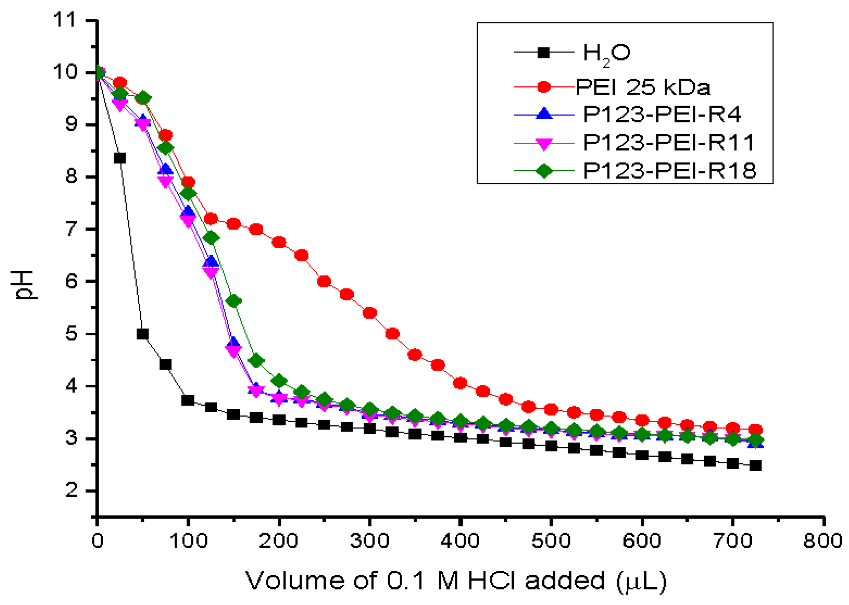
3.3. Buffering Capacity of the P123-PEI-R4 and P123-PEI-R11 Polymers
The synthesized polymer solution P123-PEI-R4 and P123-PEI-R11 were prepared in 50 mL flasks (0.2 mg/mL, 30 mL), respectively, using pure water as control. After adjusting the initial pH to 10.0 with 0.1 M NaOH, 25µL increment of 0.1 M HCl were added, and the pH of the solution was measured with a pH meter after each addition.
3.4. P123-PEI-R4/DNA and P123-PEI-R11/DNA Complexe Formation
Different charge ratios (w/w) of P123-PEI-R4/DNA complex were prepared as the ratio of the weight of P123-PEI-R4 and DNA. The ratio of P123-PEI-R11/DNA complex was expressed in the same way. An appropriate polymer solution (PBS 0.1 mol/L, pH 7.4) was added into the aqueous solution with a fixed amount of plasmid DNA (50 ng/µL) with the equal volumes. Then the complexes were incubated at room temperature for 30 min.
3.5. Particle Size and Zeta Potential Measurement and Morphologic Observation
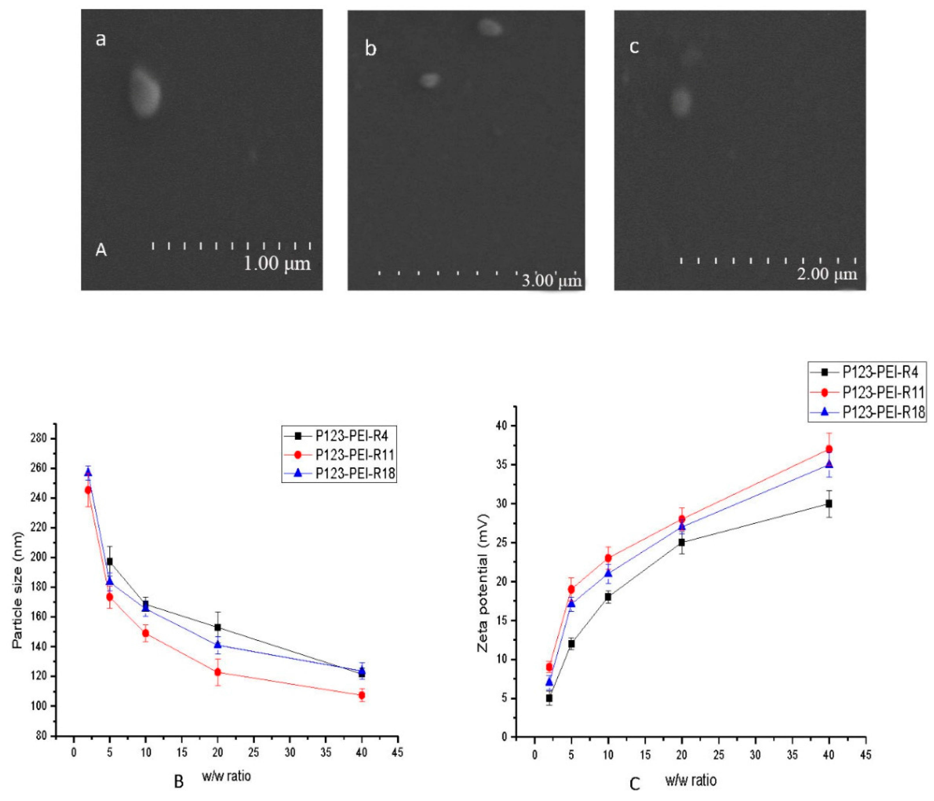
Particle size and zeta potential of the polymer/DNA complexes were measured using an electrophoretic light-scattering spectrophotometer (Zetasizer Nano ZS90, MAN0317 Issue 5.0, Malvern Instruments Ltd., Malvern, UK), with a 90° scattering angle in PBS buffer at room temperature. The complexes were prepared at desired w/w ratios ranging from 0 to 40, and then incubated at 37 °C for 30 min, and measured for size and zeta potential. All the experiments were performed in triplicate in order to ensure the accuracy of the results.
Morphology of the polyplexes was observed by transmission electron microscopy (JEM 2100F, JEOL Ltd., Tokyo, Japan). The complexes P123-PEI-R4/DNA and P123-PEI-R11/DNA were prepared according to 3.4 at the w/w radio of 5 and 2, respectively. A drop of the DNA/polymer polyplexes suspension was placed onto a copper grid. The excrescent solution was wiped off with lens wi** paper and the grid was dried at room temperature for several minutes before observation.
3.6. Agarose Gel Retardation Assay (Binging Capacity of Polymers/DNA Complexes)
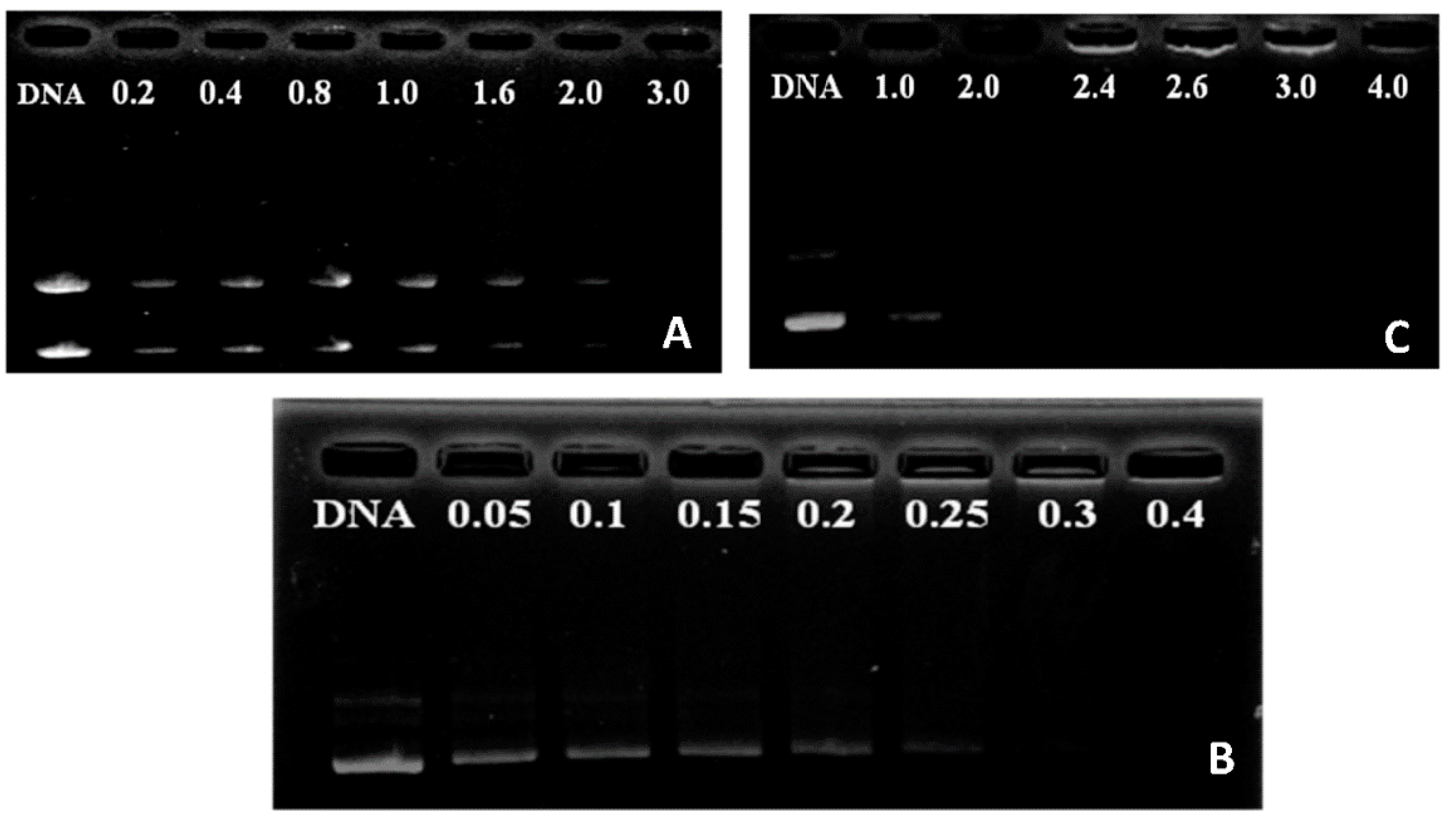
Agarose gel electrophoresis was performed to investigate DNA binding ability of P123-PEI-R4 and P123-PEI-R11. Polymer/DNA complexes at different weight ratios ranging from 0.05 to 3.0 were prepared. These obtained complex solutions were analyzed by electrophoresis with the condition of 120 V and 30 min on a 1% (w/v) agarose gel in loading buffer. Then these gels were stained with 0.5 g/mL ethidium bromide for 15 min and illuminated by a UV illuminator to show the location of DNA.
3.7. Resistance to DNaseI Digestion and Serum
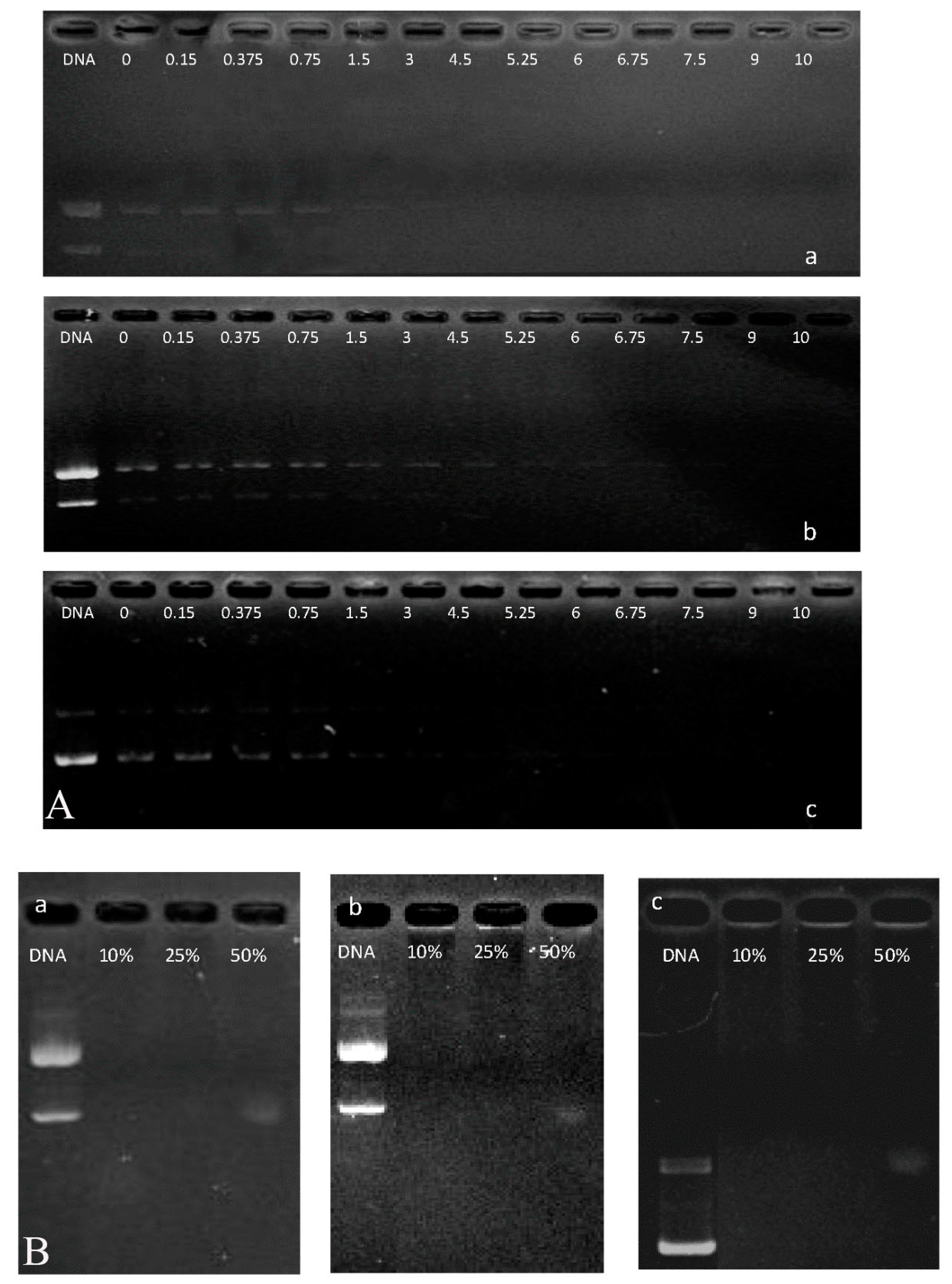
Stability of the polymer/DNA complexes was evaluated by testing the ability to protect plasmid DNA (pDNA) against DNaseI degradation. Generally, the designed amount of DNaseI solution were added to 10 µL complex solution (250 ng of pDNA) in 0.5 mL Eppendorf tubes, with the range of DNaseI doses per pDNA weight unit maintained between 0 and 10 U DNaseI/µg DNA, and incubated at 37 °C for 30 min. Then, 2 µL of 250 mM EDTA solution was added to each tube and incubated at room temperature for 10 min to inactivate DNaseI. Next, 10 µL 2 mg/mL sodium heparin was added to each tube and incubated at room temperature for 2 h to dissociate the complex completely. Stability of the complex to DNaseI digestion was further analyzed by electrophoresis.
In the experiments with serum, the complex solution (10 µL containing 10%, 25% and 50% serum) was obtained by adding different concentrations of FBS and incubating at 37 °C for 60 min. Then, electrophoresis was performed to determine the sensitivity of complex P123-PEI-R4 and P123-PEI-R11 to serum. In addition, the resistance of complex P123-PEI-R18 to DNaseI digestion and serum was used to compare with the sensitivity of complex P123-PEI-R4 and P123-PEI-R11.
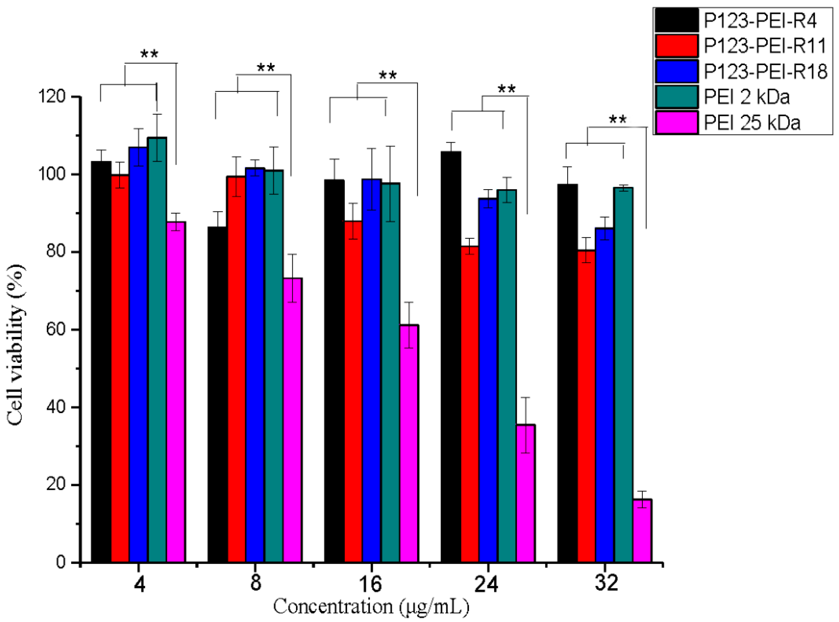
3.8. Cytotoxicity Assay
The cytotoxicity of P123-PEI-R4 and P123-PEI-R11 polymer was measured by MTT assay. B16 cells were distributed to each well of a 96-well plate for 48 h. Previous medium was abandoned. 200 µL serum-free media with different concentrations of the polymer (0, 4, 8, 16, 24 and 32 µg/mL) were added to per cell well, respectively. After incubating the plate for 4 h with 5% CO
2 at 37 °C, fresh growth medium was replaced and kept for 72 h. Thereafter, the wells were replaced with 20 µL 5 mg/mL sterilized MTT solution and 180 µL of fresh growth medium, and kept at 37 °C for 4 h. Subsequently, the MTT/growth medium was replaced by 150 µL DMSO and kept for 10 min with gentle vortexing at room temperature. The absorbance value at 570 nm was read by an ELISA plate reader with background subtraction. Cell viability was calculated using the following equation:
3.9. In Vitro Gene Transfection
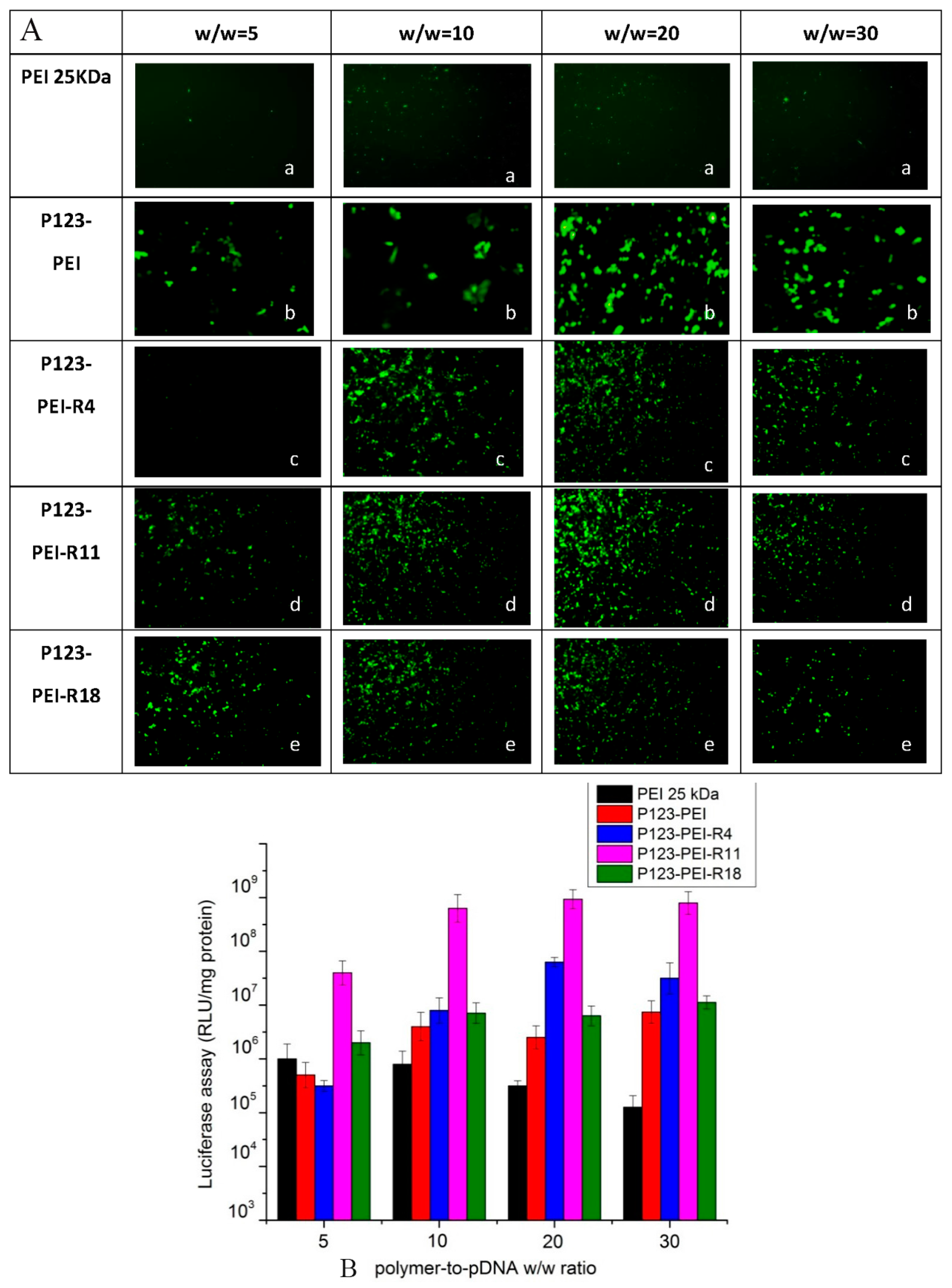
The transfection efficiency of P123-PEI-R4/DNA, P123-PEI-R11/DNA and P123-PEI-R18/DNA complexes in B16 cells was examined using the plasmid pEGFP-N2 and pGL3-Control. Cells were seeded in a 24-well plate at a density of 105 cells per well in RPMI 1640 medium containing 10% FBS and incubated for 24 h until 80% confluence. Then, 100 µL polymer/DNA complex solution containing 2.5 µg DNA at various weight ratios (2–30) were added to the 24-well plate and incubated for 4 h at 37 °C in a 5% CO2 atmosphere. After that, the medium was replaced with 500 µL medium containing 10% FBS and then incubated for additional 48 h. The pEGFP-N2 expression ware was observed with an inverted fluorescent microscope (AE-31, Motic Corporation, Wetzlar, Germany).
The luciferase assay was conducted according to the manufacturer’s specifications. The medium was replaced with 100 µL cell culture lysis reagent (CCLR) and shaken for 30 min. After mixing with substrate, luciferase activity was examined with a luminometer (Turner Designs Luminometer Model TD-20/20, Promega Corp., Madison, AL, USA) as soon as possible. A bicinchoninic acid (BCA) protein assay was used to measure protein contents. Transfection efficiency for the pGL3-Control was calculated by the relative light units (RLUs) against the corresponding protein contents.
4. Conclusions
The new polyplexes P123-PEI-R4 and P123-PEI-R11 were successfully developed by cross-linking LMW PEI with P123 and then further coupled with peptide R4 and R11, respectively. Compared with branched PEI-25 kDa, the new polyplexes both showed suitable buffer capacity. In addition, these polyplexes could efficiently condense DNA into stable nanoparticles with proper sizes and zeta-potentials. Our gel retardation experiment confirmed PEI-P123-R4 had the ability to form complexes with DNA and was able to condense DNA effectively at a w/w ratio of 3.0. More importantly, P123-PEI-R11 was able to efficaciously condense DNA and neutralized its charge at a w/w ratio of 0.4, indicating that P123-PEI-R11 has a higher DNA binding ability. Furthermore, these polymers can effectively protect plasmid DNA from being degraded by DNaseI and have distinctly serum tolerance. Moreover, the new polyplex P123-PEI-R11 showed much lower cytotoxicity compared with P123-PEI-R4 and P123-PEI-R18 in B16 cell lines, it also exhibited the highest gene transfection efficiency compared with the other polyplexes (P123-PEI, P123-PEI-R4 and P123-PEI-R18) at any ratio. These results suggest that this new polymer (P123-PEI-R11) could be used as a potential safe and efficient non-viral carrier for future cancer gene therapy due to low cytotoxicity and high transfection.


