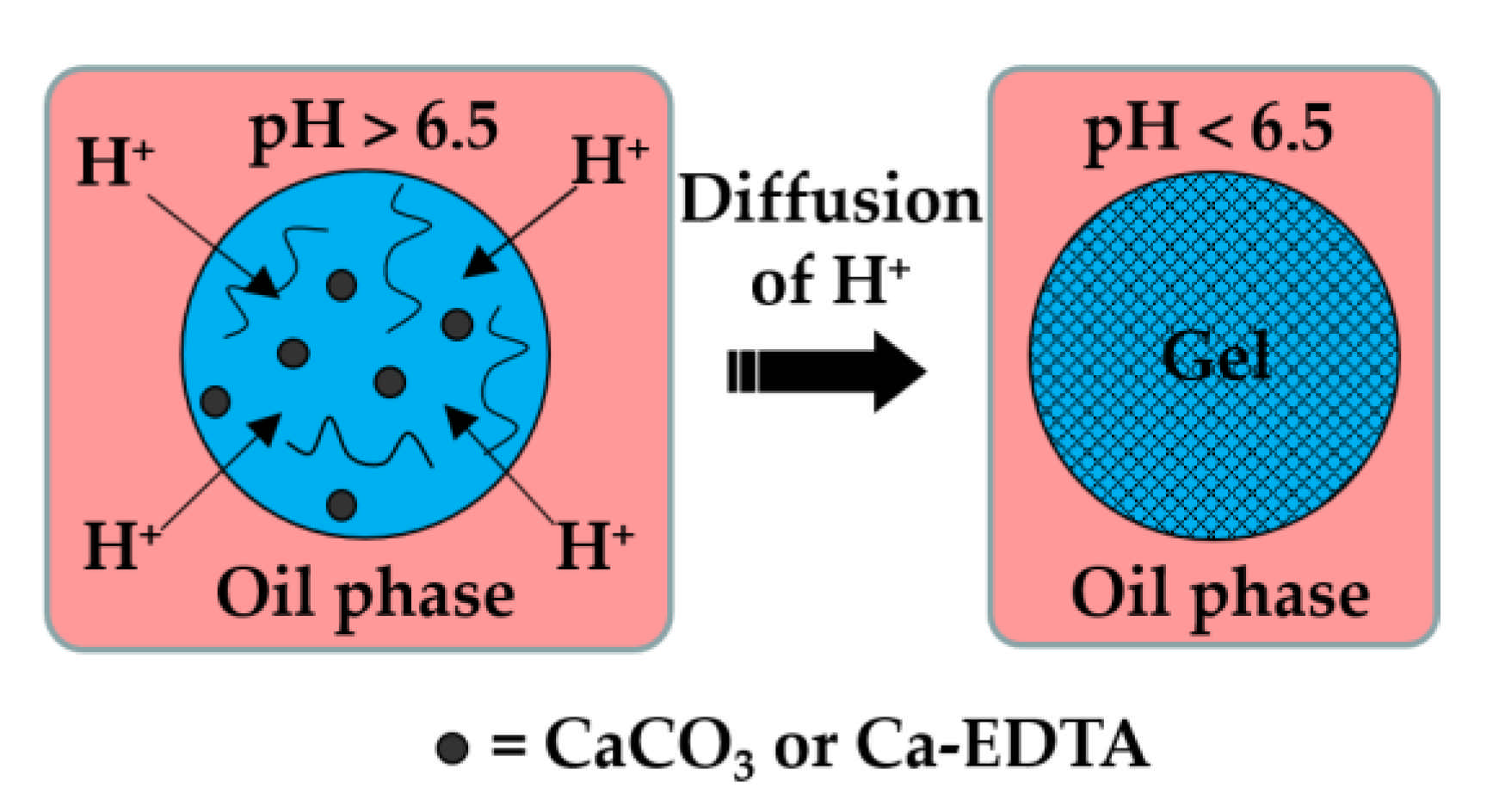
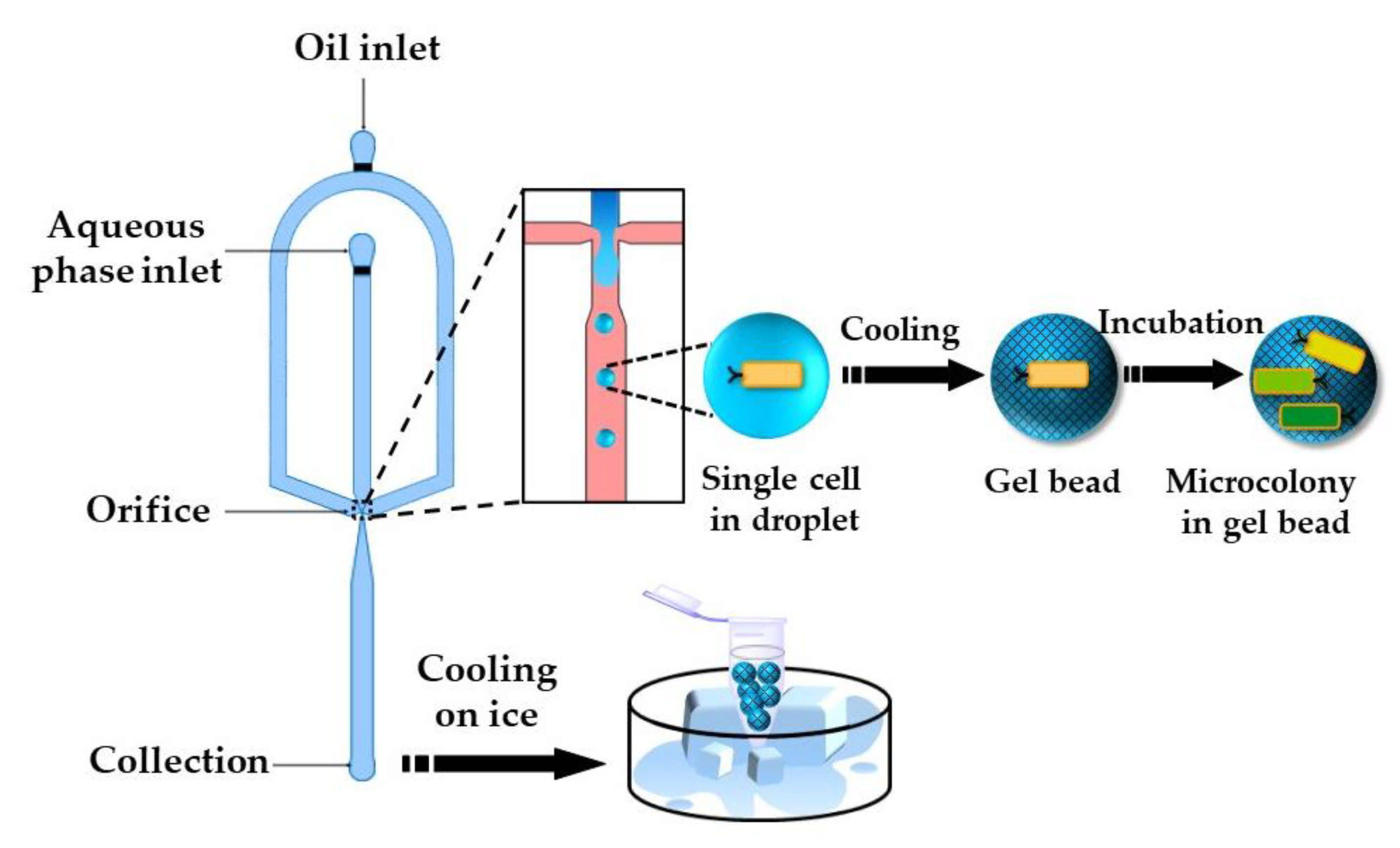
2.1.1. Internal (In-Situ) Gelation
Here, the dispersed phase is an aqueous solution of charged polymer containing an undissociated calcium or barium compound (shown as solid black circles in
Figure 1), such as CaCO
3, BaCO
3 [
27], tricalcium citrate [
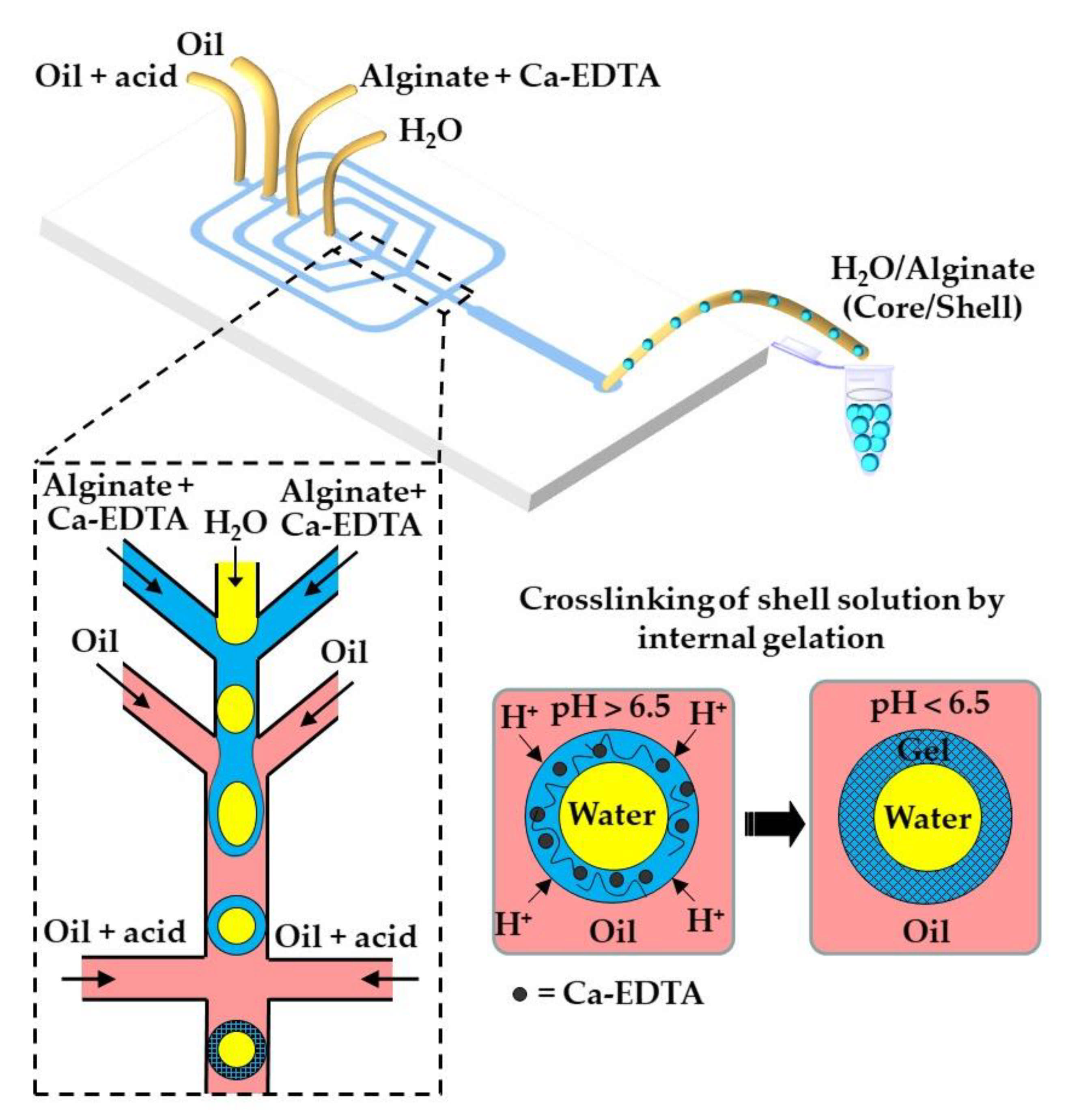
28], and Ca-EDTA [
29].
The continuous phase is a mixture of hydrophobic surfactant and organic acid, usually acetic acid (CH
3COOH), dissolved in an inert oil. The typical inert oils used in this process are vegetable oils [
30,
31,
32,
33], dimethyl carbonate [
34], fluorocarbon oils [
35], and hexadecane [
36]. After droplet generation, the organic acid diffuses through the oil phase and dissociates at the droplet surface into acetate anions (CH
3COO
−) and protons (H
+). H
+ reacts with undissociated Ca or Ba compounds inside the droplets and releases Ca
2+ or Ba
2+ ions, which crosslink the polymer. In the case of the CaCO
3/alginate/acetic acid system, the following reactions occur:
Although the method offers homogenous gelation throughout the entire droplet volume, it results in a reduction of pH below the physiological pH, which can be detrimental to cell viability [
37]. To minimize the exposure of cells to a low pH, a stoichiometric amount of acetic acid can be added off-chip after droplet collection [
38].
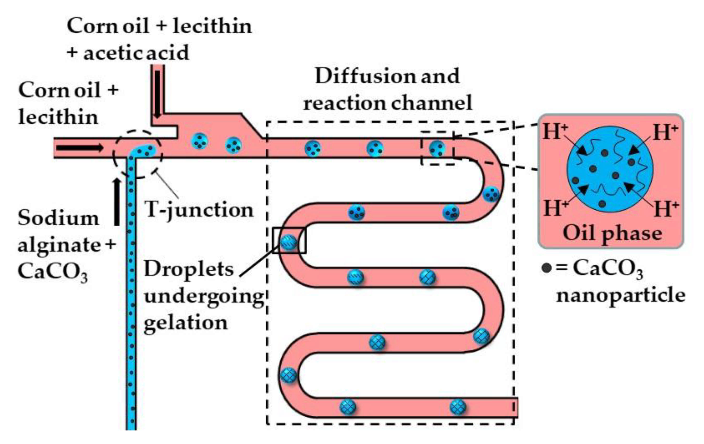
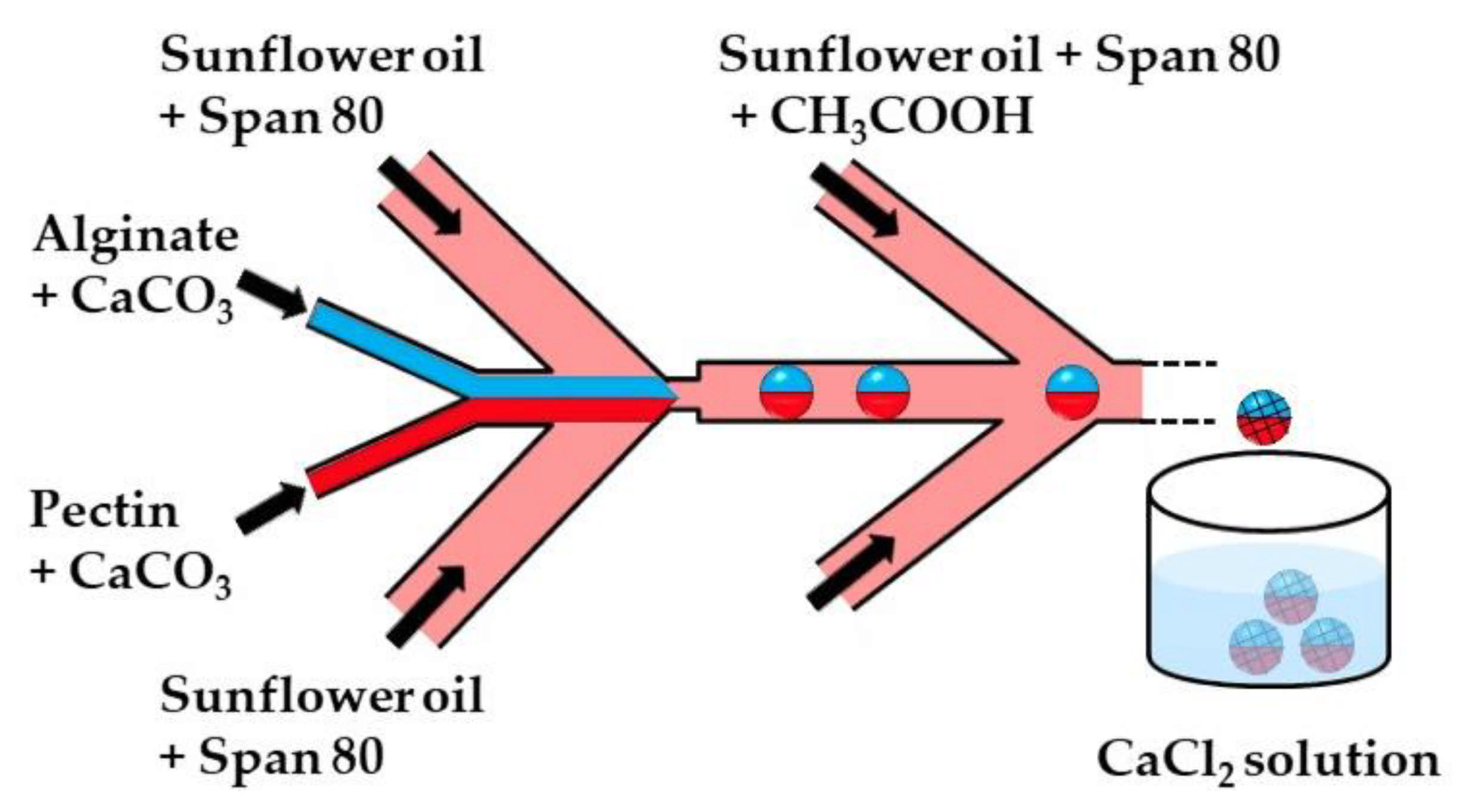
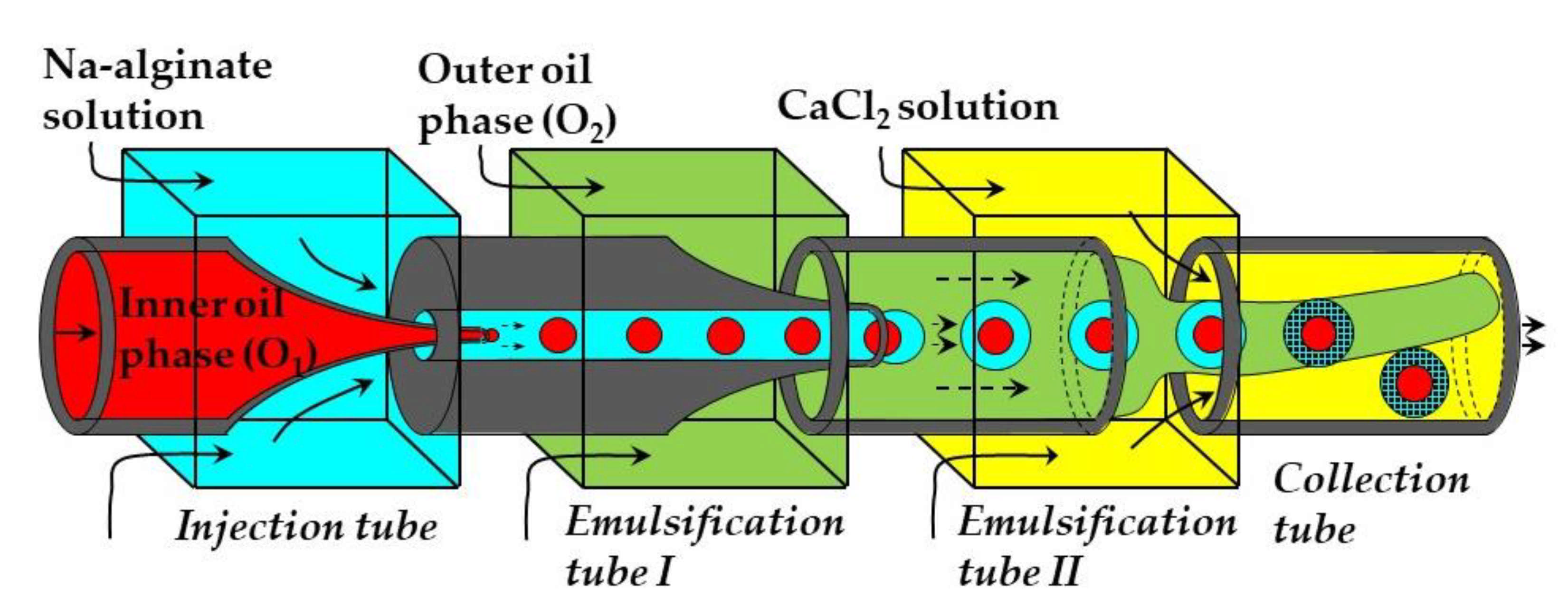
Figure 2 and
Figure 3 show the microfluidic internal gelation strategies implemented to produce homogeneous alginate beads and alginate/pectin Janus beads, respectively.
Internal gelation can also be triggered by adding a slow hydrolyzing acid or a photo-acid generator (PAG) into the polymer solution rather than supplying acid from the oil phase across the droplet boundary. Morimoto et al. [
39] added glucono-1,5-lactone (GDL), a slow-release acidifier to the dispersed phase, which hydrolyzed to gluconic acid to release Ca
2+ from CaCO
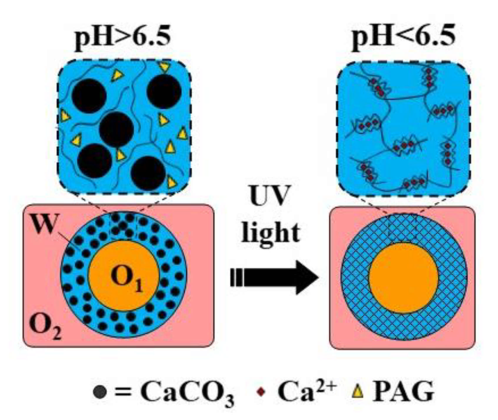
3 and to initiate alginate crosslinking without any external trigger. Photo-acid generators (PAGs) are compounds that release protons irreversibly upon illumination. Liu et al. [
40] added diphenyliodonium nitrate (DPIN) to the aqueous phase to initiate the gelation of an alginate solution from the middle phase of the O/W/O emulsion droplets upon UV irradiation. The photolysis of DPIN results in the release of protons (H
+) and hydrophobic uncharged by-products [
41]:
To prevent these by-products from precipitating within droplets and causing channel clogging, a macrocyclic compound can be added to the dispersed phase that can bind these hydrophobic species [
41]. The emulsion formulations used to produce microgels by internal gelation are summarized in
Table S1 in the supplementary material.
2.1.2. External Gelation
The external gelation in microfluidic systems can be achieved using several methods, as follows. (i) An oil-soluble crosslinking agent, such as calcium acetate, can be dissolved in the oil phase and be used for on-chip crosslinking. (ii) A crosslinking agent such as CaCl
2 can be added to the gelation bath, where the droplets are collected and crosslinked [
42]. (iii) An aqueous crosslinker solution can be emulsified in a carrier oil and this emulsion can be used for on-chip crosslinking [
43]. (iv) The same emulsion can be used as a shell liquid in core−shell droplets and can be used for on-chip crosslinking of aqueous cores [
44]. (v) An aqueous CaCl
2 solution can be emulsified in a carrier oil to produce a W/O emulsion, which can be dehydrated to form surfactant-coated CaCl
2 nanoparticles dispersed in the oil phase. This nano-dispersion can be used for on-chip crosslinking [
45,
46]. (vi) An alcoholic CaCl
2 solution can be dissolved in the oleic acid and can be used for on-chip crosslinking after alcohol evaporation [
47,
48,
49]. Finally, (vii) a powdered crosslinking medium, such as dehydrated cell culture medium, can be dispersed in a carrier oil and used for on-chip crosslinking [
50].
Table S2 provides examples of the microfluidic methods used for external gelation.
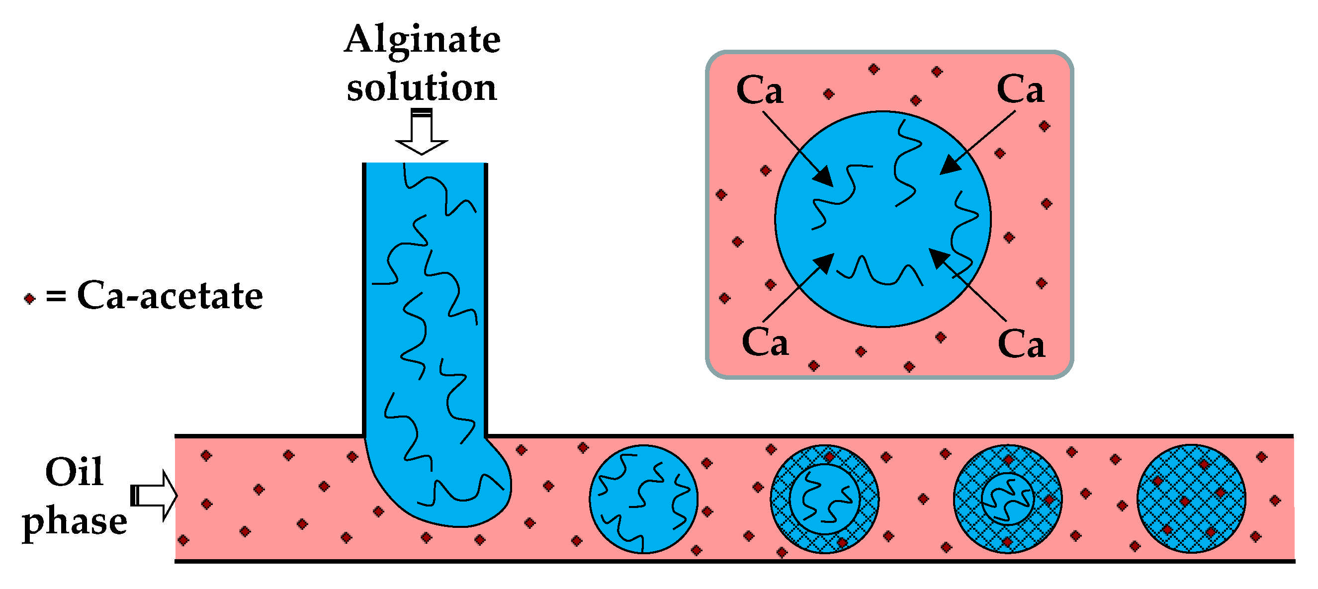
In the first method (i), the dispersed phase is usually an aqueous polyanion solution, while the continuous phase is an oil-soluble salt of divalent or multivalent cations, e.g., Ca
2+, Zn
2+, and Fe
3+, dissolved in an oil–surfactant mixture (
Figure 4). Depending of the type of oil and polymer, the oil-soluble salt can be calcium acetate [
30], Fe (NO
3)
3 [
51], CaJ
2 [
51], BaCl
2 [
52], SrCl
2 [
53], or ZnCl
2 [
54]. In the case of alginate crosslinking with calcium acetate, Ca-acetate diffuses through the oil phase to the droplet interface and dissociates in water to form Ca
2+ ions, which trigger polymer crosslinking within the droplets, according to the following reactions:
For on-chip gelation, the combined diffusion and reaction time,
, must be shorter than the residence time of the droplets in the chip, but greater than the droplet formation time, 1/
f, where
f is the frequency of droplet generation. The diffusive flux of salt across the oil/water interface depends on the salt concentration in the oil phase and its diffusivity in the oil phase. Another limiting factor is the salt solubility in the aqueous phase, which must be sufficiently high to trigger polymer crosslinking. The solubility of calcium salts of fatty acids in water decreases with increasing the number of carbon atoms in a molecule [
30]. For example, calcium butanoate and calcium 2-ethylhexanoate cannot be used for external gelation, because no gelation occurs within 7 days when the concentration of either salt in the oil phase is 0.5 wt% [
30]. The salt solubility in the oil phase could also be a limiting factor. A mixture of CaCO
3 and acetic acid can be used instead of Ca-acetate to increase its solubility in oil [
55].
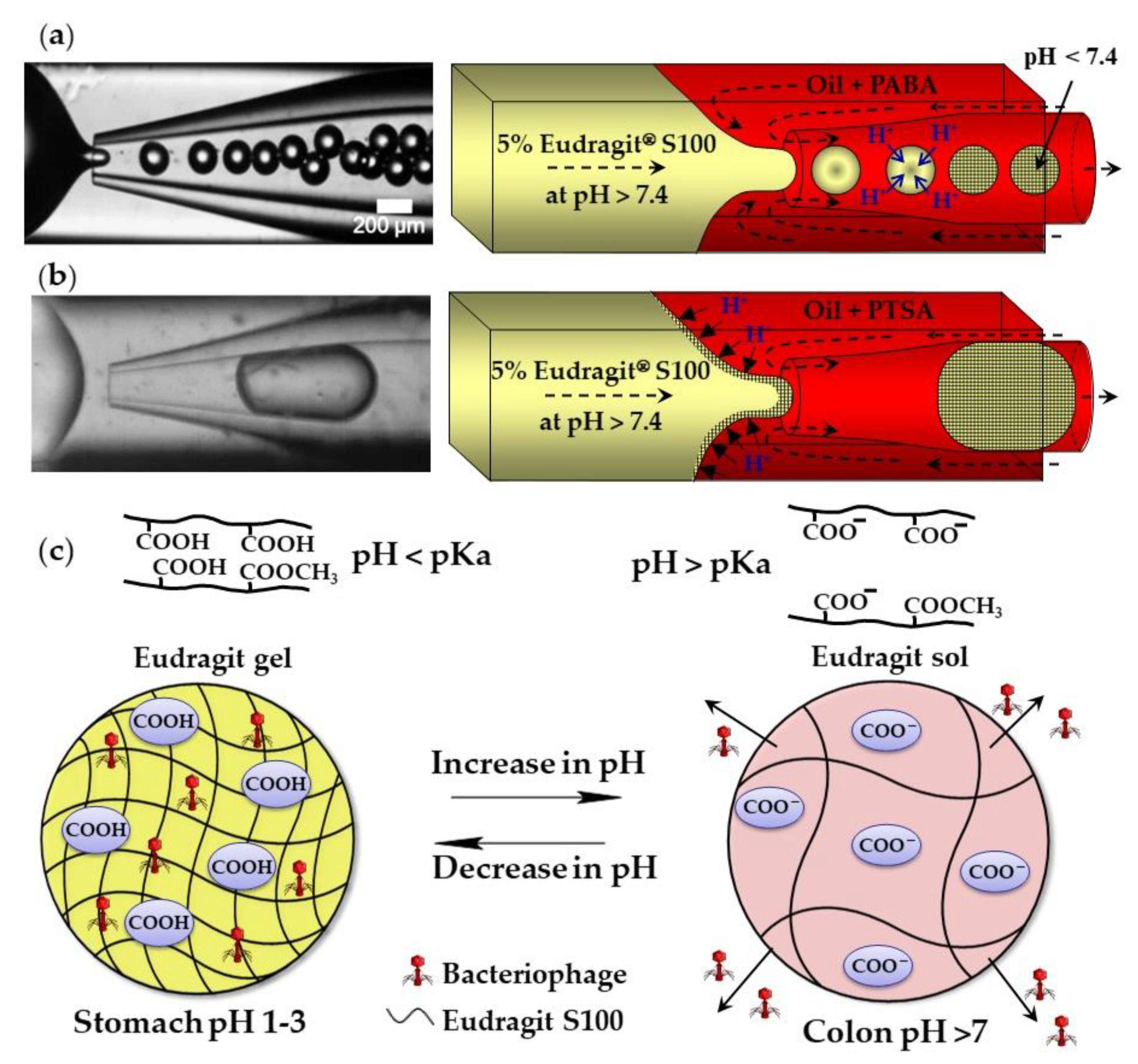
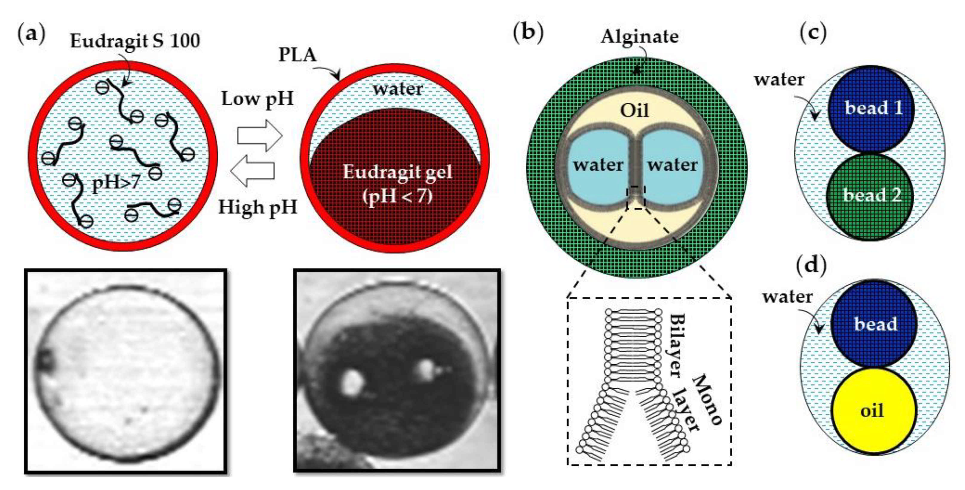
The external gelation of pH sensitive Eudragit
® polymers can be triggered by the exchange of H
+ ions between an organic acid dissolved in the carrier oil and the polymer dissolved in aqueous droplets. Above pH 7.4, Eudragit S100 is soluble in water because of the dissociation of carboxylic acid of methacrylic acid monomer units. A sol–gel transition occurs at pH < 7.4 because of charge neutralization (
Figure 5c). Monodispersed gel beads were produced when the sol–gel transition was triggered by p-aminobenzoic acid (PABA), a weak acid with a pKa of 2.38 (
Figure 5a). However, when the gelation was triggered by p-toluenesulfonic acid (PTSA), a strong acid with a pKa of −2.8, the beads were large and polydisperse, because of the premature polymer gelation at the oil/water interface (
Figure 5b) [
56]. Eudragit microgels can be loaded with
Clostridium difficile bacteriophages at a low pH and can be used for the treatment of
C. difficile infections as an alternative to conventional antibiotic therapies. The beads are stable in the acidic environment of the stomach and release the phages at the infection site in the colon (
Figure 5c). Similarly, chitosan beads can be produced by exposing droplets of acidified chitosan solution to OH
− ions [
57]. The gelation of chitosan chains occurs as a result of the deprotonation of amine groups (NH
3+) above pH 6.2–6.5.
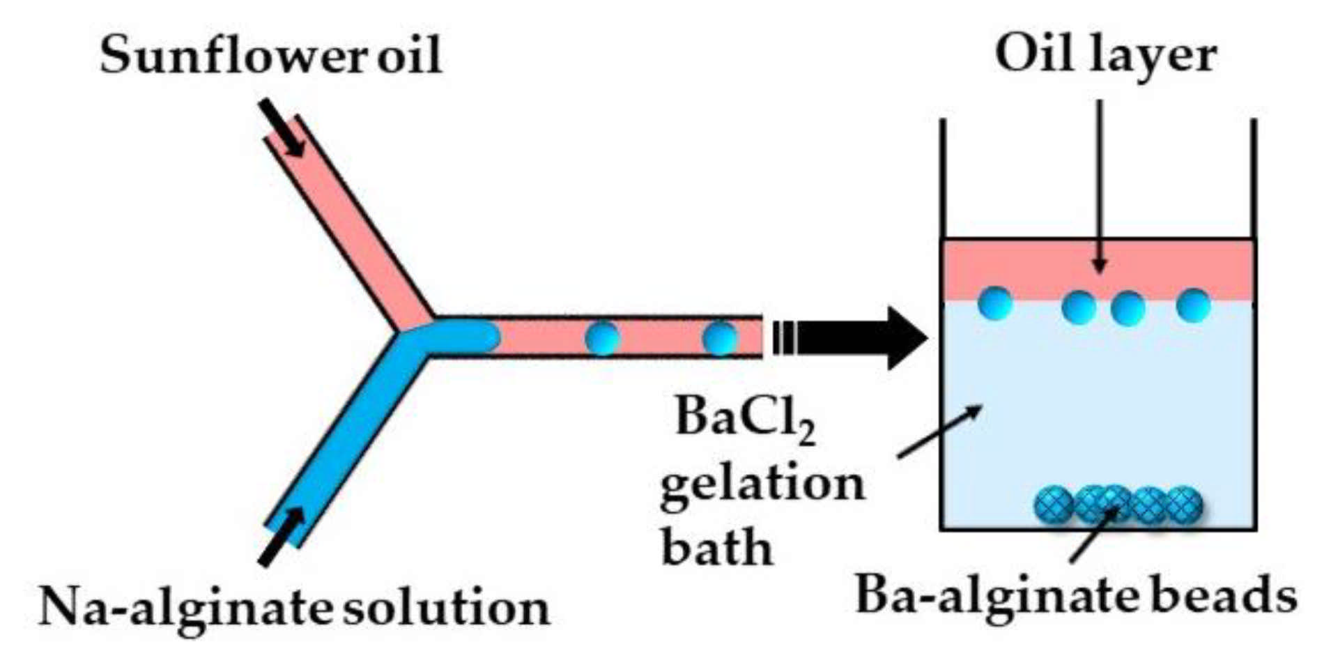
In the second method (ii), microgels are formed by off-chip polymer crosslinking in the gelation bath. Capretto et al. [
27] produced barium alginate beads in a microfluidic Y-junction by collecting the generated W/O emulsion droplets in a gelation bath containing 1.5 wt% BaCl
2 (
Figure 6). Potential issues with this technique are the accumulation of droplets at the oil/water interface as a result of the small density difference between the alginate droplets and BaCl
2 solution, and the formation of tail-shaped beads as a result of droplet deformation during settling in the oil phase. The problems can be minimized by pouring a low viscosity oil above the BaCl
2 solution in the gelation bath to decrease the shear force from the oil phase, and by adding glycerol to the Na-alginate solution to increase the density of the dispersed phase [
27].
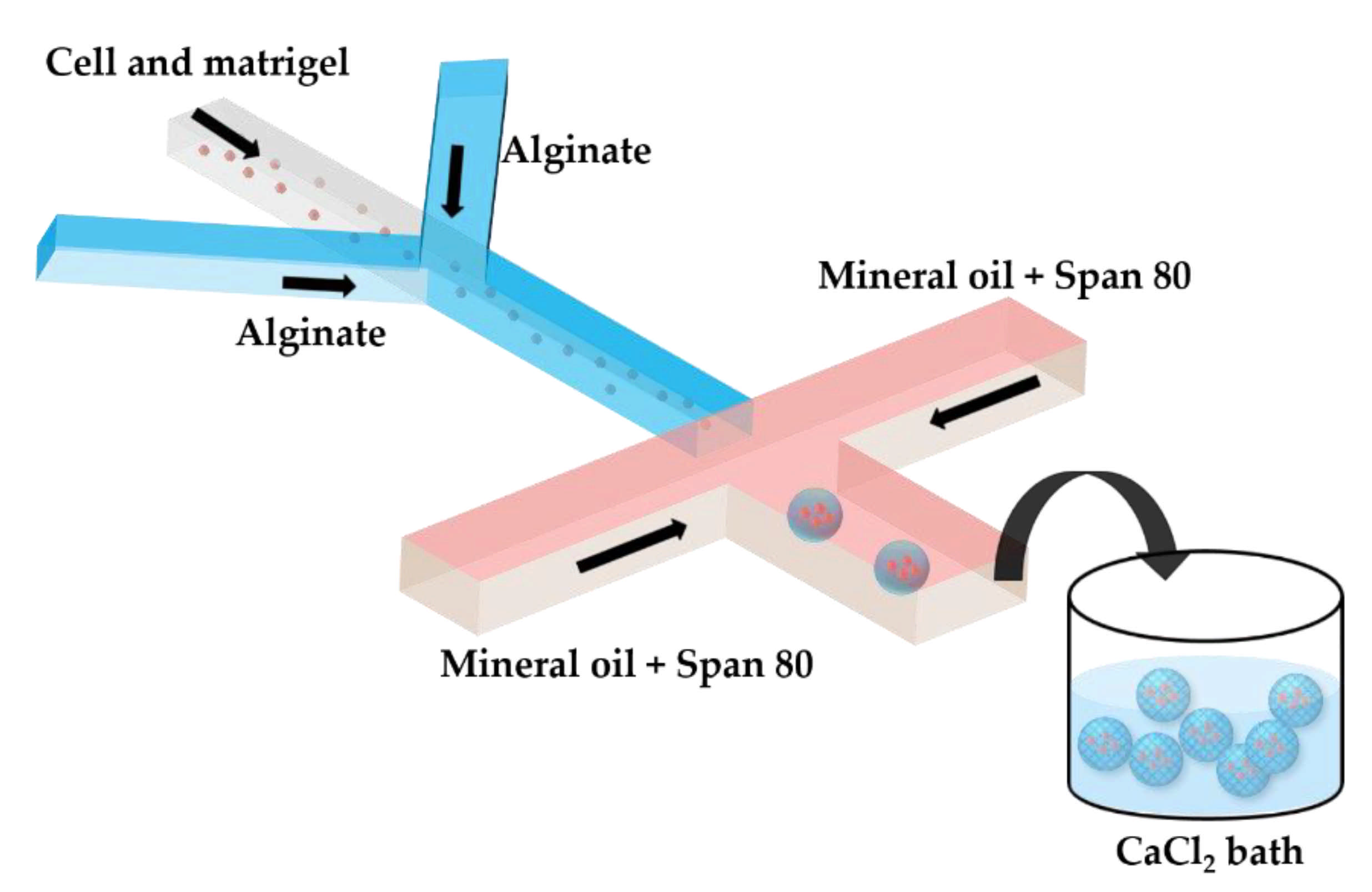
Two serial Y-junctions are useful for the preparation of mixed gel beads composed of two different polymers, e.g., Matrigel™ and alginate. Matrigel™ is a mixture of extracellular matrix proteins extracted from mouse sarcoma, composed of ~60% laminin, ~30% collagen IV, and ~8% entactin. Matrigel™ is liquid at 4 °C, but gels at 24–37 °C by self-assembly of laminin and collagen IV into crosslinked networks via entactin bridges. In
Figure 7, a mixture of tumor cells and Matrigel in a cell culture medium at 4 °C is mixed with am alginate solution in the upstream junction, and this mixture is emulsified in mineral oil in the downstream junction. As Matrigel and alginate are delivered through separate inlet channels, a mixing ratio between the two polymers can be tuned on-chip. The droplets are crosslinked in a 4 wt% CaCl
2 solution to form cell-laden composite beads.
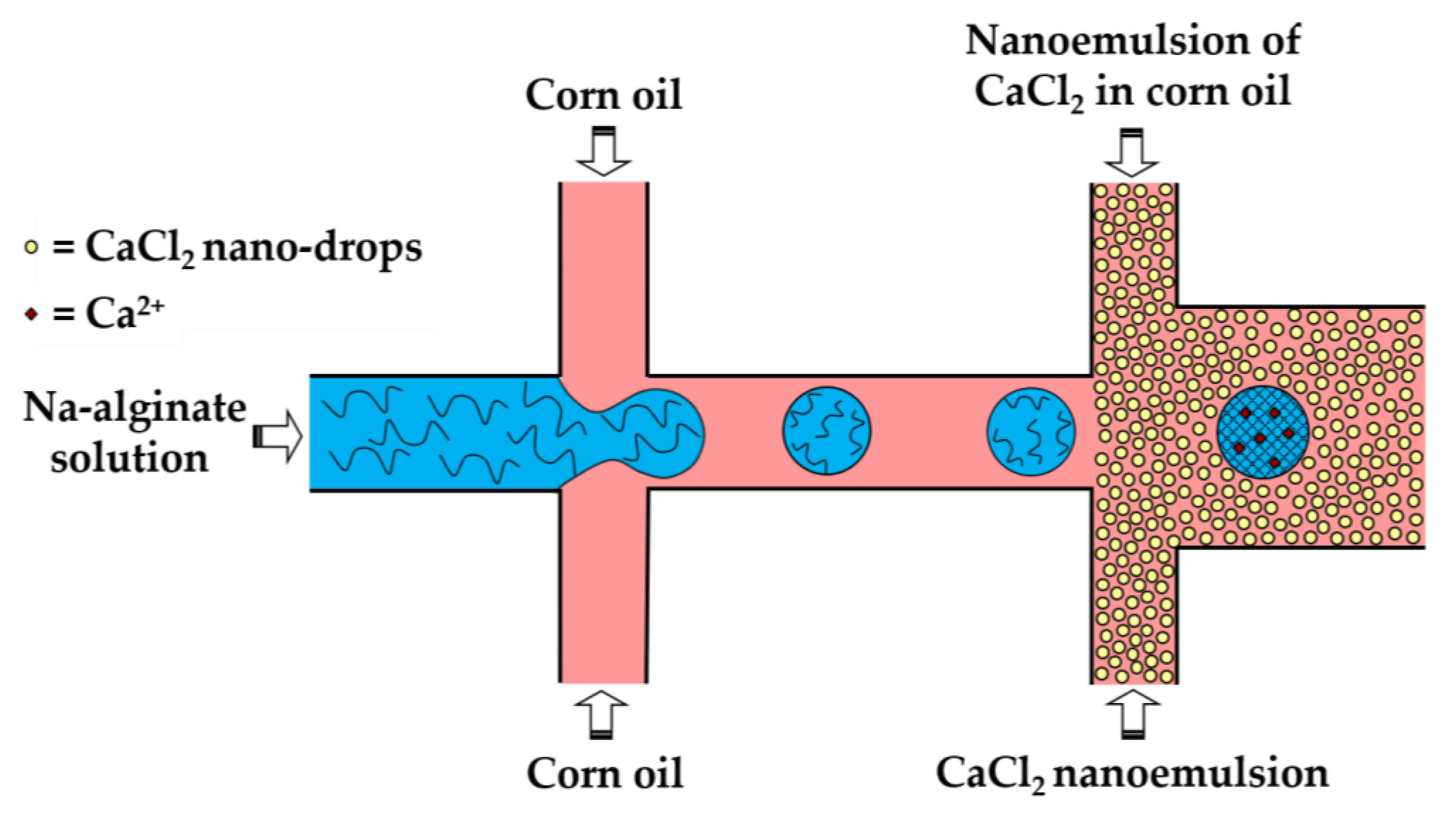
In the third method (iii), a fine W/O emulsion containing aqueous CaCl
2 droplets dispersed in corn oil is introduced through the downstream cross junction to crosslink alginate droplets formed in the upstream cross junction (
Figure 8). Using two consecutive cross junctions, droplet generation is spatially separated from the crosslinking reaction to avoid clogging of the droplet-forming channel by the gel. The channel downstream of the second junction has an increased width in order to reduce the oil velocity and prevent shear-induced deformation of droplets during gelation [
43]. The concentration of lipophilic surfactant used to stabilize the CaCl
2 droplets should be sufficiently low to allow for their merging with alginate drops. The optimum concentration of Y-Glyster CRS-75 was found to be only 0.1 wt% [
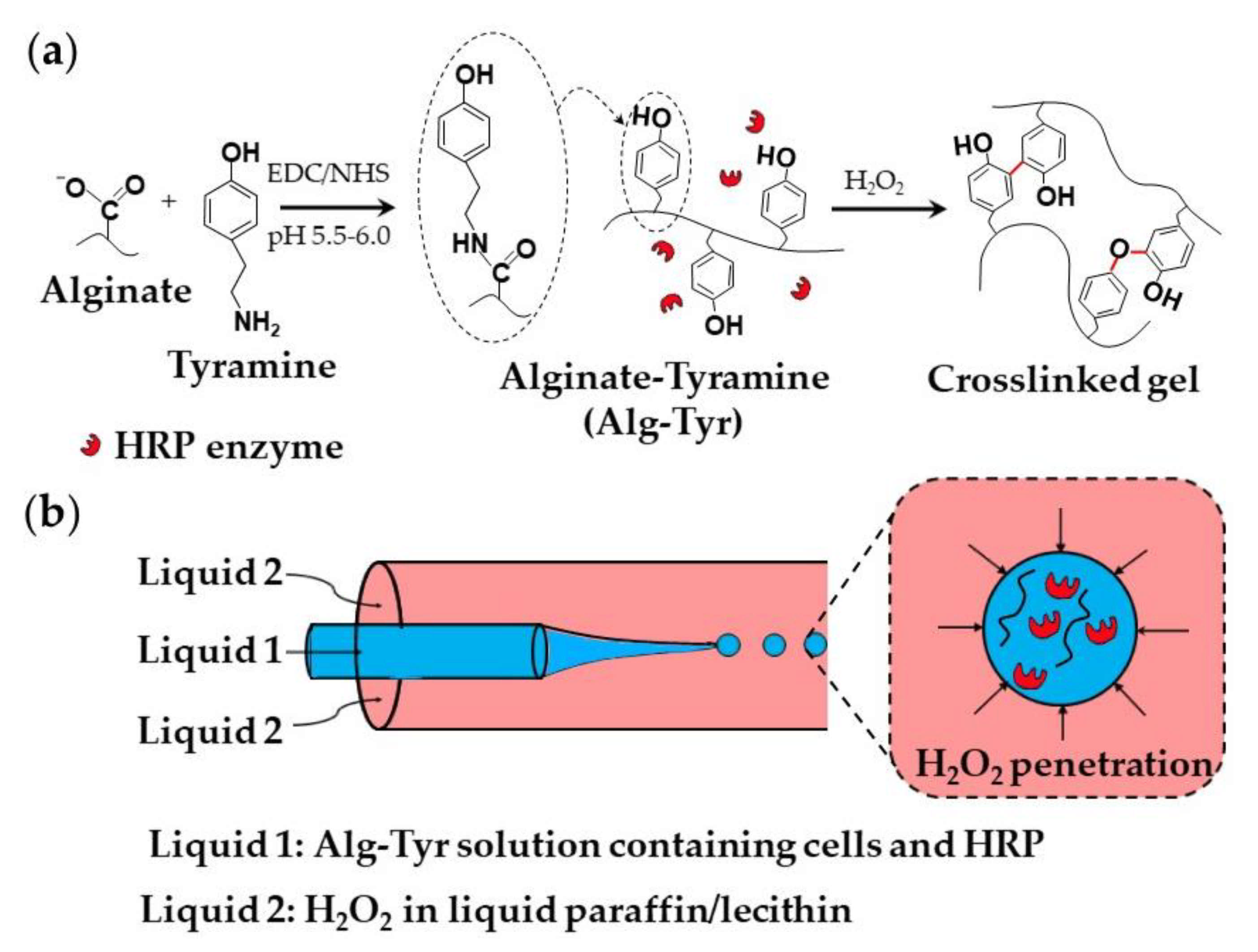
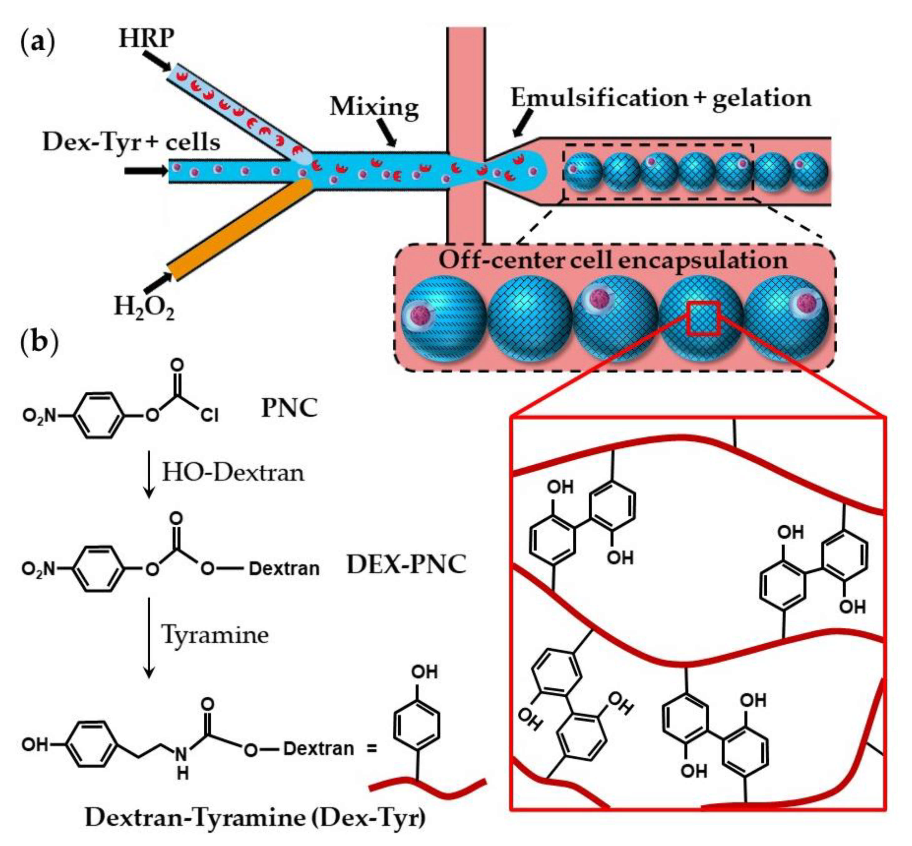
43]. A similar method can be used for the oxidative covalent crosslinking of modified natural polymers.
A slow gelation by CaCl
2 nanodroplets may cause the coalescence of alginate droplets and clogging of the outlet channel. A possible solution could be to increase the length of the downstream channel, which may lead to excessive pressure buildup in the chip, particularly because of the high viscosity of the nano-emulsion. To prevent droplet coalescence, the single emulsion method can be replaced by the double emulsion method, as shown in
Figure 9.
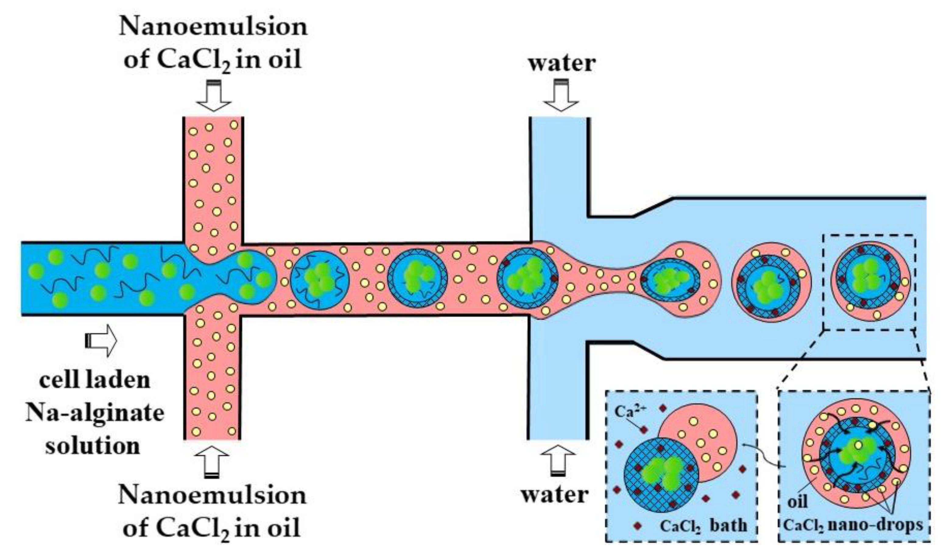
In the method shown in
Figure 9, individual alginate droplets are coated by a thin layer of CaCl
2 nano-emulsion to form core−shell droplets dispersed in the outer aqueous phase. The outer aqueous phase plays the following important roles: (i) prevents fusion of partially gelled alginate droplets; (ii) decreases viscosity in the downstream channel, which improves the chip functionality; and (iii) facilitates the separation of beads from the oil phase and minimizes cell exposure to the oil and surfactant, which increases biocompatibility of the process.
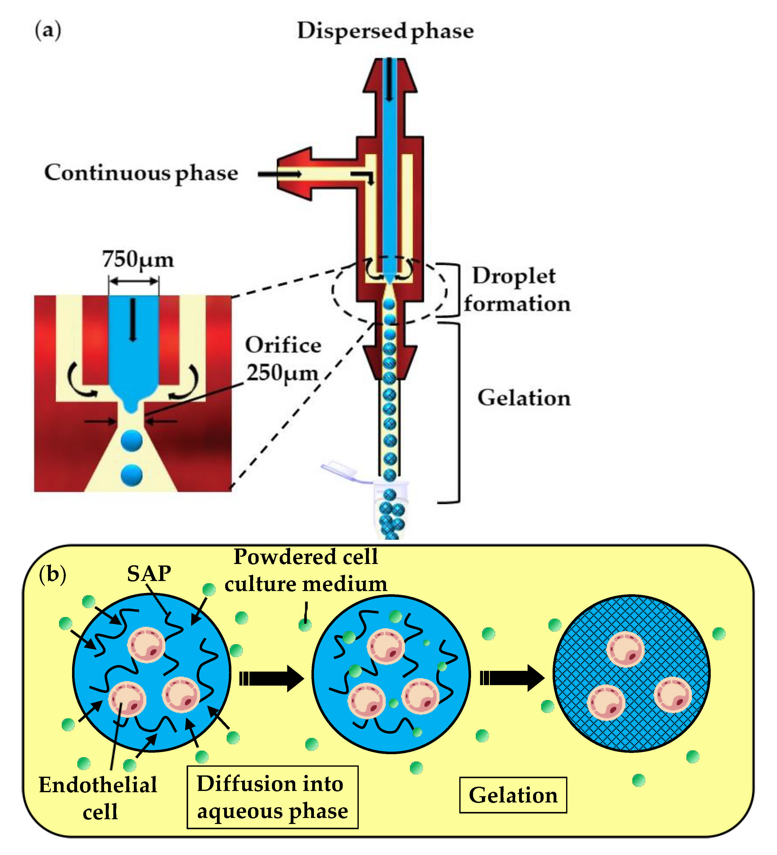
In the fourth method (vii), a self-assembling peptide (SAP) solution containing mammalian cells is emulsified in the oil phase, composed of a powdered cell culture medium dispersed in mineral oil (
Figure 10a). In the downstream channel, particles of the cell culture medium collide with the droplets and become dissolved in the SAP solution. The dissolution of low molecular weight compounds, such as inorganic salts and amino acids, leads to an increase in the ionic strength within the droplets and triggers the gelation of SAPs, as shown in
Figure 10b. Over 93% of the cells survived the microfluidic process and the fabricated microgels allowed for the diffusion of nutrients, as well as cell growth and differentiation [
50].
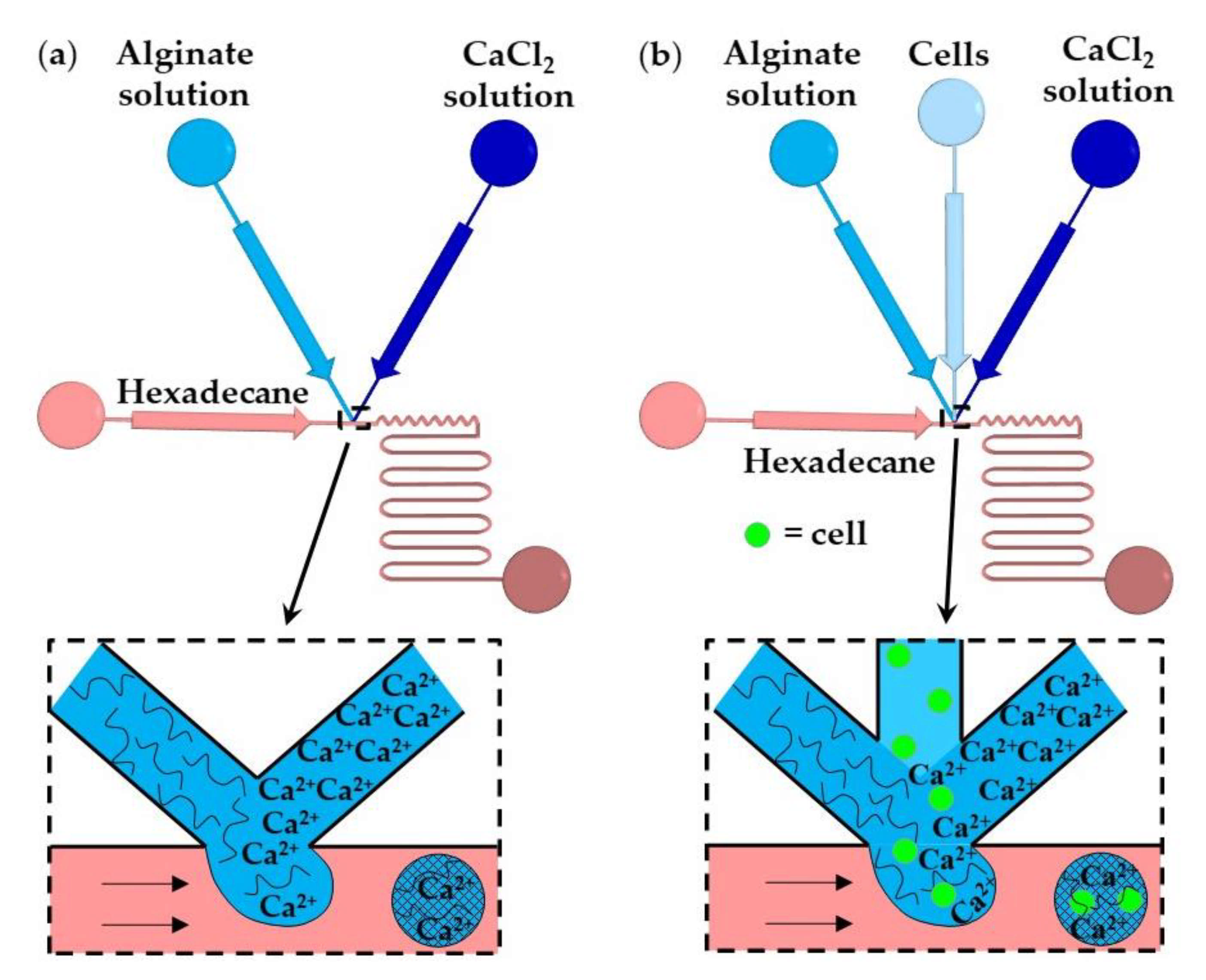
2.1.3. Rapid Mixing of Fluid Streams within Droplets
In this method, gelation is achieved by rapidly mixing two aqueous streams, usually a polymer solution and crosslinking solution (
Figure 11a), or three aqueous streams, e.g., polymer solution, crosslinking solution, and a cell suspension (
Figure 11b), immediately before droplet formation [
58]. The number of cells per bead and the crosslinking density can be controlled by adjusting the flow rate ratio and composition of the inlet aqueous streams. The examples of fluid compositions and channel geometries used in this method are provided in
Table S3 in the supplementary material.
In
Figure 11a, aqueous droplets comprising alginate and CaCl
2 are formed in an immiscible continuous phase, and the gelation is achieved by chaotic advection within the droplets. Chaotic advection is caused by hydrodynamic interactions between the droplets and the channel walls. Winding collection channels provide more efficient internal fluid circulations than straight channels. The T-junction shown in
Figure 11b is composed of three converging inlet channels and can be used for the encapsulation of cells within alginate microgels [
60]. The time required for crosslinking (gel formation time) should be longer than the droplet formation time to prevent premature gelation and channel clogging. For constant geometry of the microfluidic channels, the gel formation time primarily depends on the concentration of the reagents, while the droplet formation time mainly depends on the fluid flowrates and channel size [
59]. The gelation of alginate was also achieved by in situ mixing a solution of slow hydrolyzing acid and alginate/CaCO
3 solution [
59].
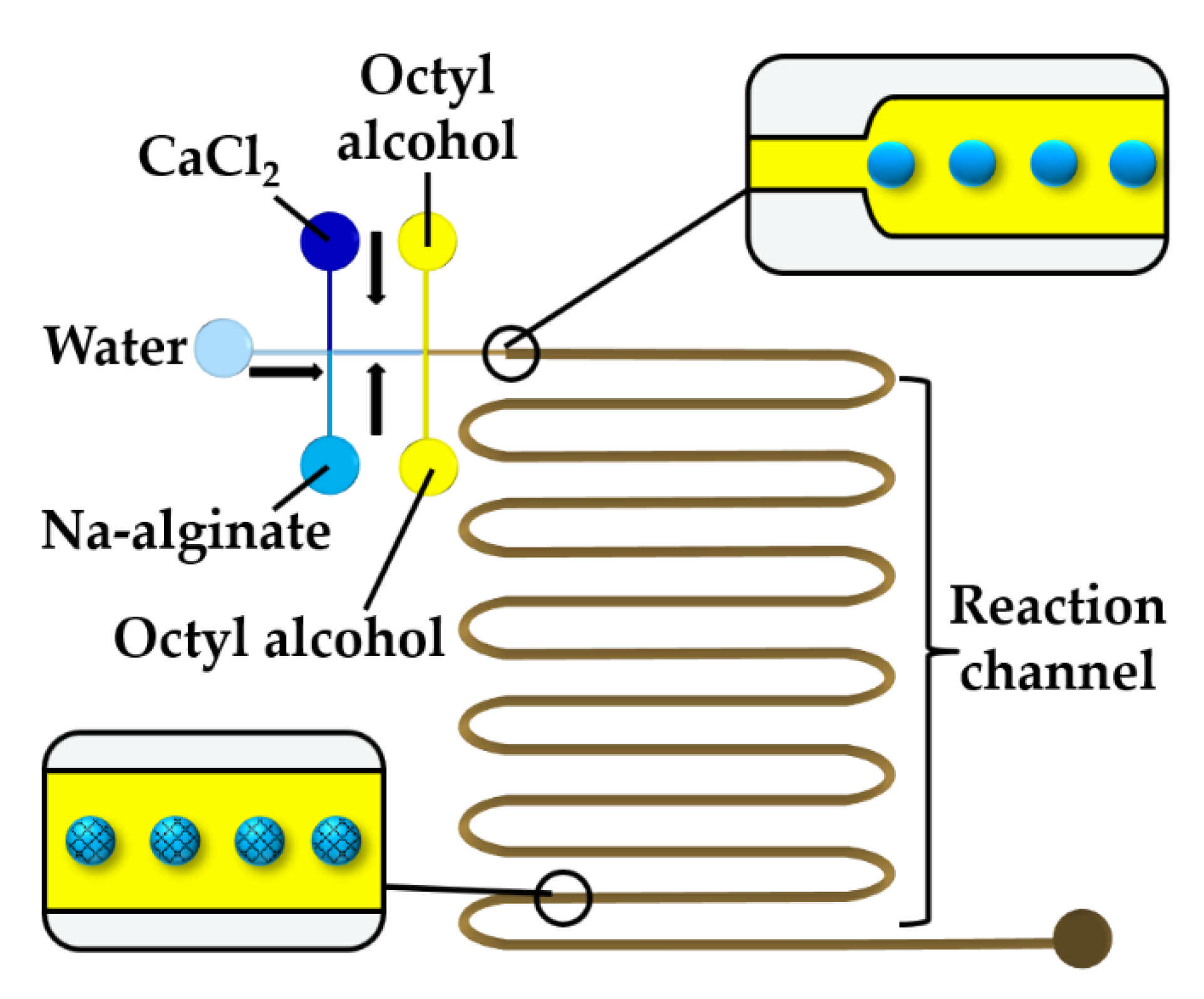
To prevent premature alginate crosslinking inside a droplet, a stream of water can be injected between CaCl
2 and alginate solutions, as shown in
Figure 12. At the upstream junction, the two side streams, namely the sodium alginate and CaCl
2 solutions, are separated by deionized water injected through the middle channel to prevent a crosslinking reaction before the droplets are formed in the downstream junction. The droplets are solidified in the wavy reaction channel. To prevent the diffusive transport of Ca
2+ ions through the thin layer of the middle stream, the thickness of the middle stream must be greater than the diffusional distance,
, given by
, where
is the diffusion coefficient of Ca
2+ and
is the contact time of the two laminar streams [
61]. The concentration of the CaCl
2 and alginate solutions plays an important role. The optimum CaCl
2 concentration was found to be 0.5–1.0 wt% [
61]. At a CaCl
2 concentration below 0.5 wt%, droplets cannot polymerize, and the jetting regime occurs at the CaCl
2 concentration of 2 wt% because of fast gelation.
2.1.4. Merging of Polymer and Crosslinker Droplets or Injection of Continuous Stream of Crosslinking Solution into Polymer Droplets
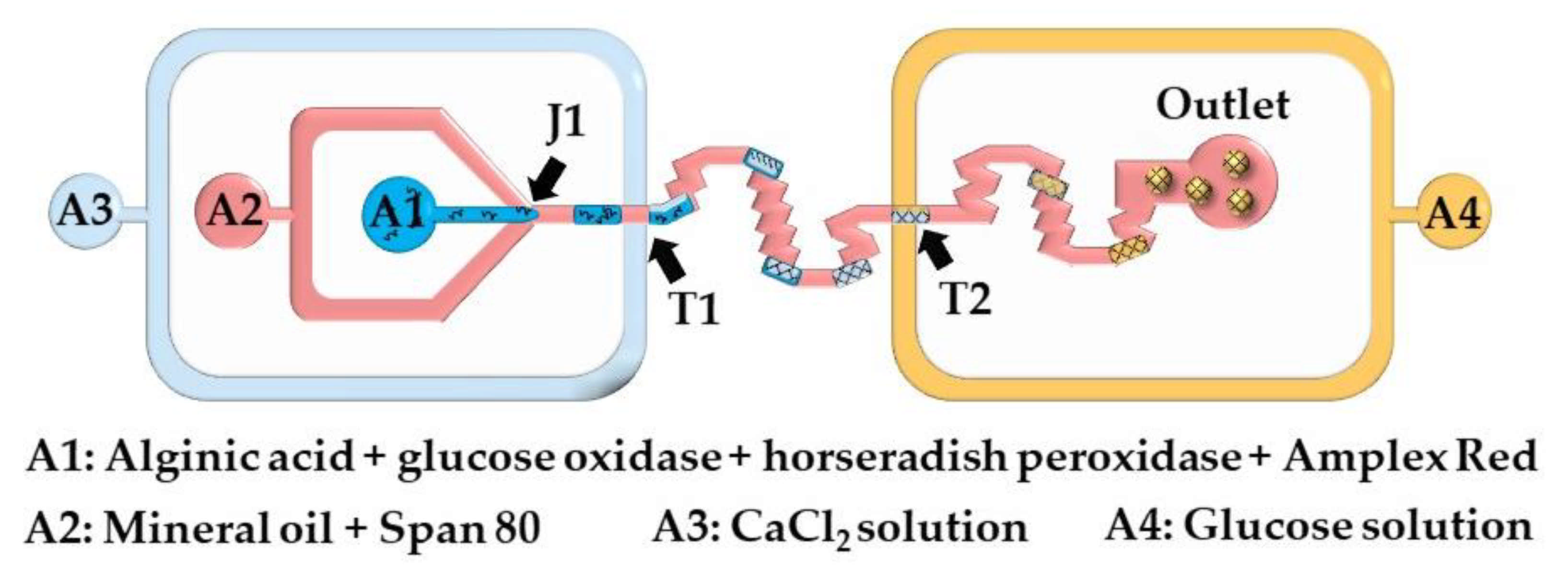
Here, microgels are produced by injecting a crosslinking solution into the polymer droplets or by merging the polymer droplets and crosslinking solution droplets. The chip shown in
Figure 13 consists of two double T-junctions for the encapsulation of glucose oxidase (GOx), horseradish peroxidase (HRP), and Amplex
® Red within alginate beads (T1), and for the colorimetric detection of glucose (T2). Aqueous alginic acid droplets loaded with GOx, HRP, and Amplex
® Red are generated at the junction J1 and are merged with a stream of CaCl
2 solution injected from inlet A3. The fused droplets generated at the junction T1 turn to solid hydrogel particles as they move through the serpentine channel between T1 and T2. A glucose-containing sample supplied from the inlet A4 is injected into the beads at junction T2. In the presence of hydrogen peroxide, released upon enzymatic oxidation of glucose within the beads, Amplex
® Red reagent is transformed into highly fluorescent resorufin. The merging efficiency of double T-junctions was over 90% under optimal conditions, and was higher than the merging efficiency of single T-junctions [
63].
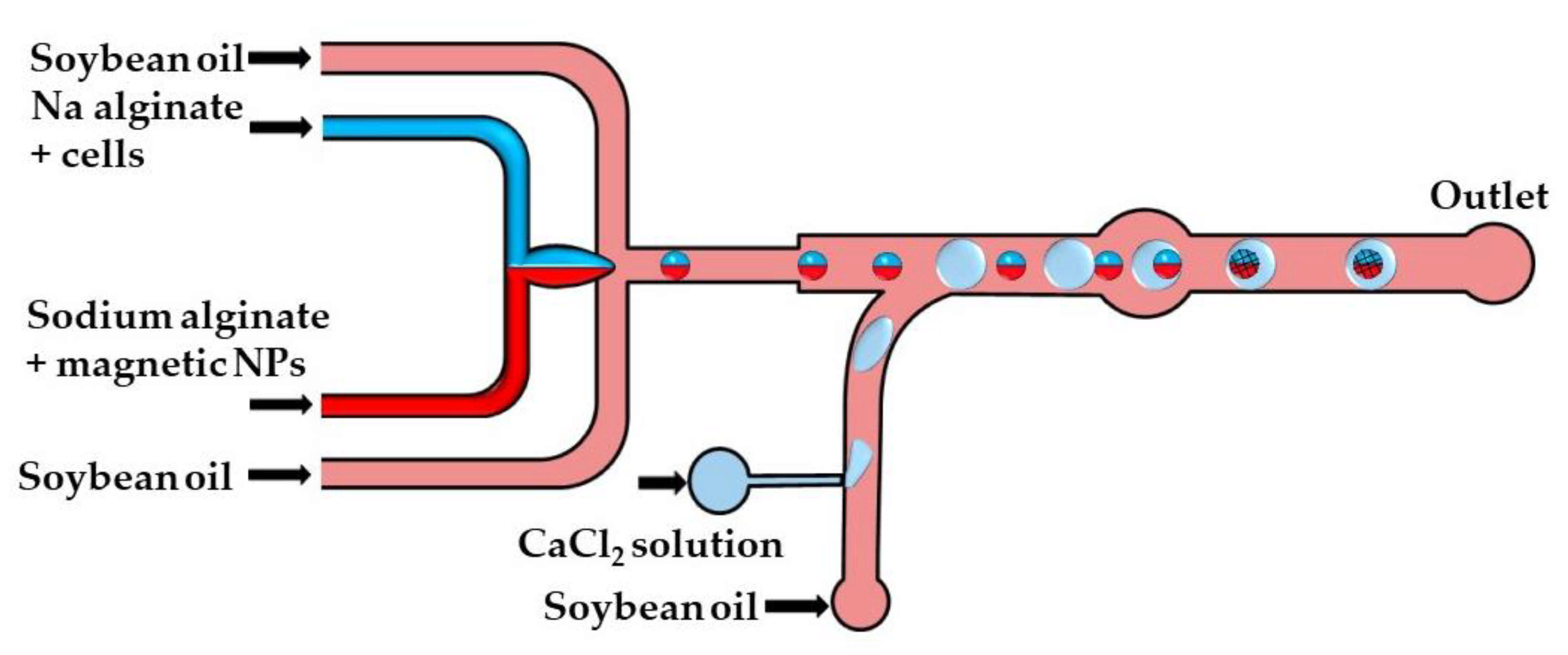
The crosslinking strategy shown in
Figure 14 is based on the fusion of alginate and CaCl
2 droplets in a cylindrical fusion chamber. A cell-laden alginate solution and an alginate solution loaded with magnetic nanoparticles were injected through two separate inlets of the head-on junction to create a biphasic flow. Further downstream, this bicolored stream was split into Janus droplets by flow focusing with an oil phase. To inhibit the mixing of the two solutions within a droplet, the diameter of the Janus droplets was limited to 80% of the channel width [
64]. The CaCl
2 droplets were delivered to the main channel from the side channel, and their production was synchronized with the production of Janus droplets to place CaCl
2 droplets between each two adjacent Janus droplets. The Janus droplets were merged with CaCl
2 droplets in a cylindrical chamber to form Janus particles with a magnetic anisotropy. The magnetic halves allow for particle manipulation by a magnetic field, while the alginate halves provide an optimum microenvironment for cell growth.
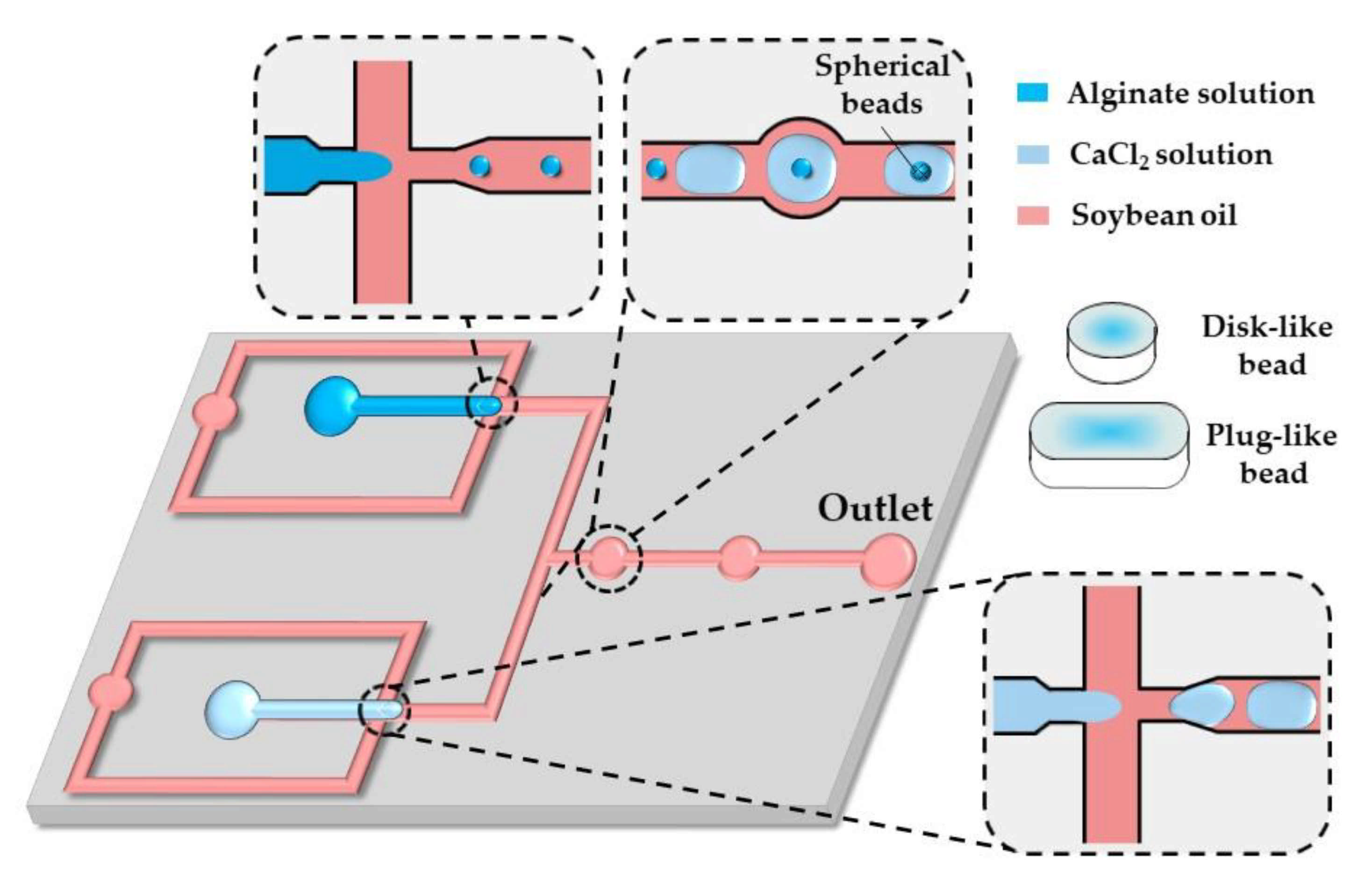
A similar strategy was applied in the chip shown in
Figure 15. Here, alginate and CaCl
2 droplets formed in separate cross junctions were alternately fed to the expansion chambers where they merged and formed microgels.
As shown by others [
65,
66,
67], in situ gelling of droplets whose dimensions are beyond the height and/or width of a microfluidic channel can be utilized to form non-spherical microgels, such as disks, rods, and threads.
By increasing the flow rate of the continuous phase or decreasing the flow rate of the dispersed phase, the shape of the beads can be changed from threads to rods to disks to spheres, reflecting different droplet volumes [
65]. The resultant droplet volume depends on the competition between the viscous forces, tending to stretch the dispersed phase into a long jet, and the interfacial tension, acting in the opposite direction. Spherical beads are formed if the droplet volume,
, is smaller than
, where
is the channel height, which is smaller than the channel width. Non-spherical beads are generated from confined droplets [
66]. Plugs (rods) and threads are formed from droplets, which are confined in two directions and have a roundness,
greater than unity, where
is the projected surface area of the beads and
is the projected bead perimeter [
68]. For spherical and discoidal beads,
and
, and the roundness is
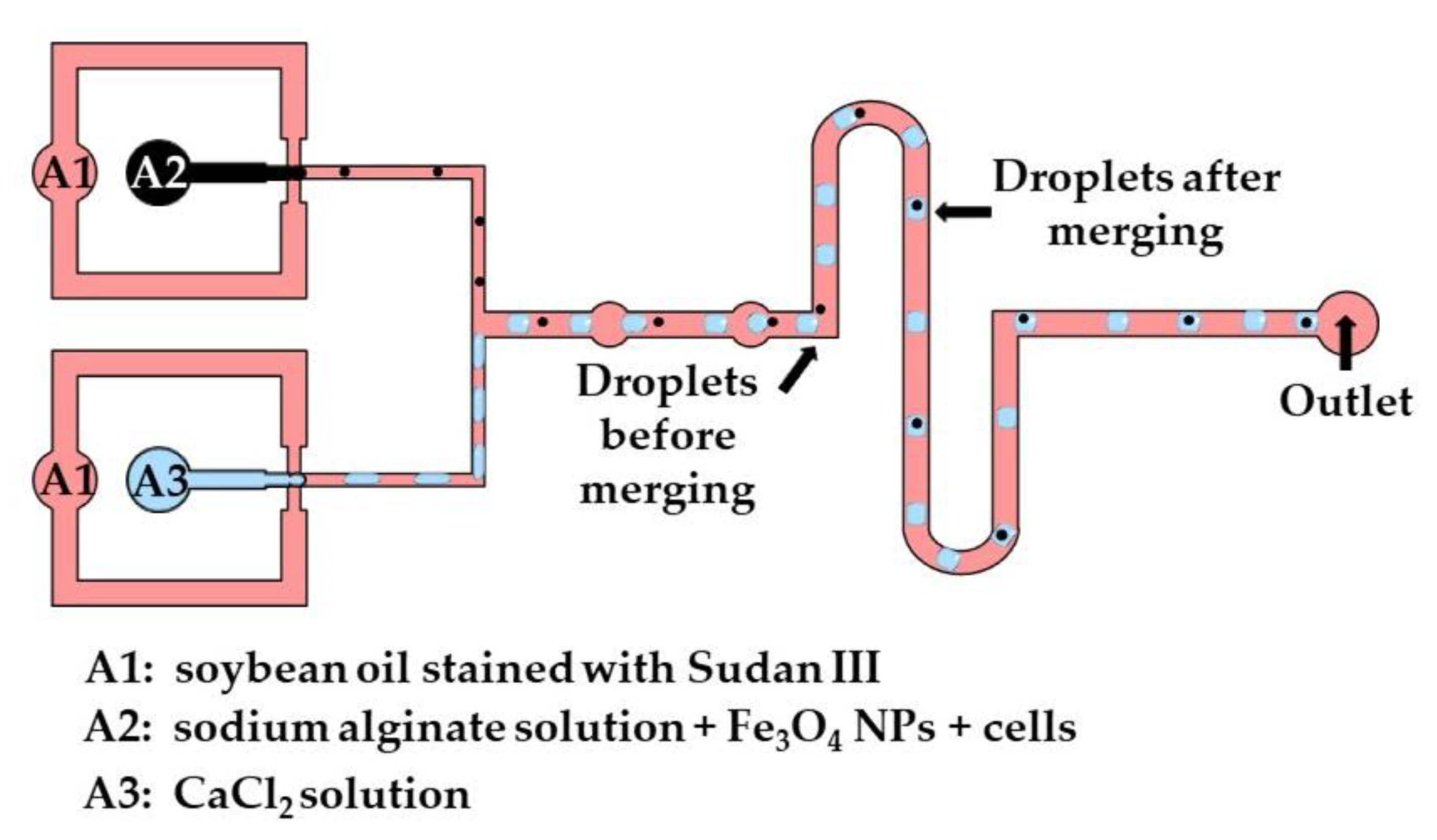
The production of disk-like magnetic alginate beads loaded with cells is shown in
Figure 16. The size of the beads and the number of cells encapsulated per bead can be adjusted by controlling the fluid flow rates. Disk-like beads with flat top and bottom surfaces are formed due to vertical droplet confinement in the serpentine channel, as the equivalent droplet diameter is greater than the channel height, and the channel width is sufficiently large to prevent the formation of plugs. A disk-like shape allows for the cell division process to be monitored without image distortion [
67]. Different strategies used for the production of microgels by droplet merging are summarized in
Table S4.
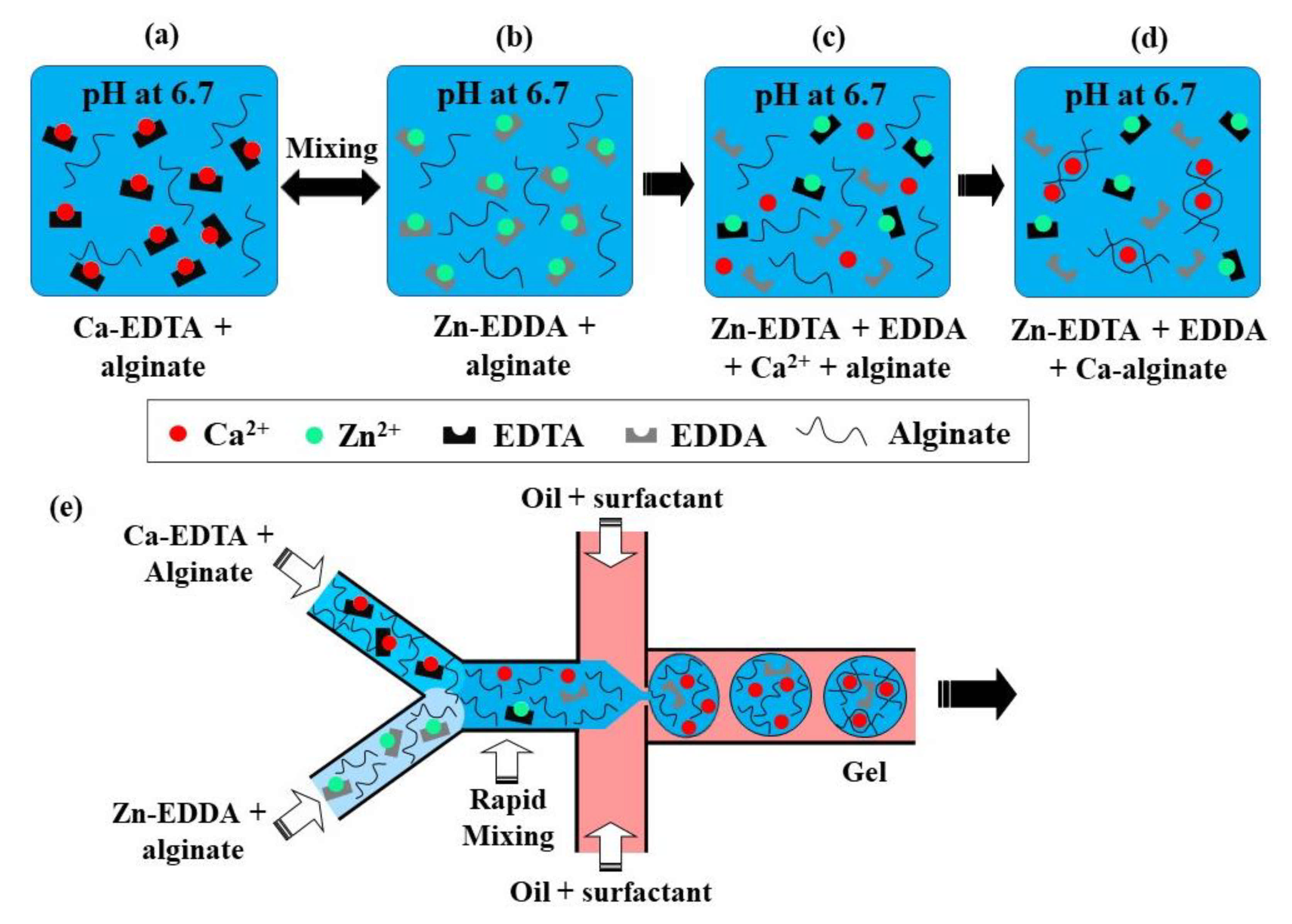
2.1.5. Competitive Ligand Exchange Crosslinking (CLEX)
This crosslinking method is based on the competition between a gelling ion (Ca
2+) and an exchange ion (Zn
2+) for binding sites on chelating agents (EDDA and EDTA) and a charged polymer [
69]. At pH 6.7, a mixture of Zn-EDDA and alginate will not gel, nor will a mixture of Ca-EDTA and alginate, because both complexes do not dissociate at this pH (
Figure 17a,b). However, upon mixing, Zn
2+ will be exchanged between EDDA and EDTA because of their higher affinity to EDTA, compared with Ca
2+. It will result in the release of Ca
2+ ions (
Figure 17c), which will then crosslink the alginate, because of the higher binding affinity of Ca
2+ to alginate than EDDA (
Figure 17d). At pH > 7.2, the amount of Ca
2+ is insufficient for gelling, as the competition between alginate and EDDA for Ca
2+ is shifted towards Ca-EDDA at such a pH. On the other hand, at pH < 6.7, the gelation is too fast because of the high concentration of crosslinking Ca
2+ ions, which leads to clogging at the junction where the aqueous phases meet.
The chip for producing alginate beads by CLEX is comprised of the following three inlet channels: one inlet is for the carrier oil phase and two inlets are for alginate solutions containing Ca-EDTA and Zn-EDDA, respectively (
Figure 17e). Alginate should be added to both aqueous streams to balance the hydrodynamic resistance in the two inlets, as well as to avoid polymer dilution after mixing. The kinetics of the ion exchange process and the amount of released Ca
2+ ions depend on the pH and the type of chelators used, which can be used to control the gelation kinetics and gel strength [
37]. The gelation time can be controlled in the range from seconds to minutes while maintaining the pH within the physiological range [
37]. It ensures enhanced cell survival rates compared with the internal gelation approach, where Ca
2+ ions are released from Ca-EDTA or solid CaCO
3 using an acidified oil phase, which inevitably results in a pH drop well below the physiological range.


