1. Introduction
Molecular imprinting is a technique for creating artificial recognition sites on polymer matrices that complement the template in terms of size, shape, and spatial arrangement of functional groups. This molecular imprinting technique uses target molecules in a synthetic polymer matrix by selectively binding [
1,
2]. Recently, molecular imprinting technology has been used to create biometric surfaces in biosensors. There are many molecular imprinting technologies, including bulk printing, surface printing, and epitope printing.
In the bulk printing method, the template molecules are printed on the entire polymer matrix, and at the end of the method, the template needs to be removed from the polymer. The polymer produced in this method is large or bulk, so grinding must be carried out on the polymer to obtain template-specific binding sites on the polymer [
1]. Due to the thick morphology of the polymer in the bulk printing, it causes low access for the target molecule to bind to its specific site. Therefore, another method was developed to overcome this limitation, namely the surface printing method and the epitope printing method.
In surface printing, the removal of template molecules will result in specific binding sites on the polymer surface [
3]. The binding site on the polymer surface causes this type of polymer to provide greater access to the binding target molecule than bulk imprinting [
1]. This technique has been widely used for various types of analytes such as proteins [
4], cells [
5], and micro-organisms [
6]. While in epitope printing, the target molecule is a protein and uses only a small portion or fragment of the macromolecule is printed to represent the whole molecule (epitope) as a template [
1]. In this method, the peptide epitope is covalently bonded to the silicon surface where the monomer is polymerized. In epitope printing, more specific and strong interactions can be obtained. The polymer has the ability to recognize templates as well as whole proteins very well [
7].
Molecularly Imprinted Polymer (MIP), which contains specific bonds between template molecules and polymers [
8], is an example of materials that use molecular imprinting techniques. MIP is a unique recognition system resulting from templates and functional monomers that are polymerized, enabling molecular recognition utilizing principles similar to those underlying the action of enzymes and their substrates [
9,
10]. The main advantage of MIP is high selectivity and affinity for the target molecules used in the molding process. Compared to biological systems such as proteins and nucleic acids, imprinted polymers have higher physical strength, high temperature, pressure resistance, and inertia to acids, alkalis, metal ions, and organic solvents. In addition, the synthesis cost is low, the storage life of the polymer can be very high, and the recognition capability can be maintained for several years at room temperature.
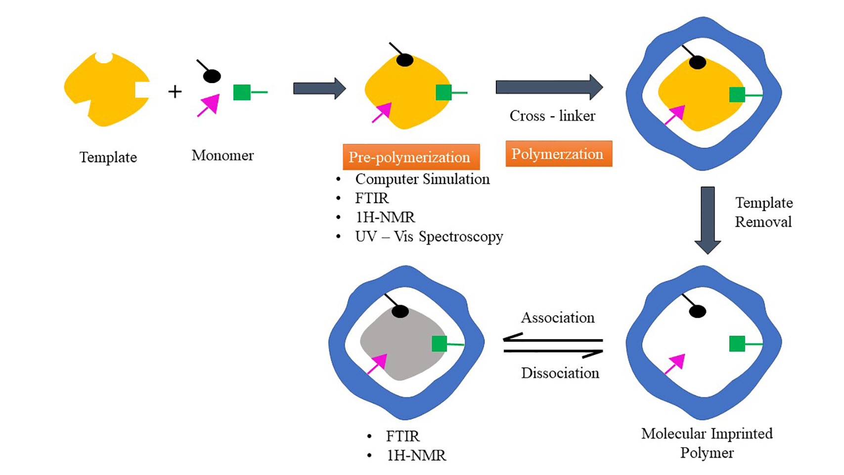
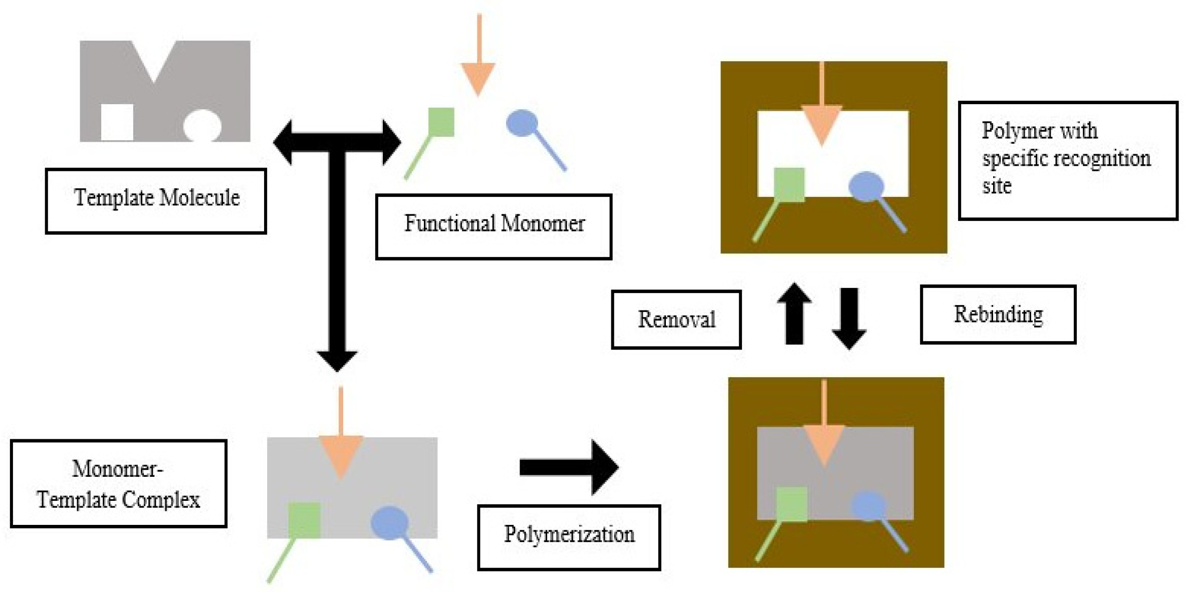
Templates, functional monomers, solvent, initiator, and cross-linker are MIP components [
11]. MIP is based on the formation of complexes between analytes (templates) and monomers of functional compounds. In the presence of excess cross-linking agents, three-dimensional polymers are formed [
12]. In the process of making MIP, the selection of the constituent components will affect the performance of the resulting imprinted polymer. A functional monomer is preferred over an ordinary monomer because a functional monomer contains a Y functional group that can interact with template molecule via hydrogen bonding, dipole-dipole, and ionic interaction to produce a template-monomer complex. The complex is then fixed in the presence of a large excess of a cross-linking agent, and a three-dimensional polymer network is formed. After the polymerization process, template molecules are removed from the polymer using a solvent, resulting in selective complementary polymer-template bonds [
11,
13]. The scheme of the molecular imprinting process can be seen in
Figure 1.
Several interactions occur in the molecular imprinting process, like hydrogen bonds, dipole-dipole, and ionic interactions. The interactions between the template molecule and functional groups present in the polymer matrix drive the molecular recognition phenomena. Thus, the resultant polymer recognizes and binds selectively to the template molecules. As interaction is the driving force for molecular recognition, finding the chemical interaction that happened while develo** MIP is crucial to be known. According to a literature search, no review has been made on methods to find chemical interaction between template-functional monomers before and after polymerization. For that reason, this review will discuss the factors that influence the success of making MIP, especially on choosing the suitable component to form MIP and methods to determine the type of interactions happening in MIP. We will divide the method on finding the type of interaction in MIP into two stages: pre-polymerization and post-polymerization [
14,
15,
16,
17]. Thus, we will discuss computer simulation, UV-Vis Spectroscopy, Fourier transform infrared spectroscopy (FTIR), Proton Nuclear Magnetic Resonance (
1H-NMR) for methods on finding the type of interaction in the pre-polymerization stage. In contrast, as a method for detecting the type of interaction after polymerization, we will discuss Suspended State Saturation Transfer Difference High Resolution/Magic Angle Spinning (STD HR/MAS) NMR, Raman Spectroscopy, Surface-Enhanced Raman scattering (SERS), and Fluorescence Spectroscopy.
4. Template-Monomer Interaction
A study has shown that physical properties and recognition of MIP depend on the successful interaction between template and monomer. This process occurs in the pre-polymerization stage [
55]. The selected functional monomer will interact with the template, producing a stable template-monomer complex [
17]. Therefore, functional monomers and templates must complement each other [
59].
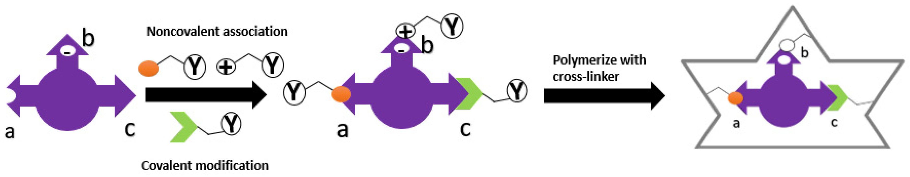
There are currently two strategies used in MIP technology based on the nature of the template-monomer interaction. Two types of molecular imprinting strategies have been set by covalent or non-covalent interactions between the template and functional monomer [
74]. An example of interaction can be seen in
Figure 3. These two strategies are:
1. Self-assembly approach, which uses non-covalent bonds between monomer and template, such as hydrogen bonds, Van der Waals forces, ionic or hydrophobic interactions, and others [
11]. The functional monomers are regularly positioned around the template molecules during the self-assembling process due to different exchanges [
75];
2. Pre-organized approach, which uses reversible covalent bonds between the functional monomer and template. This strategy will reduce non-specific sites on MIP [
11,
76].
The technique most widely used in the manufacture of MIP is the self-assembly approach. In this technique, template-monomer complexes are formed in situ by non-covalent interaction [
77]. Hydrogen bond, hydrophobic, and electrostatic interactions are the most widely used bonds for manufacturing MIP due to their excellent adaptability [
65].
MIP manufacturing techniques using covalent and non-covalent bonds have their respective advantages and disadvantages, as listed in
Table 1.
The interaction between the template molecules and functional monomer and the formation of the polymerization mixture can lead to three different results [
81]:
1. If the template is added in the pre-polymerization step, at the same time as the monomer, solvent, cross-linker, and initiator, the reaction medium will be very rich in functional monomers. The polymerization process will occur rapidly due to the dominant role of the cross-linker. A weak complex will be formed between the template and the functional monomer, producing MIP with a relatively low imprinting effect;
2. If the template is added after the start of the polymerization process, a few minutes after adding the monomer, cross-linker, initiator, and solvent, the reaction medium will contain functional macromonomers and a few monomers. Due to the nature of functional macromonomers, namely their high flexibility, they will freely re-arrange around the template to produce a MIP with a better imprinting effect;
3. If the template is added too late in the polymerization process, minutes after the optimum time of adding the template (point b), the reaction medium will be rich in pre-formed nanogel particles, which will further form a cross-linked macrogel. The condition is particularly unfavorable because the cross-linked nanogel particles are more rigid than the functional macromonomers. The MIP formed is predicted to have a low imprinting effect.
5. Analysis of Template-Monomer Functional Interactions
Intermolecular interactions between molecular templates and functional monomers will affect the selectivity and affinity of MIP [
82]. These intermolecular interactions can be analyzed in the pre-polymerization process using computer simulation, UV-Vis spectroscopy, FTIR, and
1H-NMR. Meanwhile, Suspended-State STD HR/MAS NMR, Raman spectroscopy, SERS, and fluorescence spectroscopy were used after MIP formation [
14,
16,
83,
84,
85,
86]. Hydrogen bonding interactions are the kind of interaction that focuses on all methods as this interaction strongly contributes to the affinity of molecularly imprinted polymers (MIPs), especially for low molecular weight compounds in organic, aprotic solvents [
87].
5.1. Computer Simulation
The development of in silico-based technology makes it easy to select MIP components such as templates, functional monomers, and suitable porogens [
88,
89]. In addition, it can be used to determine intermolecular interactions that occur in the pre-polymerization mixture [
88]. In the pre-polymerization process, a strong template-monomer interaction will result in a suitable MIP [
90]. A computational approach can be used to evaluate hydrogen bond interactions between functional template-monomer [
91]. In making MIP, computational studies play a role in determining the best monomer type and ratio in a shorter time than doing experimental trials to get high selectivity [
92]. Predictable functional template-monomer interactions can be found using Density Functional Theory (DFT) [
83]. The DFT method can check intermolecular interactions based on the distance between the template and functional monomers and become the most extensively utilized approach in the design of MIP [
70,
93,
94,
95,
96].
Wungu et al. [
93] used the DFT method based on Becke three-parameter Lee-Yang-Parr (B3LYP)/6-311 + G (d) to investigate the interaction between MAA as a template molecule with D-glucose as a functional monomer. This study used Gaussian 09 software to calculate electronic properties. Before optimization, the initial structure of the MAA-D-glucose complex had a distance of 6.62 Å between the O2 atoms of MAA and the C4 atoms of D-glucose. After optimization, the distance between the MAA complexes and D-glucose was reduced to 2.81 Å, indicating non-covalent interactions with hydrogen bonds [
93].
The type of interaction between the monomer templates also can be predicted using computer simulations based on the change in charge on the natural bond orbitals (NBO). NBO analysis allows for the calculation of the number of atoms in the molecule, the molecular structure, and the intermolecular or intramolecular interactions [
97]. Huang and Zhu [
98] conducted a computational model study to see the interaction between spermidine (template) and methacrylic acid (monomer). In theory, the two compounds can interact through hydrogen bonds. From the observation of the NBO charge, there was an interaction between the N13 atom in spermidine and the H5 atom in methacrylic acid, which was seen from the change in the NBO charge. Before complex formation, the NBO charge of N13 was −0.671, and H5 was 0.485. Meanwhile, after complex shape, the NBO charge of N13 became −0.707 and H5 became 0.501. This change in NBO charge indicates a charge transfer between the proton donor (H5) and proton acceptor (N13) in both molecules (template and monomer) and demonstrates that spermidine and methacrylic acid interact through hydrogen bonds [
98].
Intermolecular interactions can also be seen through binding energy (∆E), even though we can not always determine the interaction type [
93]. The initial confirmation of the respective molecular templates and functional monomers was optimized to obtain the molecule’s Gibbs free energy (∆G). The molecular templates and functional monomers are combined to produce stable complex conformations without imaginary frequencies. Functional monomers can be selected depending on the size of the ∆E [
97]. The formation of the complex will be more stable as the binding energy value decreases; the lower the value of the binding energy, indicated by the negative ∆E value, the more likely the complex formed will exist in its complex form [
99,
100,
101]. ∆E can also be used to determine the extent of the reaction, while ∆G is used to determine the spontaneity of the reaction [
97]. The ∆E values were calculated using the following equation:
where E (complex) is the total energy of the template-monomer complex, E (template) is the energy of the template, and ΣE (monomer) is the energy of the functional monomer [
102].
5.2. UV-Vis Spectroscopy
The UV-Vis spectroscopy analytical method aims to determine the strength of the intermolecular interaction between the template and the functional monomer. The strength and affinity of the template-monomer will affect the selectivity and affinity of the polymer. Therefore, it is crucial to determine the suitable functional monomer which will interact strongly with the template to form a stable complex [
52].
The interaction of template and monomer can be investigated using UV titration with nothing changes in absorbance [
103]. The titration method using UV-Vis spectroscopy can evaluate the template-monomer association constant (Ka) so that the intramolecular interaction between template and functional monomer can be determined during the pre-polymerization process and also the specificity and selectivity of the polymer [
104,
105]. The Ka is calculated using the Benesi-Hildebrand equation [
103] as follows:
where ΔY is the change in absorbance, YΔHG is the change in absorbance at the end of the titration, and [G] is the concentration of the monomer added [
106].
Hasanah et al. [
103] used UV titration to determine the interaction between atenolol (template) and itaconic acid (functional monomer). Based on the calculation results, the association constant was 542.67 M
−1 in methanol solvent. [
103]. In another study, Hasanah et al. [
107] used UV spectroscopy to determine the value of the association constant between atenolol template and Itaconic acid as a functional monomer in a mixture of methanol: acetonitrile (1:1) resulted in a constant association value of 6.277 × 10
2 M
−1. The interaction between atenolol and itaconic acid is a hydrogen bond from the amine groups, and the carboxylic group of atenolol and itaconic acid as the Ka value was increased in a more polar aprotic solvent [
103,
105].
Hasanah et al. [
108] used UV Vis spectroscopy to see the interaction between itaconic acid monomer and diazepam as a template. The value of Ka obtained using the Benesi–Hildebrand formula was 381.9 M
−1 ± 0.4. As stated by Wang and Yu [
109], a weak interaction has a Ka value less than 25 M
−1, and a strong interaction has a Ka value more than 100 M
−1. So the interaction between itaconic acid and diazepam includes a strong interaction [
108]. Fu et al. [
52] also used UV-Vis spectroscopy to see the strength of the interaction between template luteolin and three functional monomers, namely acrylamide (AM), 4-vinyl pyridine (4-VP), and 1-aryl piperazine (1-ALPP) with different concentrations. Interaction strength was detected from significant changes of absorbance on maximum wavelength. 1-ALPP was found to cause reducing absorbance on maximum wavelength due to the π-π transition of luteolin when added in higher concentration. Meanwhile, no changes were found on AM and slight changes on 4-VP [
52].
Based on the literature search, UV-Vis spectroscopy is usually used to see the strength of the interaction (from Ka value) or binding experiment. In Lulu Wang’s research in 2019 [
110], a critical study was conducted to evaluate the adsorption performance of MIP and NIP for gossypol (an acidic organic compound) made with 2-(Dimethylamino)ethyl methacrylate (DMAEMA) monomer using UV spectroscopy at 373 nm by comparing absorbance values before and after absorption. More significant differences in the value represented higher binding. The gossypol molecule has a pKa value of 6.5 [
110,
111] and six phenolic hydroxyl functional groups -OH in its structure; it can form an acid-base ionic pair interaction with the basic amino group -NH
2 of DMAEMA monomer. Theoretically, functional monomers containing basic groups can interact strongly with phenolic acid hydroxyl groups through acid-base interactions [
112]. When we want to know the type of interaction, experiments in a few different solvents need to be run.
5.3. Fourier Transform Infrared Spectroscopy Analysis (FTIR)
FTIR spectroscopy can analyze samples in many forms, including liquids, solutions, gases, powders, and pastes [
113]. FTIR is used in many MIP develo** to identify the formation of chemical bonds, especially hydrogen bonds between molecular templates and functional monomers, due to absorption of the infrared spectrum by the sample and the peak shifting [
83,
114].
** analysis, and therapeutic drug monitoring. However, there are still some problems to be explored and solved:
1. Nanostructured MIP materials often suffer from problems such as the hardenest of template removal. The condition often results in MIP nanomaterials with non-uniform recognition sites;
2. Different efficient synthesis methods to produce MIP with high binding ability and selectivity are still needed to explore to maximize commercial conversion;
3. Knowing chemical interaction that happened between MIP and template with the affordable instrument still needs to be explored shortly as an instrument available now expensive for some laboratories.


