1. Introduction
Extracellular vesicles (Evs) were first studied and described in 1946 [
1] and once were regarded as a cellular waste [
2]. These EVs gained considerable attention throughout the years [
3] due to the active mechanism of their release from the cells [
4]. These sacs were described as extracellular vesicles [
5].
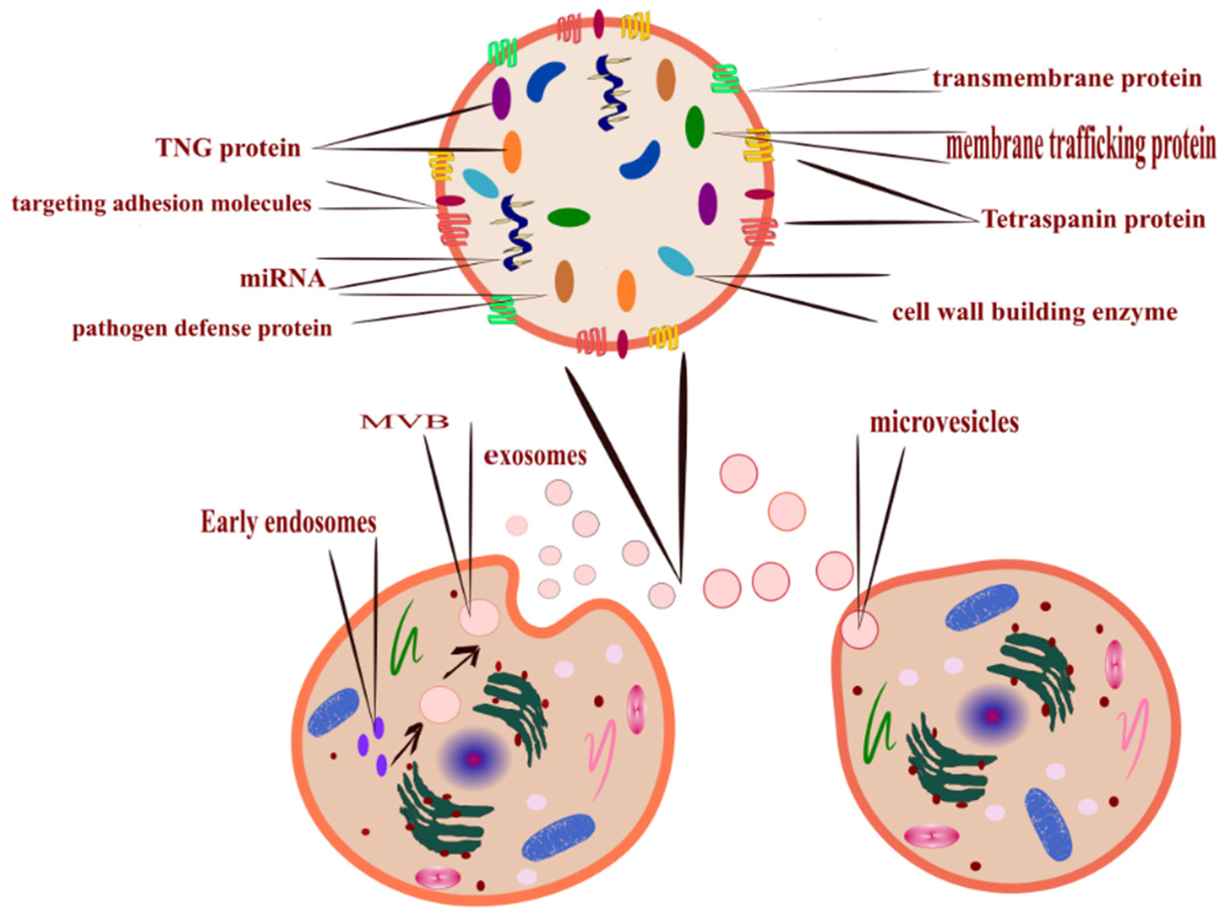
A recommended definition by the International Society for Extracellular Vesicles (ISEV) advised using the term extracellular vesicles as the characteristic label for particles released naturally from the cells, distinct by a lipid bilayer, and do not replicate. The short term is EVs [
6]. In order to study EVs, the process should undergo characterization to identify the transmembrane luminal protein; the characterization process should include but not be limited to zeta potential, electron microscopy, and flow cytometry [
7]. EVs have been described as the novel communication method between cells, either on a long or short-range signaling requirement [
8]. EVs are heterogeneous sacs of membranous vesicles released from the cells into the surrounding environment [
9].
The recognized role of EVs is to carry a molecular cargo including RNA and surface markers [
10] originating from the mother cell and reflect their status [
11] to be delivered to the receiving cells. Organisms that range from prokaryotic cells to higher eukaryotes can produce EVs. In mammals, all kinds of cells release EVs [
12]. An increasing number of studies focused their interest in plant-derived EVs as therapeutic agents since plant source materials retain less toxic effects than chemotherapeutic-based therapy [
13].
This review summarised the latest updates regarding mammalian and plant source EVs in terms of classification, cargo, uptake, and function. Furthermore, we reviewed the cutting-edge isolation and characterization techniques—their role as a diagnostic and therapeutic tool, safety assessment, and evolved results.
4. Plant-Derived EVs
Human and animal EVs were the subject of research, but plant-derived EVs and their cargo are also the focus of research (
Table 2). Plant extracellular vesicles demonstrated therapeutic functions via the oral route in animals. Additionally, plants are readily available, been consumed daily without any toxic effect or immune response in humans, and their assessment is the subject of many studies [
70]. Plant-derived EVs were evaluated for their therapeutic activities towards tumor cells and their abilities to be used as drug carriers. These EVs were extracted from edible plants [
71]. Only recently did plant-derived EVs enter the recruitment processes for clinical trials.
For example, the studied edible plants included corn, tomatoes, tobacco, sunflower, rice, and grapefruit; the latter showed an immune modulation effect in the intestine [
72].
Additionally, lemons and grapes-derived EVs were isolated, characterized, and thoroughly studied to establish their beneficial effect [
73]. Gut bacteria can take up plant exosomes like nanoparticles, contain RNA and siRNA in vitro, and modulate their communication with the host cells by altering their genetic makeup. These EVs, particularly their RNA, instigate tissue repair and antimicrobial immunity [
74].
Moreover, Wang et al. [
75] engineered grape fruit-derived nanoparticles to be delivered to tumor mice model intravenously. The nanoparticles transported chemotherapeutic agents to target cells showing enhanced efficiency and specificity. Furthermore, the safety of these nanoparticles was tested in vivo; the results showed no toxicity. Furthermore, these vesicles originated from a natural source; they provided efficient drug delivery with no toxic effect on humans [
76].
EVs derived from plant sources have a structure and cargo similar to EVs isolated from mammal cells. Furthermore, the cargo of plant EVs contains proteins, bioactive lipids, and both mRNA and microRNA, and can transfer this significant cargo to other cells, just like mammal EVs [
13]. One of the research subjects was corn, tomatoes, tobacco, sunflower, rice, and grapefruit; the latter showed an immune modulation effect in the intestine. There are also clinical trials using plant EVs to prevent cancer [
72]. Gut bacteria can take up plant exosomes like nanoparticles, contain RNA and siRNA in vitro, and modulate their communication with the host cells by altering their genetic makeup. These EVs, particularly their RNA, instigate tissue repair and antimicrobial immunity [
74].
5. EV Function
EVs can travel long distances through the blood circulation and reach various tissues affecting the signaling process [
82]. EVs can modulate several pathological and physiological functions [
83]; both actions can be used as disease biomarkers, giving them a significant advantage in the biomedical field [
84], including immune function and metastasis [
85]; this is achieved by transporting their cargo between cells [
86] (
Table 3).
The cargo within the EVs can interact with the receiving cell, causing them to function as mediators [
87]. These vesicles cannot self-replicate [
7]; their significance is presented in the RNA cargo [
54].
On the other hand, EVs play a significant role in anti-inflammatory and antiapoptotic actions [
88]. It could either work as protected vesicles that expel invading pathogen from the infected cell or as vector-transmitted virulence factors that will facilitate the infection [
33]; for example, CNS-derived EVs illustrate defensive functions by releasing anti-inflammatory reactions [
86]. In addition to its therapeutic functions, EVs released from infected cells are capable of acting similar to the mother cell, such as viruses [
89], transferring viral RNA and other pathogenic proteins [
90] that behave as barriers to prevent the immune mechanism from recognizing the virus. Thus, EVs have a vital role in the process of a viral infection, such as the case in malaria, and are involved in the communication between infected cells, viruses, and healthy cells [
91]. Cancer cells can direct EVs as a messenger to transfer aggressive traits to the neighbor cells [
84]; also, EVs can help the mobility of cancer cells, which is accomplished by forming lumps that will ensure persistence migration of the tumor [
92], several studies concluded that tumor-derived EVs are part of tumor angiogenesis [
93].
In the receptor cells, EVs can bind directly to the plasma membrane by activating the surface receptors of these cells or the endocytic membrane [
50]; this action will instigate the release of intraluminal content in the cytoplasm of the recipient cells; this step is a significant activity that leads to the release of miRNA and mRNA [
42].
Not only do mammals release EVs, a parasite, for example,
Heligmosomoides polygyrus, releases EVs upon entry to the host cells that block the pro-inflammatory TNF-α and IL-6 to deliver the virulence cargo to manipulate the immune response [
94]. Bacteria also release EVs essential for their survival and development; these bacterial EVs are referred to as outer membrane vesicles OMVs because they protrude from the cell’s outer membrane, 20–250 nm in size [
95].
6. EVs as a Disease Biomarker and Diagnostic Tool
There is an increased interest in using EVs as disease biomarkers and liquid biopsy due to their availability in all biological fluids and the ability to transfer its cargo intact to the receptor cell. EVs as a disease biomarker have been the subject of several studies, especially cancer research [
10]. EVs gained significant interest due to their relationship to pathological processes and gene delivery in short or long distances [
109]. When EVs indicate dynamic changes in their content, they are directly related to the mother cell and their pathological condition, reflecting reliable disease biomarkers [
110].
Furthermore, their non-invasive traits as disease biomarkers because of their miscellaneous content and their variety of biologically active components [
15]. They consist of overlap** content, and their communication and functional mechanisms are not entirely different amongst all EV types. Providing a comprehensive investigation tool for some diseases, including cancer, cardiovascular diseases, neurological diseases, and infection [
5]. Indicating that the detection of mutated EV-RNA is an effective tool to diagnose cancer [
111].
Since cancer cells also produce EVs rich in a mitochondrial membrane protein that can be isolated from plasma that increases in melanoma, ovarian, and breast cancer [
112]. There are specific features explicit for tumor-derived EVs that were the focal point of the focus of several studies as non-invasive diagnostic tools. These markers include epithelial cell adhesion molecules, epidermal growth factor receptors, and mucin [
113]. EV-enriched proteins play a role in cancer development and can be used to diagnose metastatic cancer with a weak prognosis [
114]. Tumor EVs are detected in almost all bodily fluids, including blood, urine, and cerebrospinal fluid. Confirms that EVs are circulating cancer biomarkers, as they represent in liquid biopsy in the breast, prostate, ovarian cancer, and melanoma [
39].
The detection of EVs and their subset can predict diseases and anticipate the risk factors or development of a pathological process. Hence, EVs have also been used in a Framingham risk score (FRS) tool, a risk assessment approach [
115].
The appropriate approach to use EVs in diagnosis is to study the nature of these vesicles and their origin. For example, analysis of plasma EVs confirmed their involvement with a specific type of cancer. Furthermore, some biomarkers are released directly from the cancer cell; others can be identified by their specific protein. All these indicators and tools can be incorporated with traditional tools to study cancer prognosis, development and, possible prediction [
116].
For example, EVs were tested as early non-invasive diagnoses of colorectal cancer (CRC); the isolated EVs expressed tumor-specific protein circulating extracellularly, which can be utilized as early stages diagnostic tools [
117]. Other than employing EVs in cancer diagnosis, some research focused on using EVs to diagnose rapidly progressed diseases that instigate a challenge for early diagnosis. Such is the case in Pleural effusion; EVs can be a promising source to detect this disease. After metabolic and lipidomic characterization of Pleural effusion-derived EVs, the results indicated that these EVs are enriched in specific metabolic cargo with a noticeable variation from tuberculosis-derived EVs and malignant tissue, indicating a promising future to use these particular characteristics as an early diagnostic tool [
118].
In a study conducted by Saenz-Pipaon et al. [
10] aiming to evaluate the potential use of EVs as a diagnostic tool for Peripheral Arterial Diseases PAD associated with cardiovascular conditions, the study concluded that EVs are likely reliable liquid biopsy to predict the PAD molecular component signature. Additionally, EVs play a significant role in the lungs by controlling the airways’ homeostasis and providing a significant circulating disease biomarker in chronic obstructive pulmonary disease and potential therapeutic due to its cellular communication abilities [
38].
Cheng et al. [
119] conducted detailed profiling of EVs collected from the frontal lope of Alzheimer’s patients. The study investigated the RNA makeup of these EVs and the changes associated with disease prognosis. The results indicated that EVs are involved in Transcriptomic deregulation of miRNA expression, achieved by horizontal transfer of the RNA via EVs. The study suggested that these early changes can be used to indicate and diagnose Alzheimer’s [
119]. EVs also helps in providing long-term disease monitoring and predicting possible relapse. Moreover. A study by Melo et al. [
120] acknowledged that the number of exosomes was significantly elevated in vivo in pancreatic cancer before the disease was detected by screening techniques [
120].
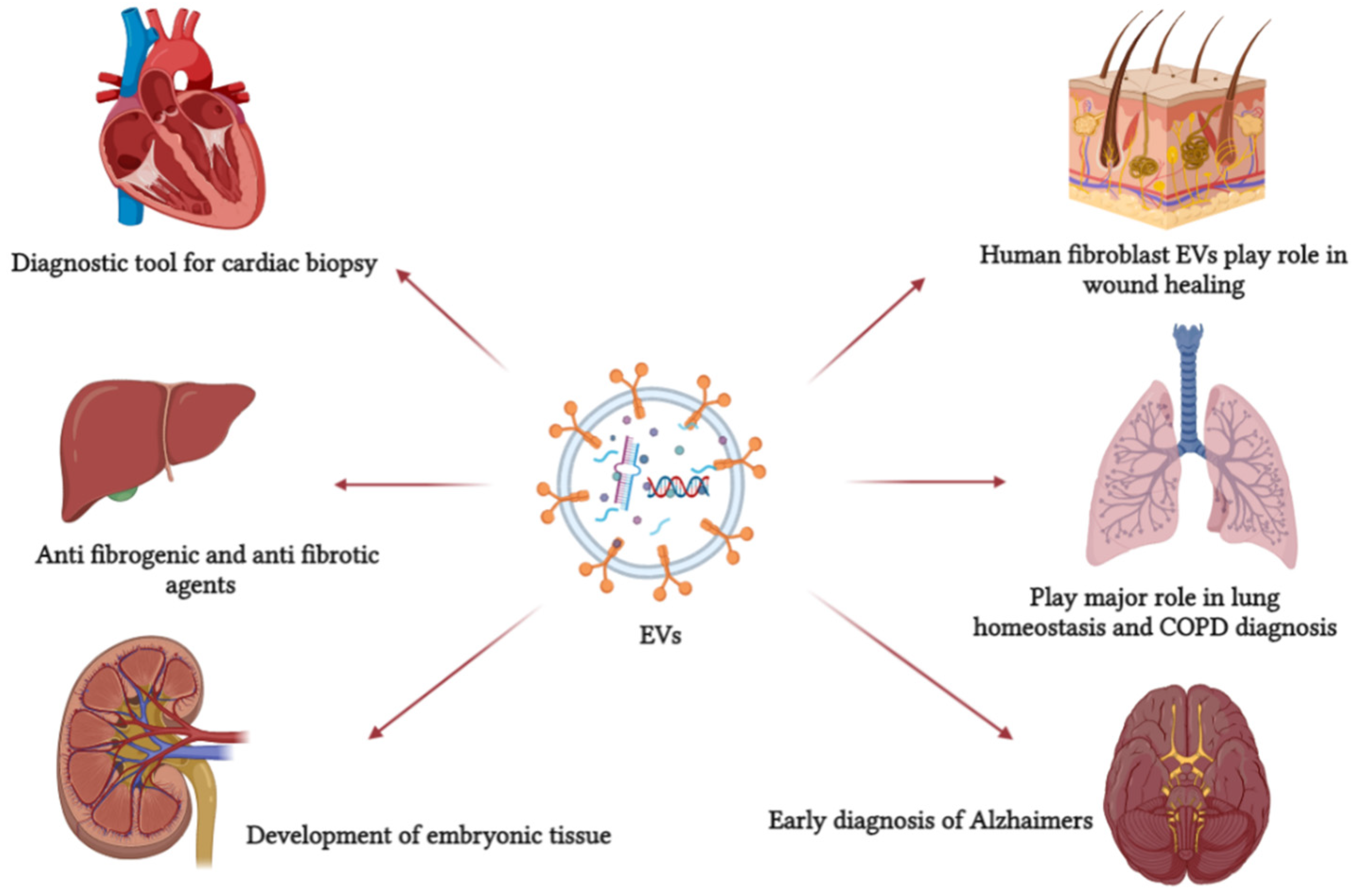
7. Application of EVs as a Therapeutic or Drug Delivery Agent
Evidence showed that EVs function systematically as a natural response and drug delivery vehicle and can be employed for target delivery (
Figure 2). EVs are detectable in most biological fluids in disease and immune response [
121]. EVs can be used as original therapeutic agents or as delivery agents. Most studies focused on the possibility of using them as drug delivery cargo to a specific target due to its membrane protein [
67]. Once the drug is encapsulated inside EVs, the advantage is that the drug works with the elements naturally present in them. In other cases, EVs only act as a drug carrier protecting the drug, passing it safely to the target location [
73].
Several studies indicated that EVs derived from stem cells could be used as a non-invasive treatment for a brain injury or brain infection, as these EVs retain the same therapeutic functions as stem cells. Furthermore, the secretomes of stem cells, including EVs, are the active compounds that attribute to their therapeutic roles; in addition, stem cell-derived
EVs can transfer the required genetic information and protein to accomplish the therapeutic process. Several studies applied stem cell-derived EVs on injured brain cells; the EVs acted as therapeutic agents on these cells [
122]. Additionally, immune cells also release EVs that act similarly to their origin and can be used in therapeutic applications. For example, dendritic cells (DC) release EVs that activate CD4+ T cells [
67] via the endocrine pathway, causing enhanced cardiac performance and reducing the time needed for wound healing in myocardial infarction. EVs derived from macrophages are used in plasmin encoding therapeutic protein and vaccination [
123]. Salivary EVs prevent the Zika virus attachment to the host cell, explaining why the virus is rarely transferred via saliva. However, the same study stated that the salivary EVs are not effective against SARS-CoV-2 that majorly communicate via salivary droplets [
124].
There are a variety of challenges facing scientists when develo** a therapeutic approach. These included toxicity, safety, target specificity, and large-scale production. Furthermore, a synthesized nanoparticle can solve the target delivery issue. However, it must undergo in vivo safety and toxicity assessment, and there is the issue with costly large-scale production. Using extracellular derived nanoparticles as drug carriers provides a possible solution since they contain lipid, RNA, and protein [
63]. As EVs transfer their content from the mother cell, it is the most critical attribute in drug delivery and cell communication [
36]. Their cargo, specifically RNA, was of particular interest due to the diverse population of miRNA with target genes that can influence and regulate the biological process in mammalian cells [
125]. Additionally, a growing body of facts indicates that cell apoptosis proteins are involved in EV formation. Unveiling the interaction pathway between EVs and autophagy that promotes cancer cell motility, providing significant advantages in early diagnosis and cancer treatment using EVs [
126]. The protein profile of EVs derived from mesenchymal cells retains a therapeutic effect observed in vivo. These proteins include osteoprotegerin and angiogenin [
127].
Drug loading into EVs can be divided into two strategies. The first is directly loading the drug into the exosomes; the second is targeting the mother cell by loading drugs during the biogenesis of exosomes [
34]. Furthermore, in the case of loading lipophilic drugs, the process is relatively easy due to the interaction between EVs lipid bilayer and the drug, which occurs via hydrophobic interaction [
128]. Several methods were utilized to achieve exogenous loading of EVs with drugs, including electroporation, incubation, sonication, and thawing. The level of success differs from one method to another; some cause degradation to either EVs or their cargo [
129].
The first time EVs were used as a drug cargo was when San et al. [
130] used EV-encapsulated curcumin fused with the plasma membrane. It was reported that EVs isolated from the plasma of humans and rats showed a protective mechanism against Acute myocardial ischemia/reperfusion [
131]. Successfully, EVs were loaded with anti-cancer and RNA-based drugs and cancer vaccination. These applications are based on cancer immunotherapy, indicating that the success of this approach is that EVs can be taken up efficiently by macrophages and tumor cells [
132]. Other drugs loaded into EVs include a lipophilic drug with a small molecular weight and RNA-based drugs, for example, small interfering RNA (siRNA) [
128]. Furthermore, the blood-brain barrier is the most selective in the human body. There are three primary methods to deliver drugs and therapeutic components into the brain, including invasive, pharmacological, and physiological approaches—the first approach is based on delivering drugs by breaching the blood-brain barrier. The pharmacological method includes modifying the active compound to enter the brain via a passive crossing. The last approach is the most efficient; it depends on the naturally present receptors found in abundance on the surface of the blood–brain barrier. These receptors provide easy and equal distribution of the loaded drugs into the brain. EVs possess two significant advantages as the usage will be non-invasive and were proven to cross the blood-brain barrier easily [
133].
Employing EVs as a drug carrier has several advantages compared to platelets. To mention a few, (1) EVs are smaller in size, which is an advantage when delivering drugs to cancer cells. For instance, EVs were more efficient in delivering the membrane protein SIRPa than ferritin. (2) EVs are originated from different sources. They are smaller and less toxic than lysosomes, with enhanced tissue biocompatibility and tolerance. (3) The cargo within EVs is protected by the double lipid layer that reduces the degradation and improves their biological stability. (4) Target cells can exclusively distinguish the specific receptors present on the EV membrane, reducing systemic toxicity and lowering drug outflow compared to conventional drug administration methods. (5) It is established that EVs could pass the BBB, giving an advantage in treating brain tumors. (6) Due to EVs’ ability to circulate the blood, the drug’s effect can last longer in the system [
71].
8. Drug Loading into EVs
Loading EVs with foreign substances for therapy continues to grow and develop [
134]., enhancing the therapeutic potentials of these particles. The loading approach can be categorized into EV alteration or cellular modification [
44]. Furthermore, it can be achieved post EV isolation (exogenous). Or during the biogenesis of EVs within the mother cell (endogenous) [
51].
EVs were tested to be used as a vehicle for drug delivery systems. One of these applications was developed by delivering systemically injected exosomes to target epidermal growth factor receptors in breast cancer; the study stated that these exosomes successfully conveyed miRNA and anti-tumor agents into epidermal growth factor receptors [
135]. Conjugating gold nanoparticles developed another system into the doxorubicin anti-cancer drugs that were physically loaded into the exosomes. The study stated that the target cell successfully took up these modified exosomes [
136]. Another modified EV consists of exosomes-liposomes fusion by repeated freeze-thawing mechanism; this method successfully altered the treated exosomes surface, resulting in enhanced colloidal stability and reduced immunogenicity [
135]. The significant advantage in using EVs as a drug delivery agent or as a therapeutic agent on their own is the ability of these EVs to control their RNA, proteins, and lipids and employ it as a tissue-specific response [
121]. Furthermore, EVs are being engineered and modified to enhance the encapsulation of the drug and the targeting accuracy [
137].
Although EVs proved to have significant targeting abilities, some studies indicated that EVs were accumulated in the spleen and liver and later removed by macrophages. Two techniques were developed to correct the off-target internalization of EVs. The first is based on modifying the surface of EVs using polymers, increasing the duration of EV blood circulation and possibilities of target accumulation. The second approach is based on modifying the surface of EVs by adding a part of a molecule that has an affinity to the target cell or tissue [
138].
Advantages of EV-Drug Delivery Technique
The internal cargo of EVs is protected by their membrane, likely by encapsulation, primarily originating from the cytosol of the mother cell, which will be transferred to the recipient cell upon fusion. Since the intraluminal vesicles are originated in the acidic condition of the multivesicular body, EVs and their protein should be resistant to pH. Additionally, upon entering the endosomal pathway, EVs will also undergo progressive acidification [
139].
In addition, since EVs are encapsulated phages, some demonstrate higher tolerance for harsh conditions, providing higher efficacy when orally administered. The pH conditions in tumor cells may range from 6.0 to 6.8. Initial data concluded that cancer cells cultured in a physiological pH 7.4 released a significantly lower concentration of EVs than those cultured in 6.4 pH. Concluding that, cells release EVs at low pH to avoid toxin accumulation [
140].
Ogawa et al. [
139] studied the stability of EVs under different pH conditions. The study stated that EVs are stable in functionality and shape under acidic conditions for at least 3 h. In case of EVs rupture, the action can be due to membrane penetration by protease. The process is facilitated by fatty acid cleavage mediated by phospholipase A2, leading to EV surface protein degradation [
139]. The surface protein profile on the EVs’ membrane is important due to proteases damaging the extracellular environment. For example, the IgA present on the surface of EVs protects these vesicles from breaking down [
141].
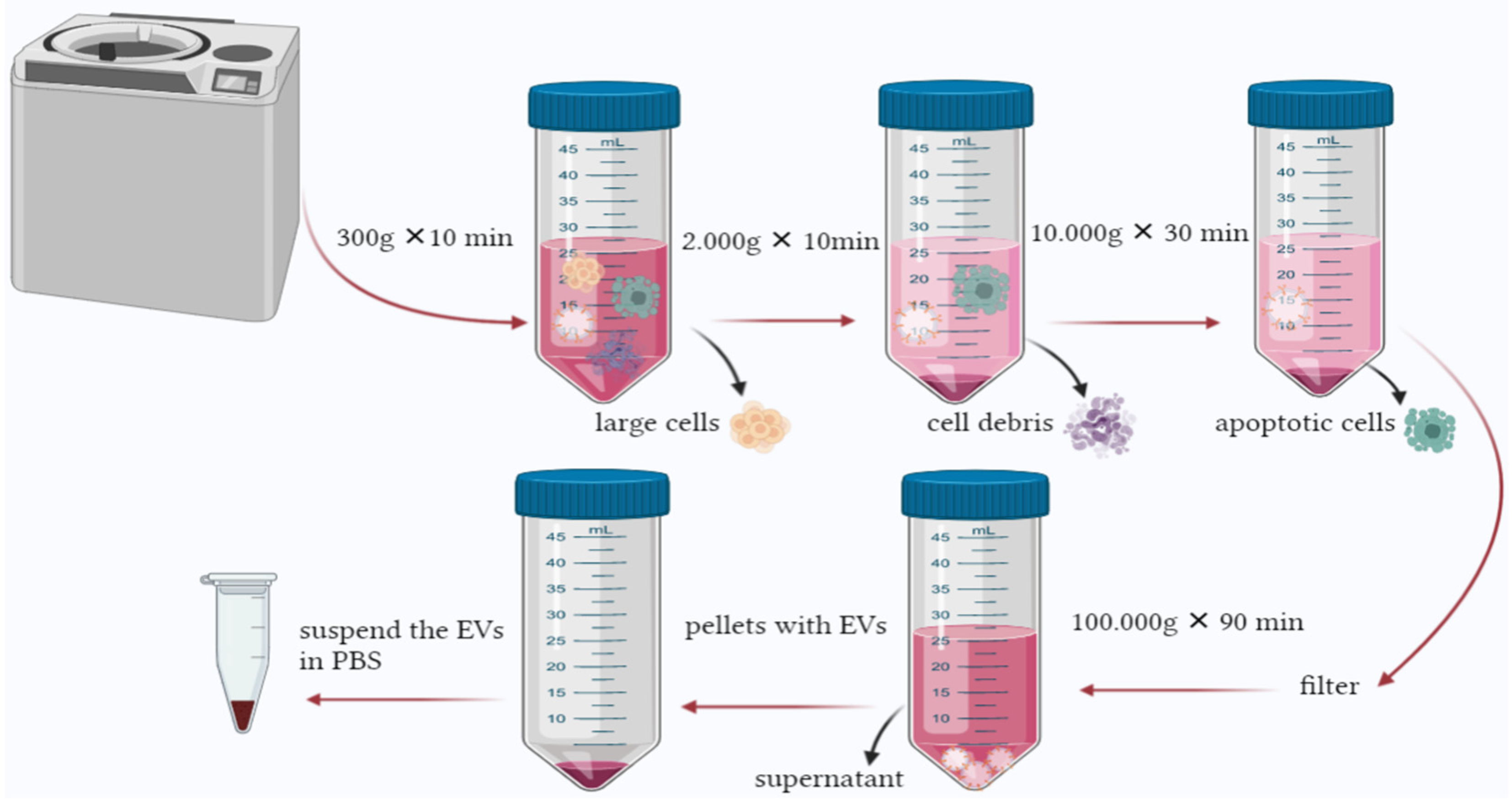
10. Characterization of EVs
The size of EVs in general and exosomes, in particular, is so small, making accurate characterization of EVs very challenging. For instance, nanoparticle tracking analysis will not detect particles smaller than 50 nm; on the other hand, flow cytometry will only detect particles larger than 300 nm. Dynamic light scattering will detect particles varied in size between 1 and 6000 nm with overestimating the particle count. Another option is using atomic force microscopy and electron microscopy. However, these methods are time-consuming, costly, and require professionals to operate the device. For example, if the sample concentration is high, the support film will be damaged, preventing EV counting and imaging since the characterization methods are not standardized and integrated. The data available in the literature are diverse and conflicting [
71].
Physicochemical and biochemical characterization of EVs aims to identify the isolated population and elucidate their profile in terms of lipidomics, proteomics, and gene expression analysis [
154]. Physicochemical characterization of EVs studies their size [
155], morphology [
156], cargo, or the number of vesicles; the latter can be achieved by zeta potential analysis [
157]. Size characterization is commonly achieved using dynamic light scattering techniques [
158], light microscopy, and electron microscopy [
21]. Vesicle concentration and size distribution are studied using nanoparticle tracking analysis [
159,
160] and flow cytometry [
105].
Cry electron microscopy characterizes the particles without adding heavy metals to the sample as fixative agents. This method was used in a study to characterize single cell EVs in terms of their morphology, structure, and lipid layer. The cry electron microscopy provided information confirming that single-cell EVs showed diverse characteristics [
26].
Flow cytometry has advantages and disadvantages. The functioning concept is detecting the vesicles’ fluorescence and light scattering signal individually, but the technique does not detect all EV subclasses. Therefore, the quality of the results depends on the device’s sensitivity since EVs concentration increases when their size decreases [
161].
Additionally, proteomic characterization is a practical approach to detect common EV markers. The most common proteomic characterization is conducted by mass spectrometry [
101]. Label-free quantitative proteomics is a mass spectrometry approach that detects common EV protein markers regardless of their morphology, origin, or cellular strain. This method can detect plasma membrane protein, tetraspanin, and GTPase [
162]. Liquid chromatography-tandem mass spectrometer (LC-MS/MS) analysis also characterizes EV protein concentration and cargo [
150].
Characterization of EVs based on their genetic makeup can be achieved by running a quantitative real-time PCR; the analysis takes place after extracting and amplifying the RNA to detect the genes present and their respective metabolic process [
105]. Ex vivo behavior and communication of EVs, especially tumor-derived EVs, are investigated by applying 2D and 3D cell culture models; these two techniques are significant for evaluating RNA-derived EVs. Although, the 3D cell culture model is more competent when evaluating the shape of the vesicles, their behavior, and their extracellular communication [
49].
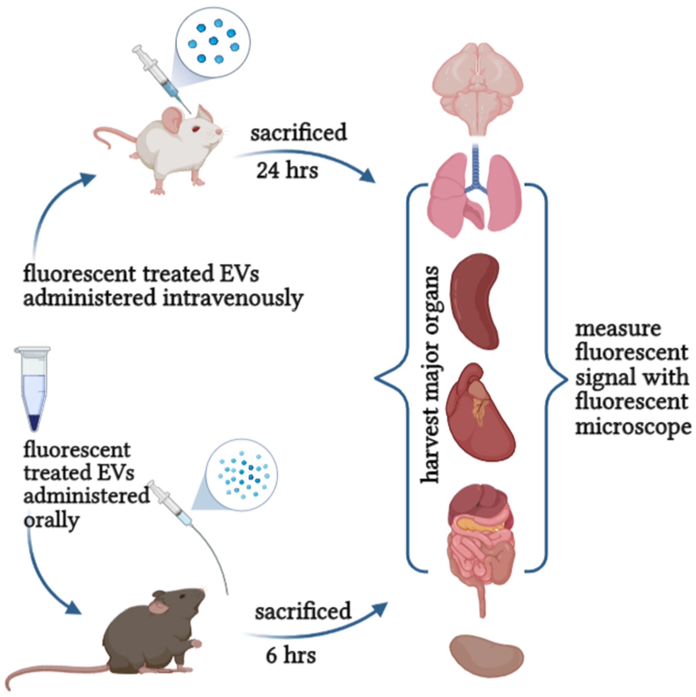
11. Toxicity and Safety Assessment of EVs
Due to the endogenous properties of EVs, they migrate between cells and deliver their biological cargo upon crossing the membrane [
86]. EVs can travel long distances through the blood circulation and reach various tissues affecting the signaling process [
82], mediating several pathological and physiological functions [
83]. On the other hand, EVs play a significant role in anti-inflammatory and antiapoptotic actions [
88]. It could either work as protected vesicles that expel invading pathogens from the infected cell or as vector-transmitted virulence factors facilitating the infection [
163]. Meaning that the function of EVs upon entering the body depends on the biological status of the host and the receiving cell. There are cases when EVs act as immune responses against infection. Furthermore, it worked as a signaling pathway for pathogens [
164].
Studying the efficacy of EVs in therapy is significant, especially in preclinical development. However, evaluating their risk and toxicity is essential in develo** their therapeutic performance. Attention must be directed towards the possibility of immunogenicity or toxicity of therapeutic EVs [
165]. Therefore, an in-depth understanding of EVs’ overall toxicity and biodiversity following in vivo studies is required before using them as therapeutic agents or drug carriers, primarily if the EVs are originated from xenogeneic sources [
166].
Somiya et al. [
101] evaluated the safety of milk-derived EVs; the paper stated that no systematic toxicity or immunogenicity was detected. The mice were given an intravenous injection containing EVs derived from milk. A blood sample analysis confirmed the safety of these EVs, and no toxicity markers from the kidney nor the liver were observed.
A study conducted in vitro evaluation of the toxicity of mesenchymal and bovine milk-derived EVs stated that there is no genotoxic response to either EVs, but collagen-induced platelet aggregation in a dose-related manner was recorded. In vivo investigation was conducted to assess the safety of engineered HEK293T cell-derived EVs via intravenous administration for 21 days. The results provided no signs of toxic effect, immune changes, and no alteration of the EVs [
167].
According to a study conducted by Roccaro et al. [
168]. EVs were isolated from Mesenchymal stem cells obtained from multiple melanoma patients. The results indicated that EVs promote tumor growth by dissemination and metastasis to distant multiple melanoma niches. Besides, EVs are associated with stimulating or suppressing the immune response and play a role in regulating the suitable microenvironment for the tumor’s biological process [
163].
Plant-derived EVs pose several advantages over mammalian-derived EVs. Therefore, the field experts recommended an accurate assessment of their toxicity and safety for clinical application [
169]. Engineering EVs into a therapeutic tool is valuable, ensuring quality and safety. in the time being, most available data on the safety and toxicity of EVs in vitro and in vivo are using mesenchymal stromal cells-derived EVs. Not enough attention was dedicated to other EVs from different sources [
166].
Therefore, evaluating the toxicity and safety of EVs in vitro and in vivo will set the dose for future clinical use and safe consumption recommendations for human application. It provides fundamental insight into pharmacodynamics, therapeutic benefits, purity, ideal storage, shelf-life, and quality control (
Figure 4). These quality and safety control processes will innovate the possibilities of applying plant-derived EVs in therapy and establish regulations and standards for production and development.
Although, there are no set standards or parameters to evaluate the safety and toxicity of EVs. However, understanding their uptake and behavior locally and systematically is vital to assess their safety [
170].
13. Discussion and Conclusions
This review provides a concise retelling of the history, concept, development, latest findings, and recommendations related to extracellular vesicles. We recommended this paper to those new to the field needing to understand EVs and those already familiar with it but wanting to keep up with the latest findings. We listed the continuous change in technical developments, new findings, and debates along the review lines.
Starting from the early history of these riveting nanoparticles, we observed that it is well established that contrary to the previous common concept that classifies EVs as a mere cellular waste carrier, the field’s advancement unveiled that these nano-sized vesicles play an essential role in several processes [
175]. One of the most is their involvement in cellular communication, transportation of miRNA, protein, lipids [
60], and as a significant player in several pathological processes, including infection and metastasis [
176], agreeing that EVs are an appealing tool for non-invasive rapid diagnosis that can be exploited in the case of asymptomatic diseases [
177]. Another vital fact is that EVs play an essential role in embryonic growth, development, and maturation. Such as in breast milk-derived EVs, which transports EVs from the mother to the newborn [
178]—confirming their contribution to pathological or physiological processes.
The reported number of searches on EVs is elevated yearly, providing more knowledge and clues to understand these nanoparticles better.
This review focused on the most recent advancement in extracellular vesicles, their function, the latest update on EVs cargo, and their contribution and involvement in physiological and pathological processes [
179]. We highlighted some of the technical challenges, current findings and, breakthrough developments in research related to EVs isolation, characterization, and drug loading technology. That has been addressed by many papers [
180], giving hope for more findings and better results.
A fact affirms an increased count of the number of EVs circulating in the system during infection, providing early diagnostic tools significant in cancer or sudden brain ailment [
83]. Several researchers concluded that cancer cells release EVs and therefore are found circulating in the body carrying their genetic code [
181]; these EVs act as a messenger, communicating with nearby or distant cells [
182] during metastasis, making the surrounding environment ideal for the pre-metastatic niche [
183].
ISEV visited the topic in a position paper, stated that EVs had already been used in human trials to treat cancer in early 2000 [
177]. Furthermore, the available results point to the importance of the role played by plant-derived EVs in cancer studies as therapeutic and drug carriers [
71]. The cargo of plant EVs is substantial and similar to mammal EVs, not to mention their use in therapy is meaningful to those who are strictly vegan or religious individuals. All are essential contributions to the fight against cancer.
Some studies used plant-derived EVs to understand their function and potential use as a drug carrier [
184]. Plants EVs also act similarly to mammal EVs, albeit with a slight difference in the biogenesis, circulate, allocate and carry specific cargo [
185]. The significance of plant EVs is underestimated since plants are readily available from biological origins [
186]. Not to mention that the toxicity of plant source materials is not substantial, with no considerable reported side effects [
187]. As a result of this review, mammal and plant EVs are similar in function, characteristics, and cargo, and can act in physiological and pathological conditions. Using these EVs in future research can majorly contribute to the field, especially if the safety of plant EVs were to be confirmed. The already-existing body of evidence on the use of plant-derived EVs for nano-delivery offers a wide range of possibilities for their future use in health and therapeutics.
Since there are several obstacles, the researchers face this field [
188]. We highlighted the latest advancements, recommendations, and contributions from ISEV, the leaders in this field. ISEV stated in an early published position paper that EVs’ field faces continuous challenges technically, including the expanding number of EVs isolation methods with low yield [
189]. Advising that, it is essential to accurately state whether the studied EVs are used for clinical application as the active drug component, or it is simply the drug carriers following ISEV guidelines [
177].
Additionally, based on the recommendations from ISEV that were published on their position paper titled ‘Applying extracellular vesicles-based therapeutics in clinical trials an ISEV position paper’. The paper mentioned that using EVs in therapy must comply with the established regulations. Clinical investigation and quality control are crucial aspects of EV production in therapy. It is essential to accurately state whether the used EVs are the active drug component or used as a drug carrier. Their statement was:
EV-based therapeutics can be defined as biological medicine and belong to the pharmaceutical class of biologicals. Regulatory frameworks for manufacturing and clinical trials exist in Europe, Australia, and United States, but special guidelines targeting EV-based therapeutics may be needed.
However, more extensive studies are needed to fully understand the former as an active component in therapy; we recommend that added attention be directed towards plant-derived EVs, their function, cargo, and characteristics that were the subjects of a shy number of articles compared to mammal EVs. More safety assessment studies and quality control standards are additional clinical application recommendations. We observed that the lack of standard isolation, characterization protocols, and quality control hinders these studies’ advancement in either purity or yield of EVs for clinical use [
190], making it hard to link and compare the available findings. Then again, the field recognized these issues [
65] and they have been addressed on several occasions, emphasizing the need to standardize the isolation protocols. Although there is so much more to learn about EVs, what scientists have discovered so far is fascinating and promising. Nevertheless, this preliminary data offers additional insights into other possible benefits of EVs for a better future.


 ,
, 


