3.1. Optical, Structural and Morphological Characteristics of CQDs
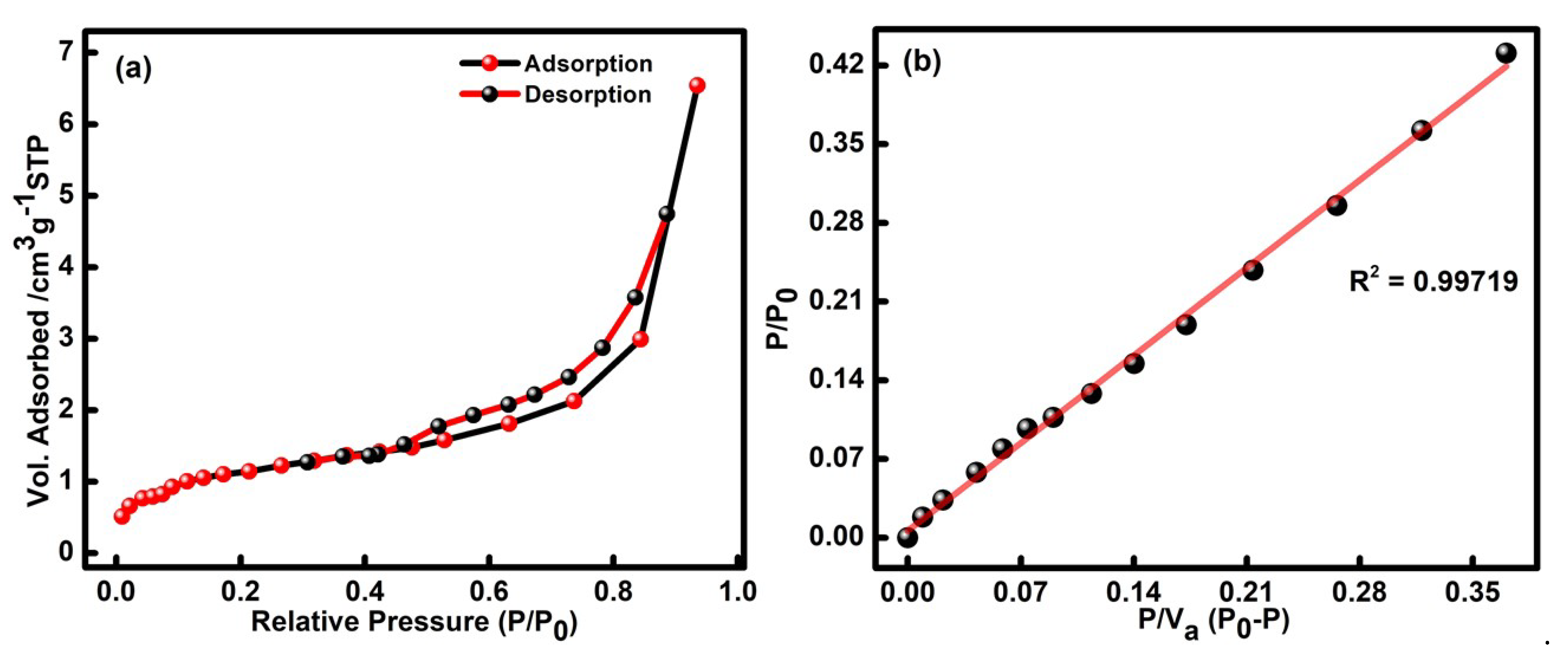
The surface area of the CQDs is examined using N
2 physio-sorption analysis in liquid nitrogen at 77 K, as presented in
Figure 1a,b. The obtained adsorption-desorption isotherm of the formed CQDs is classified as a type IV isotherm with a hysteresis loop. For the examination, the saturated vapor pressure and adsorption temperature were set up at 100.95 kPa and 77 k. The surface area of the RS@CDs was determined to be 3.95 m
2/g, as estimated in the BET plot (
Figure 1b). Moreover, the respective linear fit curves displayed the mean pore diameter in the range of 10.23 nm. The pore size volume and micropore volume of the formed CQDs were found to be 0.01012 cm
3/g and 0.9088 cm
3/g, as estimated through the BET plot illustrated in
Figure 1b. The respective outcomes have confirmed that the mesoporous nature of the developed particles can further be useful in detecting harmful pollutants to a large extent.
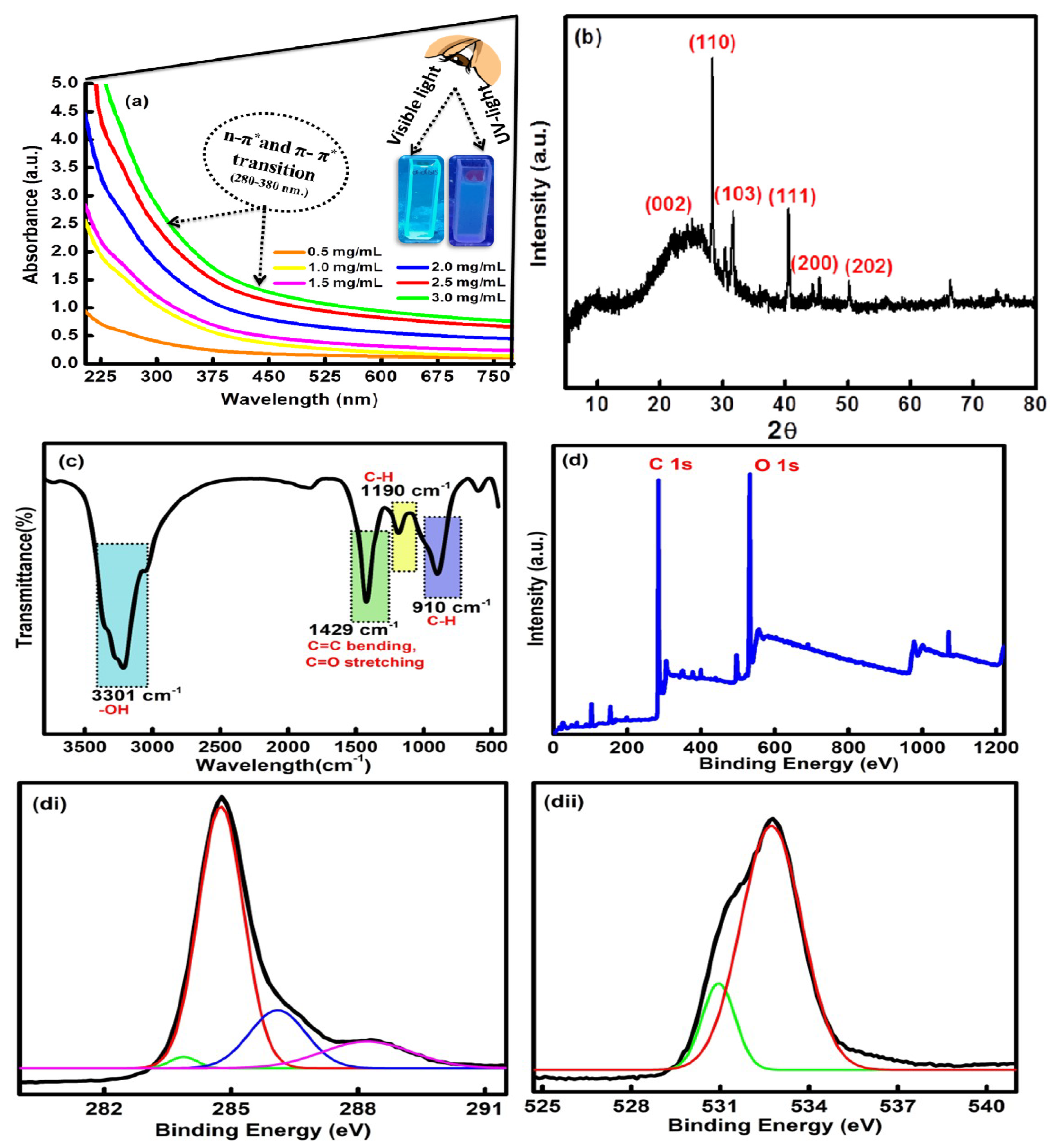
The UV-vis. spectral profile of the formed CQDs has displayed a characteristic absorption band between 300 and 400 nm (
Figure 2a). The obtained absorption hump is related to the n-π* and π-π* transition of the C=O and C=C bond of the carbonyl and carboxyl groups on the exterior facade of the CQDs [
35]. The synthesized CQDs have a reflected, milky, transparent color under the visible light, and an oceanic blue color was obtained under UV light (inset
Figure 2a). The effect of different concentrations of CQDs, ranging from 1.0 mg/mL to 4.0 mg/mL, was also measured using UV-vis spectroscopic studies (
Figure 2a). On interpreting the results, it was found that, with an increase in the concentration, three bands at wavelengths of 320 nm, 350 nm and 370 nm were observed, accompanied by a progressive increase in the absorbance of the samples. The results supported the idea that the absorbance of the sample was dependent on the concentration of the formed CQDs. The absorption band located around 320 nm is ascribed to the core state of the π-π* transition. The edge band of the n-π* transition was mainly seen at 350 nm in the prepared CQDs. However, the band at 370 nm mainly arises due to the existence of different types of functional groups such as carboxyl, carbonyl and hydroxyl bonded on the exterior edge of the CQDs [
36]. The X-ray diffraction spectra of the fabricated CQDs have shown the existence of major peaks at 2θ = 23.5°, 27.29°, 29.8°, 40.1°, 45.2° and 49.9° corresponding to the (002), (110), (103), (111), (200) and (202) diffraction planes in the CQDs [
37]. The presence of a broad peak at 23.5° indicates the carbonaceous core of the CQDs (
Figure 2b). The outcomes have given direct evidence of the effective synthesis of CQDs from rice straw waste with a high purity and a good crystalline size ranging between 8 and 9 nm [JCPDS NO. 26-1076].
The surface composition of the CQDs has further been assessed by using FTIR analysis (
Figure 2c). The spectra displayed three main sharp peaks at 3301 cm
−1, 1429 cm
−1 and 910 cm
−1. However, two minor peaks were found at 1190 cm
−1 and 610 cm
−1. The high intense peak at 3301 cm
−1 is mainly due to the presence of O-H stretching vibration from the hydroxyl groups over the exterior facade of the CQDs [
38]. Further, the strong characterization peaks at 1429 cm
−1 arose due to the presence of C=C bending and C=O stretching vibration in carbonyl and carboxyl functionalities on the surface of the CQDs. Another sharp peak at 910 cm
−1 has been assigned to the C-H moieties in the CQDs [
39]. The absorption peaks at 1190 cm
−1 and 610 cm
−1 appeared due to the C-H bending vibrations in the CQDs. The presence of hydroxyl, carbonyl and carboxyl functional groups on the outer surface of the CQDs has further enhanced their potential in sensing harmful toxic elements and has strengthened their aqueous solubility to a large extent [
40].
The elemental configuration and content were further studied using XPS analysis. The high-resolution XPS survey has shown two typical peaks at 284.20 eV and 530.45 eV, which are further associated with the characteristic binding energy signals of C 1s and O 1s, respectively (
Figure 2d). The core-level XPS resolution of C 1s indicates the existence of peaks at 284.84 eV, 285.50 eV, 286.54 eV and 287.12 eV, which were assigned to the C=C, C-C, C=O and C-O bonds (
Figure 2di) [
41]. The high-resolution XPS of O 1s was further deconvoluted into two peaks at 532.33 eV and 534.44 eV, which may be attributed to the C-O-C and C-OH functional groups in the as-synthesized CQDs (
Figure 2dii) [
42]. The results of the FTIR and XPS studies have further validated the existence of carbon and oxygen due to the presence of carboxyl, hydroxyl and carbonyl functional moieties over the exterior façade of the formed CQDs.
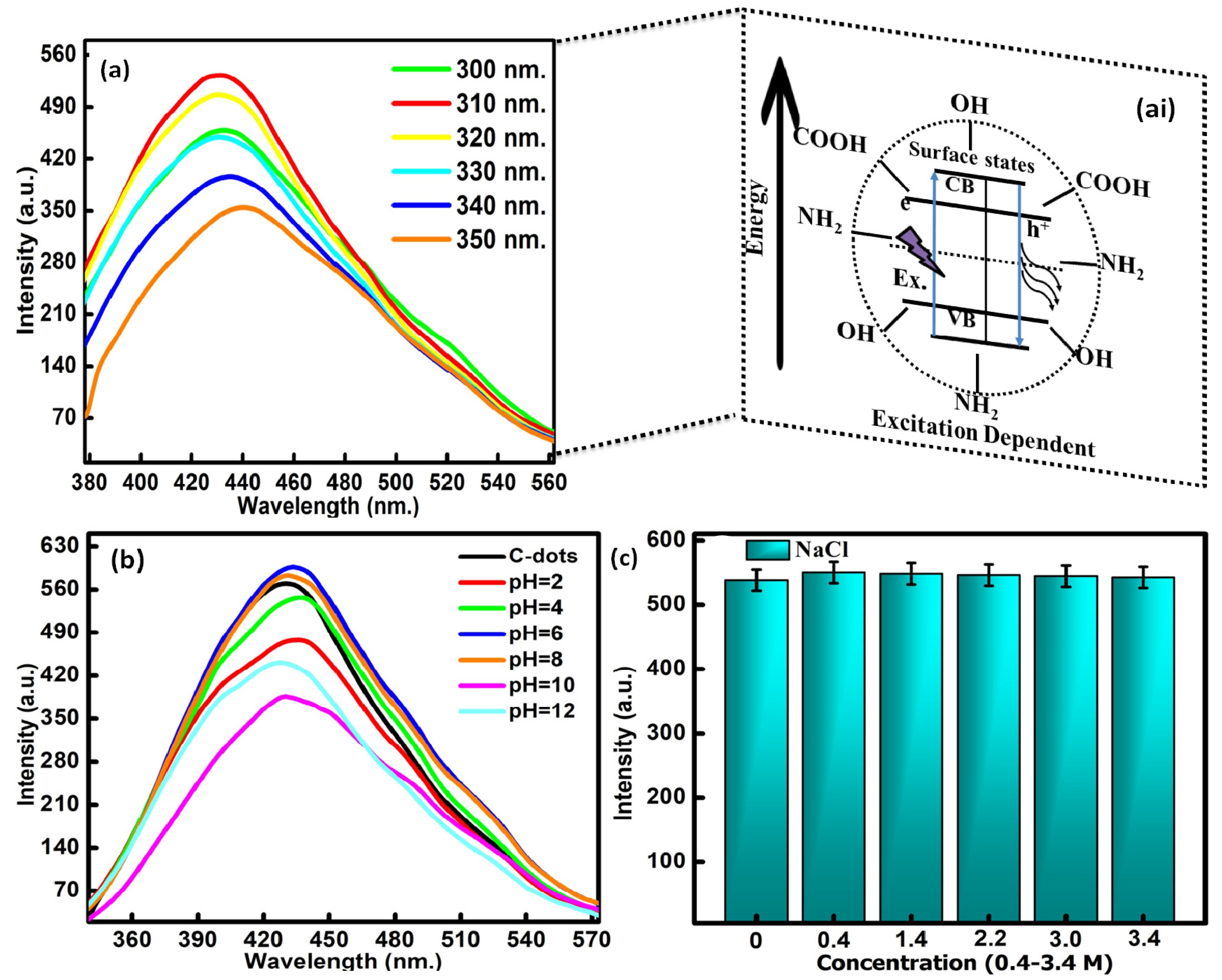
The excitation-dependent emission properties of CQDs were assessed to evaluate the stability of the formed particles (
Figure 3a). The measurements were taken out as a function of the excitation wavelength in the range of 300 to 350 nm. The fluorescence spectrum has displayed the maximum emission intensity at 535 nm with the excitation wavelength of 310 nm. Therefore, all the fluorescence emission studies were carried out at a wavelength of 310 nm. These optoelectronic properties of the CQDs were associated with carbon core defects. The significant distribution of emissive trap sites in the CQDs is also responsible for the emission properties in the formed particles [
43]. These surface defects have the ability to capture the epicenters of excitons due to the presence of oxygen and nitrogen-containing functional groups on the exterior facade of the CQDs. It will further enhance the surface-stated interrelated fluorescence in the developed CQDs. The quantum yield calculated using the reference standard method came out to be as high as 66% in reference to the quinine sulfate dye. The stability of the CQDs was further examined by varying the pH range from 2 to 4, 6, 8, 10 and 12 in the reaction media (
Figure 3b). It was observed from the spectra that the emission intensity gradually increased in the pH range of 2 to 8, with the maximum emission at pH = 6. The results therefore supported the stability of the prepared particles in the slight alkaline medium. Upon enhancing the pH value of the reaction media above 8, a significant decrease in the peak position with the blue shift was observed in the fluorescence intensity (
Figure 3c). This mainly arises due to the alterations in the fluorescence behavior of the acidic nature of the CQDs in the basic environment. The observed outcomes can be explained by the removal and addition of the proton part of the hydroxyl and carboxyl functionalities that exist on the exterior facade of the CQDs, which leads to the further modification of the electrostatic interacting behavior of the formed particles. Furthermore, the influence of ionic strength on the fluorescence potential of the CQDs has also been evaluated in the presence of different concentrations of NaCl solutions (0.4–3.4 M). The resultant graphs indicated that very high concentrations of NaCl produced a negligible effect on the fluorescence emission intensity of the formed particles (
Figure 3c). The above outcomes have supported the stability of the formed particles in the presence of high-ionic-strength solutions. The outcomes have further supported the aptness of the formed particles in the field of toxin sensing because of their high emission and exceptional stability in different physiological and ionic strength media.
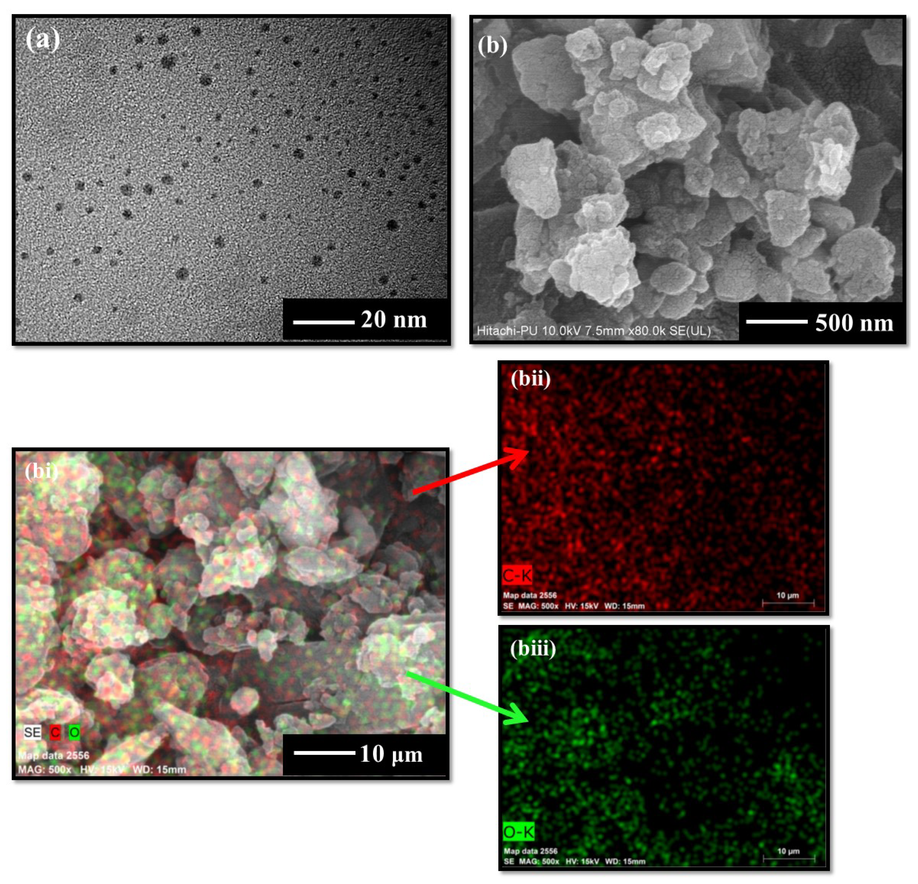
The morphological studies using HRTEM analysis have shown the existence of unevenly spherical and well-diffused particles with an average size range of 5–10 nm (
Figure 4a). The FESEM images have further shown the existence of the circular lamellar porous nature of the CQDs (
Figure 4b). However, the particles appeared to be in an agglomerated state in solid form, as shown in
Figure 4b. The respective elemental analysis represents the main constituents of the formed particles containing the carbon (C) and oxygen (O) elements in the major atomic percentage, as illustrated in
Figure 4bi-biii. These findings indicate that the developed CQDs have only specified elemental peaks, and the lack of any other peak indicates the high purity of the synthesized particles.
3.2. In Vitro Stability and Colloidal Assay of CQDs over Blood Components
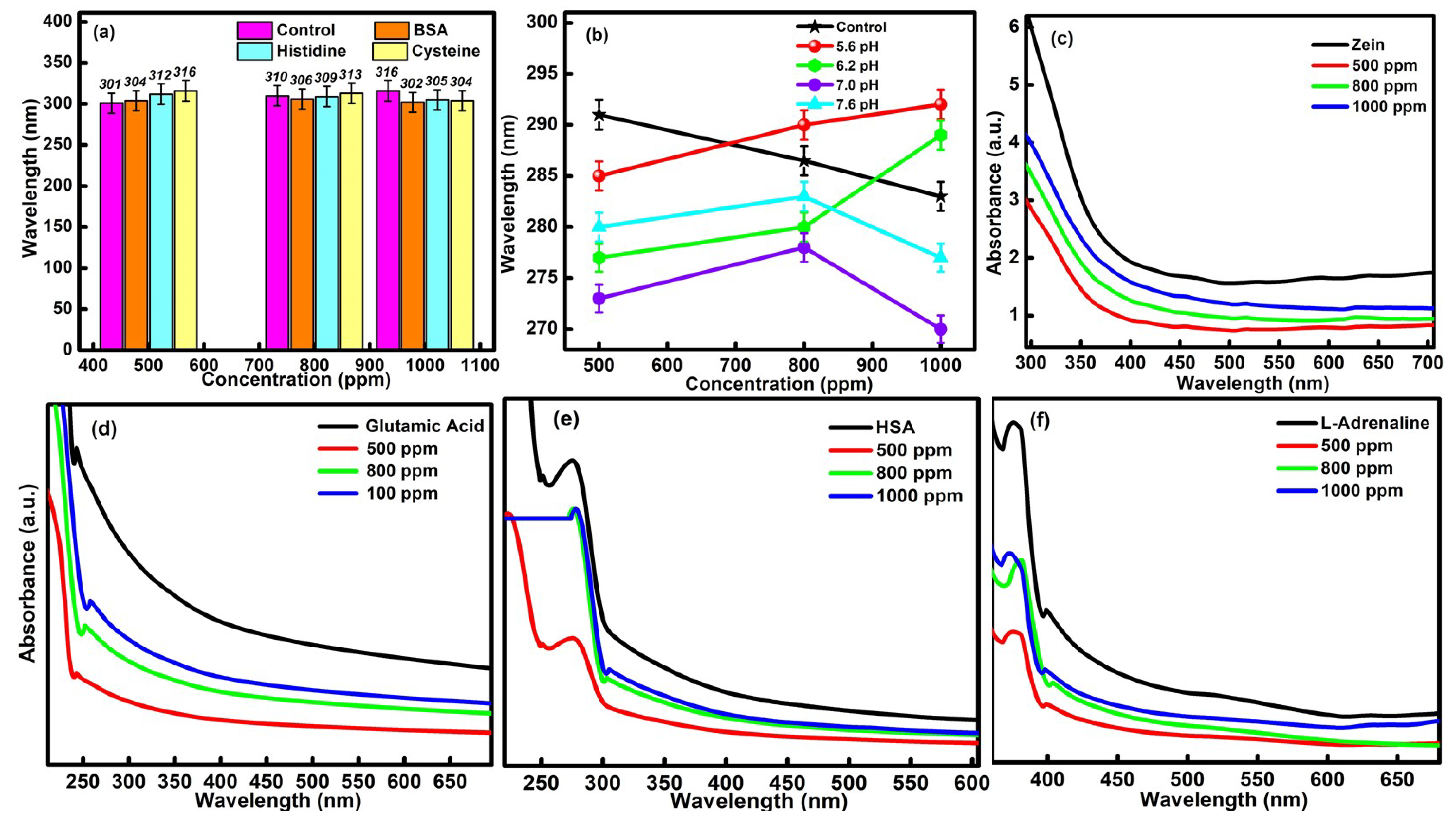
The prime requisite condition before executing the practical and in vivo application of formed CQDs is to evaluate their in vitro and colloidal stability with essential blood protein components. The respective study was performed by determining the absorption efficiency of the different types of blood components (BSA, HSA, cysteine, histidine, L-adrenaline, zein and glutamic acid) in the presence of different concentrations of CQDs (i.e., 1000 ppm, 800 ppm and 500 ppm). The corresponding effect of different pH conditions on the various biological components has also been tested for the developed CQDs. Upon interpreting the data, it was found that the high concentrations of CQDs have not produced any kind of visual or aggregation changes in the chosen blood components and supported the in vitro stability of particles in biological media. It was also observed that the wavelength of CQDs was only shifted in the permissible range of 14 nm in the presence of different blood components, which further confirmed the in vitro stability of CQDs (
Figure 5a). The outcomes have further been compared with the buffer solutions of different pH values ranging from 5.6 to 7.6, (
Figure 5b). The observed changes in the absorbance values were quite comparable with the earlier results. In addition, the slight hypochromic and bathochromic shift in the data is mainly attributed to the bio-compatible nature of the developed CQDs.
The colloidal stability of the CQDs was further assessed by carrying out the absorption studies in the presence of zein, glutamic acid, HSA and L-adrenaline (
Figure 5c–f). The ascorbic acid has been used as a control during the measurements. The absorption peak at λ = 250 nm, in the case of glutamic acid, and the characteristic peak at λ = 310 nm, for HAS, have been checked in order to interpret the results. In the case of zein, the characteristic absorption peak was observed at λ = 350 nm. The UV-vis peak at λ =360 nm was checked in the presence of L-adrenaline. Upon increasing the concentration of CQDs from 500 ppm to 1000 ppm, a slight increment in the absorbance values was observed in the presence of all the chosen biological components. However, the highest absorbance value was discovered in the case of 1000 ppm of CQDs. This has been explained due to the maximum agglomeration of the particles of essential blood elements over the surface of CQDs. In addition, no negative effect of CQDs on the essential biological components has been observed, which further reflects the biocompatible nature of the prepared particles and made them useful in the biomedical field. Furthermore, the structure of synthesized particles does not show any kind of variations in the presence of the chosen blood components, which, in turn, supports their high in vitro stability and colloidal stability.
3.4. Sensing Aptitude of CQDs
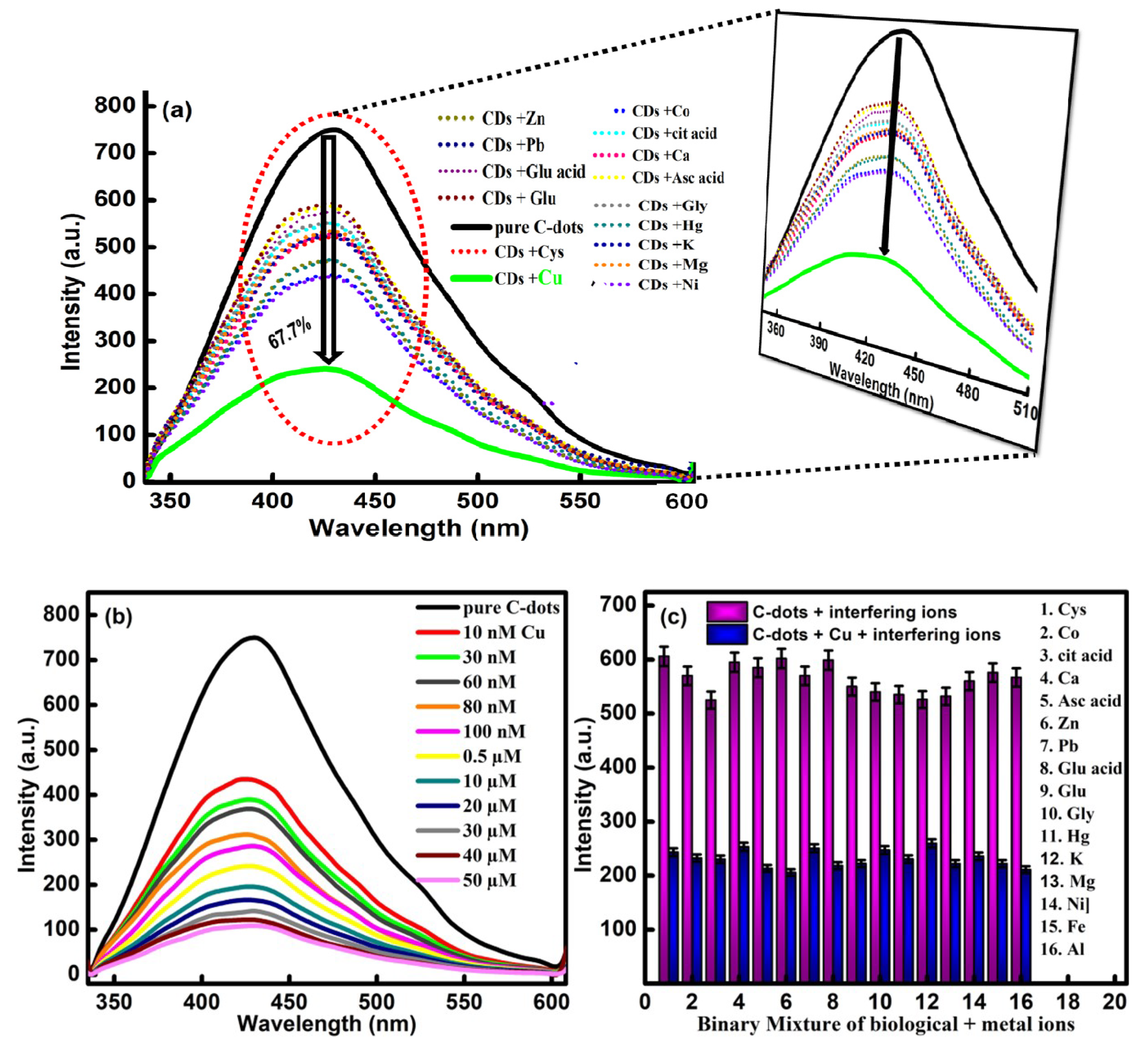
The well-defined emission properties of CQDs were checked against various types of metal ions to enhance their scope in order to develop an easy-to-sense platform without any catalytic-mediated process. For the analysis, the changes in the fluorescence behavior of the developed CQDs have been compared in the presence of 15 different types of biomolecules and metal ions including Ca
2+, Hg
2+, K
+, Mg
2+, Cu
2+, Ni
2+, Pb
2+, Co
2+, Zn
2+, glutathione, ascorbic acid, glutamic acid, citric acid, glycine and cysteine (
Figure 7a). Upon comparing the data, it has been observed that the fluorescence intensity of the CQDs have generated small variations in the emission intensity. However, the presence of Cu
2+ has produced maximum quenching in the emission intensity of CQDs. Therefore, the developed particles are found to be selective for Cu
2+ ions. The quenching in the fluorescence emission intensity is explained by the presence of different types of functional groups over the surface of the CQDs, which further affected the optoelectronic properties via the non-radiative electron transfer mechanism. The turn-off fluorescence signal of the CQDs in the presence of Cu
2+ ions is further explained by the splitting of the d-orbital of Cu
2+ during the complex formation with the carboxyl and hydroxyl groups of the CQDs and Cu
2+ [
44]. As a result, electrons present in the excited state of the CQDs are transferred to the d-orbital of Cu
2+. Additionally, the paramagnetic nature of the Cu
2+ ion leads to the restraining of the transition of the electrons in the form of radiation emission, which further results in the fluorescence turn-off [
45].
Furthermore, the sensitivity of the developed sensor has been probed by varying the concentration of Cu
2+ (50 μm to 10 nm) via the titration-metric method (
Figure 7b). The quenching efficiency of the CQDs has been enhanced significantly with the increase in the concentration of Cu
2+ ions in the reaction media. The designed sensory probe has the potential to turn off the fluorescence intensity of the CQDs in the presence of Cu
2+ ions, which further supports the high sensitivity and accuracy of the developed sensor.
To enhance the detection response of the CQDs, the selectivity response of the developed sensor has further been verified in the presence of different types of biomolecules and metal ions such as Ca
2+, Hg
2+, K
+, Mg
2+, Ni
2+, Pb
2+, Co
2+, Zn
2+, Fe
3+, Al
3+, glutathione, ascorbic acid, citric acid, glutamic acid, glycine and cysteine. The analysis has been performed by mixing the different metal ions with a 60 μM concentration of the Cu
2+ ion. From the data, it has been observed that the fluorescence response of the sensor remains unaffected in the presence of different types of metal ions (
Figure 7c).
However, the presence of the Cu
2+ ion has the potential to enhance the degree of quenching in the emissive response of the CQDs. The outcomes have suggested the high selectivity and sensitivity of the formed particles over any other metal ions and biomolecules. The complex formation between the carbonyl, carboxyl and hydroxyl groups of the CQDs with the Cu
2+ led to the transference of electrons in the form of non-radiation recombination to achieve the selective quenching of Cu
2+. The results gave direct evidence of the high selectivity and superior sensitivity of CQDs against Cu
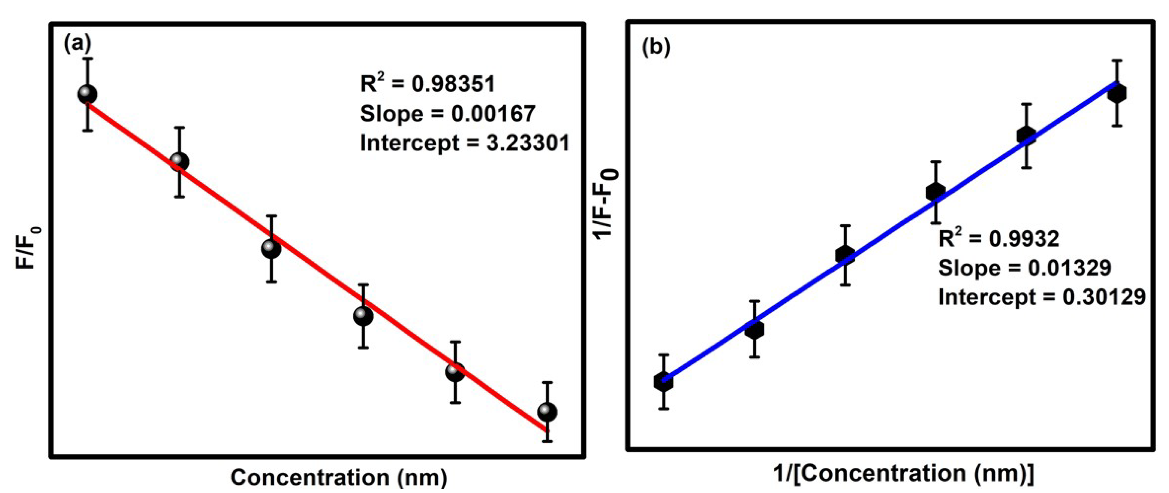
2+ ions. Furthermore, a better understanding of the quenching mechanism has been accessed by using the Stern–Volmer relationship [
46].
Here, F
0 is the initial fluorescence intensity of the CQDs in the presence of the Cu
2+ ion, whereas F represents the fluorescence intensity of the CQDs in the absence of the quencher Cu
2+. Q refers to the concentration of Cu
2+ in the solution, and K
SV is the Stern–Volmer constant. Significantly, the limit of detection was calculated by the standard deviation rule given below:
where SD indicates the standard deviation, and P is the slope of the Stern–Volmer plot (
Figure 8a). Moreover, the binding efficiency of the developed sensory probe was estimated from the Benesi–Hildebrand plot (
Figure 8b). The obtained values of the detection limit, binding constant, quantization constant and fluorescence quenching factor have been given in
Table 1. The limit of detection of the sensor was calculated to be 0.31 nm.


 ,
, 


