Article

Polyoxa- and Polyazamacrocycles Incorporating 6,7-Diaminoquinoxaline Moiety: Synthesis and Application as Tunable Optical pH-Indicators in Aqueous Solution

by
Igor A. Kurashov
1,
Alisa D. Kharlamova
1,
Anton S. Abel
1,*,
Alexei D. Averin
1,* and
Irina P. Beletskaya
1,2
1
Department of Chemistry, Lomonosov Moscow State University, Leninskie Gory, 1-3, Moscow 119991, Russia
2
Frumkin Institute of Physical Chemistry and Electrochemistry, Russian Academy of Sciences, Leninsky Pr. 31, Moscow 119071, Russia
*
Authors to whom correspondence should be addressed.
Molecules 2023, 28(2), 512; https://doi.org/10.3390/molecules28020512
Submission received: 15 December 2022 / Revised: 27 December 2022 / Accepted: 28 December 2022 / Published: 4 January 2023
(This article belongs to the Special Issue Design and Synthesis of Macrocyclic Compounds)

Abstract

:
Synthetic approach to fluorescent polyaza- and polyoxadiazamacrocycles comprising a structural fragment of 6,7-diamino-2,3-diphenylquinoxaline has been elaborated using Pd-catalyzed amination providing target compounds in yields up to 77%. A series of nine novel N- and N,O-containing macrocyclic ligands differing by the number of donor sites and cavity size has been obtained. These compounds possess well-pronounced fluorescent properties with emission maxima in a blue region in aprotic solvents and high quantum yields of fluorescence, while in proton media, fluorescence shifts towards the green region of the spectrum. Using macrocycles 5c and 5e as examples, we have shown that such compounds can serve as dual-channel (colorimetric and fluorimetric) pH indicators in water media, with pH transition point and response being dependent on the macrocycle structure due to different sequences of protonation steps.

Graphical Abstract

1. Introduction

Elaboration of efficient synthetic approaches to novel macrocyclic compounds of various types (porphyrins, calixarenes, crown ethers, tetraazamacrocycles, cryptands) is of great interest due to their unique properties. They find wide applications in supramolecular chemistry [1,2,3,4,5,6], in the construction of molecular machines [7,8,9], in coordination chemistry [10,11,12], for medical applications [13,14], and as new components of advanced materials [15]. Macrocyclic ligands are used as universal building blocks in the design of receptors [16,17,18] and optical sensors [19,20,21]. Substituted polyazamacrocyclic ligands are widely used as chelators for lanthanide ions for the design of various luminescent and photoactive materials [22,23,24].
Quinoxalines and their derivatives are important structural fragments of various chemosensors due to their optical properties. They are applied as signaling groups for detecting metal cations [25,26,27,28], anions [29,30,31,32,33] and as pH detectors [34,35,36]; quinoxalines are also used in the fabrication of novel organic light-emitting diodes (OLEDs) [27,37,38]. The ability of quinoxalines to sustain fluorescent properties in the solid phase allows the manufacturing of fluorescent materials using this fluorophore which also can be used for detection needs [39,40]. Wide possibilities of quinoxaline functionalization with electron donor and electron-withdrawing substituents in different positions open the way to the compounds of various architectures like linear molecules, macrocycles, and polymeric chains. A combination of the quinoxaline fragment and macrocyclic receptor unit in several cases was shown to be efficient for the synthesis of sensing molecules. Thus, a series of quinoxaline-containing derivatives of calixarenes [41,42,43] and porphyrinoid-based macrocycles with quinoxaline groups [44] were described as anion-sensing chemosensors. Substituted quinoxalines containing macrocyclic moieties (MC) were reported (Figure 1), allowing the detection of Hg(II) cations (MC1) [45] and acetate anions (MC2) [46]. Tritopic ligand featuring two quinoxaline moieties and annelated 18-crown-6 ether (MC3) produces a different response to Zn(II) cations in the presence and in the absence of K+ ions serving as a logic gate [47].
Earlier, we demonstrated that Pd-catalyzed amination of halogenoarenes could be used for one-step synthesis of various macrocycles in good yields [48,49,50,51]. Such macrocycles can be further developed into colorimetric [52,53] and fluorescent [54,55,56] chemosensors for metal cations detection, as well as for enantioselective recognition of chiral organic molecules [57,58]. In this work, we propose an approach to macrocyclic ligands on the basis of 6,7-annelated 2,3-diphenylquinoxaline and diazacrown ethers (Figure 1) and show the results of the preliminary studies of their optical and sensing properties.

2. Results and Discussion
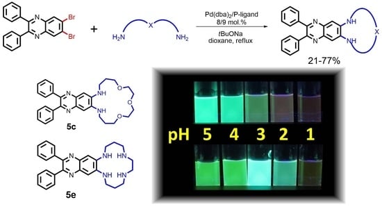
2.1. Synthesis

An available 6,7-dibromo-2,3-diphenylquinoxaline (1) was used as a starting compound. It was obtained by the reaction of benzil with 4,5-dibromo-1,2-diaminobenzene [59]. Possibilities of the Pd-catalyzed diamination of 1 were studied using a model 2-methoxyethylamine (2) (Scheme 1).
The reaction was carried out in the presence of the catalytic system Pd(dba)2/BINAP (8/9 mol%) (dba—dibenzylideneacetone, BINAP—2,2′-bis(diphenylphosphino)-1,1′-binaphthalene) in dioxane (0.1 M) under reflux for 24 h in the presence of 3 equiv. tBuONa as a base. Usually, the Pd-catalyzed diamination of the ortho-dihalogenoarenes is hindered because the introduction of the first amino group decreases the reactivity of the resting halogen atom [60,61]. In our case, the application of 4 equiv. 2-methoxyethylamine led to a 69% isolated yield of the target compound 3. This favorable result can be explained by a higher reactivity of the halogen atoms in the starting compound 1, an electron-deficient heterocycle.
Encouraged by this result, we studied the possibility of the synthesis of oxaazamacrocycles 5ad using a series of oxadiamines 4ad (Scheme 2).
The reaction conditions were adjusted to the synthesis of macrocycles by diminishing the concentration of the reagents to 0.02 M in view of reducing the oligomerization processes. Initial experiments were carried out with oxadiamine 4a. In the case of applying 1 equiv. of dioxadiamine 4a, the conversion of the starting dibromoquinoxaline 1 reached only 50%. After the increase in the reaction time up to 48 h, the conversion reached 75%, and the application of 1.5 equiv. dioxadiamine 4a resulted in full conversion of the starting compound and 46% isolated yield of the target macrocycle 5a. Cyclic dimer 6a was also obtained, though in a tiny yield of 3%. Note compound 5a is a cyclic analog of the diamino derivative 3, which is important for further coordination studies. In the reaction of trioxadiamine 4b, a full conversion of 1 was achieved in 24 h, and the yield of the corresponding macrocycle 5b was 30%. In the case of 1 equiv. of dioxadiamine 4c, the reaction provided a 34% yield of the macrocycle 5c and the application of 1.5 equiv. raised the yield to 48%. Compound 5d was equally obtained in 48% yield under optimized conditions, and the cyclic dimer 6d was isolated in 7% yield.
In order to furnish polyazamacrocyclic compounds, the reactions with a number of linear polyamines 4eh were carried out under similar conditions (Scheme 3). The much higher reactivity of the primary amino groups than that of the secondary amino groups in the Pd-catalyzed amination reactions is well established and ensures the formation of the corresponding macrocycles [62]. However, polyamines are better chelators for palladium than oxadiamines and can lead to a decrease in the efficiency of the process and diminish the yield of the target products [61].
The reactions conducted with the tetraamine 4e showed that Pd(dba)2/BINAP catalytic system was inefficient even at a higher concentration (0.06 M). Only products of C-Br bond reduction and oligomers were observed in the reaction mixture. The application of the Pd(dba)2/DPPF (8/9 mol%, DPPF—1,1′-bis(diphenylphosphino)ferrocene), which previously was shown to be optimal for the diamination of 1 with 1,3-diaminopropane [36], provided the desired macrocycle 5e in 48% yield. Earlier JosiPhos ligand ((R)-1-[(SP)-2-(diphenylphosphino)ferrocenyl]ethyldi-tert-butylphosphine) and ligands of this type were noted for its good performance in the Pd-catalyzed amination of halogen heteroarenes [63,64], and its application in our case increased the yield of 5e to 77%. Under analogous conditions, tetraazamacrocycles 5f,g were obtained in 56 and 50% yields, respectively, while the reaction with the pentaamine 4h was more difficult and afforded 5h in only 21% yield. The use of a less expensive Pd(dba)2/BINAP catalytic system was successful only in the synthesis of the macrocycle 5g (43%), the result being quite comparable with that obtained with the JosiPhos ligand. The structure of oxadiamines and polyamines strongly governs the catalytic conditions and demands fine-tuning of the reaction conditions in each case. Thus, we obtained a series of nine macrocyclic ligands differing by the number of donor sites and the macrocyclic cavity size.

2.2. Optical Properties

Initially, the spectral properties of the obtained macrocycles were studied in acetonitrile solutions, and the main spectral data are given in Table 1. The compounds 3 and 5ah possess well-pronounced photoluminescent properties, which are characteristic of the electron push-pull systems of quinoxaline [36,65,66]. The absorption spectra possess strong bands at 260–280 nm corresponding to π–π* transitions and broad bands in the 404–410 nm region corresponding to ICT [67]. Measured quantum yields range from 44 to 68% providing high brightness of the compounds.
The majority of compounds (3, 5bd, 5gh) possess emission maxima at 464–466 nm (Figure 2), while some macrocycles with small-size macrocycles (5a, 5e, and 5f) have emission maxima at a greater wavelength (485, 480, and 486 nm, respectively). It might be explained by the fact that higher conformational rigidity of these macrocycles hinders full conjugation of the amino groups at positions 6 and 7 with the aromatic system. To a certain extent, it can be supported by the values of the chemical shifts of the protons at positions 5 and 8 (Figures S47–S64). In the non-cyclic compound 3, the chemical shift of these protons is 7.15 ppm. In the macrocycles 5bd, it is shifted to 7.05–7.09 ppm; in the macrocycle 5e, these protons lay at 7.01 ppm, while macrocycles 5g,h are characterized by substantially upfield chemical shifts (ca 6.85 ppm). In the macrocycles 5a and 5f, with the smallest macro ring sizes, these protons are downshifted to 7.27–7.29 ppm. Thus, the rigidity of the macrocyclic ring influences spectral properties of the compounds. Analogous interdependence of the chemical shifts, size of the macrocyclic ring and photophysical data was observed by us earlier for the quinoline-containing macrocycles [55].
The influence of the solvent polarity on the optical properties of the compounds was studied using exemplary 3, 5c and 5e. Absorption and fluorescence spectra were registered in toluene, dioxane, dichloromethane, acetonitrile and methanol. The data are presented in Table S1. The dependence of solvent polarity on the fluorescence of the ICT systems is well established [69]. The excited state of the fluorophore is characterized by high polarity because of charge separation, and this leads to a strong interaction with the polar solvent. This interaction energy strongly depends on the polarity of the solvent and influences the energy gap between the ground and excited states. The polar solvents cause a diminishing of the gap. In our case, the increase in the solvent polarity leads to a bathochromic shift of the emission maxima of compounds 3 and 5c: in toluene and dioxane it lies at 450 nm, in dichloromethane and acetonitrile at 462–465 nm, more polar methanol shifts it to the 482–485 nm region. Compound 5e features a similar tendency, but in polar solvents, emission maxima are more red-shifted compared to 3 and 5c (Figure 3). The influence of the solvent on the quantum yields is indefinite. It may be due to various specific interactions of the molecules of the solvent with the coordination sites of the compounds tested, leading to non-emissive relaxations of various intensities.
In order to study the spectral properties of 3, 5c and 5e in water, their solubility was preliminarily investigated. The first two compounds are insoluble in water; thus, we used a 1 vol% solution of MeOH. In the 0.5–8 µM range, the Beer–Lambert law was shown to be observed (Figures S7 and S10). For A408 < 0.1 (Figures S8 and S10). Polyazamacrocycle 5e is sufficiently soluble in water without methanol additive due to partial protonation of the dialkylamino groups, and the Beer–Lambert law is observed for a wider range of concentrations, likely it was the case with a non-cyclic analog [36]. Compounds 3, 5c and 5e possess fluorescence in the green region, their emission maxima being 507, 510 and 526 nm, respectively (Table S1). Water is considered to be one of the most polar solvents (1.3 times more polar than methanol), which leads to such a strong shift in the emission maximum. As expected, quantum yields in water are lower than in organic solutions (27, 34 and 36%, respectively). However, they still provide enough high emission intensity, which can be observed by a naked eye (Figure 3), which is important for the construction of sensor devices.

2.3. Detection of Metal Cations in Acetonitrile

Compounds 3 and 5ad were evaluated for their possibilities of metal detection in acetonitrile. Absorption and fluorescence spectra of these compounds were registered in the presence of 18 metal perchlorates (Figures S13, S17, S21, S25, S29). Every N,O-ligand showed almost full emission quenching in the presence of such cations as Cu(II), Hg(II), Al(III). Partial quenching of fluorescence was noted in the presence of Pb(II) and Zn(II). A bathochromic shift of the absorption band corresponding to ICT was also observed. The lack of selectivity allows only for the use of these ligands as fluorescent molecular probes for the mentioned cations.
More detailed investigations were carried out with Cu(II) salt in order to elucidate the influence of the nature of the receptor unit on the composition and stability of complexes. For this purpose, spectrophotometric and spectrofluorimetric titrations were fulfilled (Figures S14–S16, S18–S20, S22–S24, S26–28 and S30–32). As an example, Figure 4 depicts the changes in the absorption and fluorescence spectra of the macrocycle 5c in the presence of different amounts of Cu(II). Detection limits (LODs) determined for 3 and 5ad using fluorescence spectroscopy (3σ-method) do not substantially depend on the nature of the ligand and are in the range of 4–5 µM.
At the first stage of the titration experiments, as it was expected, each ligand showed the formation of the complex of 1:1 stoichiometry with the bathochromic shift of the absorption maximum and fluorescence quenching. Further addition of copper salt led to a gradual decrease in the intensity of the red-shifted absorption band, indicating the formation of the complexes with another stoichiometry.
Using the HypSpec program, spectral data for spectrophotometric titrations were subjected to the factor analysis, which provided the composition and binding constants of the complexes, which are collected in Table 2. In the case of fluorescent titrations, the formation of 1:1 complexes can be observed, but as all forming complexes are non-emissive, it is impossible to reliably identify them using this kind of titration. All N,O-ligands form [CuL]2+ complexes of medium stability, the most stable complex (logβ = 7.57(7)) was observed for a non-cyclic ligand 3, possibly due to conformational lability of its receptor part. Oxygen atoms and nitrogen atoms of the heteroaromatic moiety probably participate in the formation of such complexes.
Macrocycles 5a and 5b both form the complexes [CuL]2+ and [Cu4(L)3]8+, their stabilities being similar, independent of the fact that these macrocycles differ by the cavity size and the number of oxygen atoms. As macrocycles with enough short linkers cannot form polynuclear complexes, the formation of [Cu4(L)3]8+ could be explained by the association of three [CuL]2+ complexes around one copper(II) center via coordination employing heterocyclic nitrogen atoms. As macrocycles 5c and 5d possess larger cavities with longer polymethylene chains between atoms, they can promote the formation of the complexes of another composition. Indeed, with these two macrocyclic ligands, besides [CuL]2+, complexes featuring two macrocycles and four Cu(II) cations [Cu4(L)2]8+ were observed. Thus, the size and flexibility of the macrocyclic cavity influence stability and composition of the complexes. The studied N,O-ligands can form mono- and polynuclear copper complexes.

2.4. Protonation Studies

The protonation of the macrocycles in the aqueous media was studied using oxygen-containing compound 5c and tetraazamacrocycle 5e. Quinoxaline derivatives were described as convenient platforms for the development of pH sensors [34,35]. As macrocycle 5c is insoluble in pure water, 0.5–1 vol% methanol solutions in water were applied, which allowed the concentrations of 5c up to 8 µM. Upon the addition of the acid to an aqueous solution of 5c notable changes both in absorption and fluorescence spectra take place, like the bathochromic shift of the absorption maximum from 420 nm to 480 nm and partial fluorescent quenching with the shift of the emission maximum from 510 to 570 nm (Figure 5).
Further diminishing pH value gives rise to a novel absorption band centered at 536 nm resulting in a rose color of the solution visible by a naked eye (Figure 6). Thus, macrocycle 5c can be proposed as a dual-channel pH sensor providing monitoring of the acidity of the media using both absorption and emission spectra.
A different response to the change of pH is manifested by the tetraazamacrocycle 5e (Figure 7). A decrease in pH leads to a small hypsochromic shift in the absorption spectrum (from 407 to 395 nm) which is accompanied by a partial emission quenching with a simultaneous shift of the maximum from 528 to 502 nm. A naked-eye note of the change of the fluorescence color from green to blue is possible (Figure 8).
Further addition of acid leads to more pronounced emission quenching with a bathochromic shift of the maximum from 502 to 575 nm, also visible by a naked eye, which coincides with the bathochromic shift of the absorption band from 395 to 475 nm. It was impossible to increase pH above 8.5 due to the formation of the precipitate (Figures S45, S46), which can be explained by the low solubility of non-protonated 5e. Thus, this macrocycle also can be put forward as a pH sensor like compound 5c, but the transition points and color responses of these two compounds are different.
The acquired data of spectrophotometric and fluorimetric titrations for the macrocycles 5c and 5e were processed with the HypSpec program. Calculated absorption and emission spectra for all species present in the solution and speciation diagrams are given in Figures S33–S44. Protonation constants compared with the known compounds 7 [36] and 89 [70] (Figure 9) are collected in Table 3.
Earlier, our colleagues studied in detail the protonation of a linear quinoxaline-containing tetraamine 7 using NMR spectra and DFT calculations [36] which provided us with the grounds to put forward the protonation hypothesis for our macrocycles.
The proposed protonation sequence is given in Scheme 4. It is assumed that the dialkylamino groups which are not conjugated with the aromatic system are first protonated, and their protonation leads but to a small hypsochromic shift of the ICT band. Such response was noted for 6,7-diaminoquinoxaline [36] and for 6-aminoquinoline derivatives [71]. In a more acidic media, the protonation of the nitrogen atoms of 6,7-diaminoquinoxaline takes place, and the bathochromic shift of the ICT band in absorption and emission spectra is observed. This assumption is in agreement with the calculations provided in [36].
In the case of the macrocycle 5c, the molecule does not contain amino groups which are not conjugated with the aromatic system, and the protonation of the nitrogen atom of the diaminoquinoxaline fragment takes place accompanied by a bathochromic shift of the ICT band in the absorption and emission spectra. The calculated pK1 constant equals 4.10(5) and 3.96(5) (from UV and fluorescence titrations, respectively). These values are in agreement with the protonation constants of the aminoquinoxalines reported earlier [72] but are higher than those reported for compound 7 (2.61 and 3.07). It can be explained by the presence of the protonated dialkyl amino groups and electron-withdrawing COOMe groups in compound 7. The second protonation step is associated with pK values of 0.6(1) and 1.20(5) (from UV and fluorescence titrations, respectively), which corresponds to the protonation of the second aromatic nitrogen atom. A notable difference between the two values calculated from spectrophotometric and spectrofluorimetric titrations stems from the difference in the basicity of the molecule in the ground and excited states [73].
Macrocycle 5e possesses two dialkylamino groups. One of them obviously is protonated at the first step. It seems reasonable to compare this macrocycle with the non-cyclic analog 7. It is known that the conformational effects in the macrocyclic systems lead to a wide variation in the basicity constants of polyazamacrocycles [70,74,75]. Unfortunately, there are no data in the literature on the protonation constants of benzocyclam. Therefore, for comparison, we selected tetraazamacrocycles 8 and 9, which are the closest in their structures and in which two neighboring dialkylamino groups connected by an ethylene linker undergo protonation (Figure 9). An attempt to accurately measure the pK1 value for compound 5e failed because when pH 8.5 was reached, precipitation was observed, and the fluorescence of the solution disappeared (Figures S45, S46). This observation confirms the fact that enough good solubility of 5e in water is due to its protonation. The curve depicting the changes in absorption and fluorescence spectra allows only an approximate estimate of the pK1 value ca 9.5, which is consistent with the data for similar macrocycles 8 and 9 [70]. In the molecule of compound 7, two alkylamino groups are distant from each other, as a result, their protonation occurs independently and the values of the amino group protonation constants do not differ (Table 3). In macrocyclic molecules, due to electrostatic repulsion, the protonation constant of the second nitrogen atom is lower than the first one by several orders of magnitude, and this difference strongly depends on the flexibility of the macrocycle [74]. In the case of compound 5e spectrophotometric and spectrofluorimetric studies upon the addition of acid gave the values for pK2 3.69(5) and 4.01(5), respectively. These values correspond to the analogous protonation constant of compound 9. A small hypsochromic shift of the ICT band and the blue shift of the emission maximum observed in the spectrum are consistent with the hypothesis that this constant corresponds to the protonation of the alkylamino group, which is not conjugated with the heterocyclic system. At the next step, the nitrogen atom of the heterocyclic nucleus is protonated, which is accompanied by a characteristic bathochromic shift of the ICT band in the absorption spectrum. A similar situation was described for molecule 7 [36]. The pK3 values obtained from UV and fluorescence spectroscopy data equal 2.36(5) and 2.46(5), respectively, and are also close to those for molecule 7. In the [5eH2]2+ structure, charged protonated aliphatic nitrogen atoms are positioned inevitably closer to the heterocyclic core due to the macrocyclic structure, which leads to lower pK3 5e values compared to 7. Upon further addition of acid new changes are observed in the fluorescence spectrum, which allows us to estimate the value of pK4 1.0 (Figure S43). However, in the absorption spectrum, the appearance of the next bathochromic shift (similar to that noted for compound 5c) is observed only in strongly acidic media (pH < 0), which can be explained by a notable difference in the basicity of the [5eH3]3+ particle in the ground and excited states.

3. Materials and Methods

3.1. Reagents

Unless otherwise noted, all chemicals and starting materials were obtained commercially from ABCR (Karlsruhe, Germany) and Sigma-Aldrich (Merck Co., Rahway, NJ, USA) and used without further purification. Preparative column chromatography was carried out using Silica gel 60 (40–63 µm) from Merck Co. Dioxane was distilled successively over NaOH and sodium under argon, CH2Cl2 and CH3CN were distilled over CaH2, chloroform was distilled over P2O5, MeOH was used freshly distilled. 4,5-Dibromobenzene-1,2-diamine was obtained according to the described 3-step procedure [76]. Pd(dba)2 was synthesized according to a known method [77] and used without recrystallization. N1-(2-aminoethyl)-N2-(2-(2-aminoethylamino)ethyl)ethane-1,2-diamine (4h) was prepared by treatment of the corresponding pentahydrochloride with a 2.5 M solution of KOH in methanol.

3.2. Apparatus

The 1H and 13C NMR spectra were registered with a Bruker Avance-400 spectrometer (Bruker Daltonics, Bremen, Germany) in chloroform-d1 or methanol-d4, using the residual signals of chloroform or CHD2OD as internal standards. MALDI-TOF mass-spectra were registered on a Bruker Daltonics Autoflex II mass-spectrometer (Bruker Daltonics, Bremen, Germany) in positive ion mode with a dithranol matrix and polyethyleneglycols as internal standards. FTIR spectra were registered on a Nicolet iS 5 (Thermo Fisher Scientific, Waltham, MA, USA) with iD3 ATR accessory (ZnSe).
The pH measurements were carried out using the «Mettler Toledo» apparatus (Greifensee, Switzerland) with a combined electrode LE438 in a glass cell. The electrode was calibrated with commercial buffers (pH = 4.01 and 7.00). UV-vis spectra were registered with a Hitachi U-2900 device (Tokyo, Japan) in a quartz cuvette (Hellma, l = 1 cm). Fluorescence spectra were registered with Horiba Jobin Yvon Fluoromax-2 apparatus (Edison, NJ, USA) in a quartz cuvette (Hellma, l= 1 cm). Luminescence quantum yields were determined relative to quinine sulfate solution in 0.05 H2SO4 (Φ = 0.53(2)) according to a standard procedure [78].

3.3. Synthesis

6,7-Dibromo-2,3-diphenilquinoxaline, [59] 4,5-Dibromobenzene-1,2-diamine (2.55 g, 9.6 mmol), benzyl (1.98 g, 9.4 mmol), and isopropanol (18.8 mL) were placed in a flask equipped with a condenser and a magnetic stirrer. The mixture was refluxed for 3 h. The mixture was allowed to cool to room temperature. The brown precipitate was separated on a glass filter. Yield 3.4 g (83%), brown powder, m.p. 166 °C. 1H-NMR (400 MHz, CDCl3) δ 7.35 (t, 4H, 3J = 7.3 Hz, H(Ph)), 7.40 (t, 2H, 3J = 7.0 Hz, H(Ph)), 7.49 (d, 4H, 3J = 6.7 Hz, H(Ph)), 8.48 (s, H(Qx)). 13C-NMR (100.6 MHz, CDCl3) δ 126.5 (2C, C-Br), 128.3 (4C, CH(Ph)), 129.3 (2C, CH(Ph)), 129.8 (4C, CH(Ph)), 133.1 (2C, CH (Qx)), 138.2 (2C, Cquat(Ph)), 140.3 (2C, Cquat(Ph)), 154.5 (2C, Cquat(Ph)). HRMS (MALDI-TOF): m/z [M + H]+ calcd for C20H12N2Br2: 438.944; found: 438.946.
Palladium-Catalyzed Amination of 6,7-dibromo-2,3-diphenilquinoxaline—General Procedure. A two-neck flask equipped with a condenser and a magnetic stirrer, flushed with dry argon, was charged with corresponding 6,7-dibromo-2,3-diphenilquinoxaline (132 mg, 0.3 mmol), Pd(dba)2 (14 mg, 8 mol%), phosphine ligand (9 mol%), and absolute dioxane (3–15 mL). The mixture was stirred for 2–3 min, then corresponding amine (0.45–1.2 mmol) and tBuONa (87 mg, 0.9 mmol) were added, and the reaction mixture was refluxed for 24–48 h. After cooling it down to ambient temperature, the reaction mixture was diluted with CH2Cl2, the solution filtered and evaporated in vacuo, and the residue was chromatographed on silica gel using a sequence of eluents: CH2Cl2, CH2Cl2/MeOH 200:1–3:1, CH2Cl2/MeOH/NH3aq 100:20:1–100:30:5. In the cases of the compounds 5a and 5b, the residue before column chromatography was dissolved in CH2Cl2 (20 mL) and washed with deionized water (2 × 20 mL) to eliminate sodium salts. The organic phase was dried over 3 Å sieves and concentrated in vacuo, and only then subjected to column chromatography according to the general procedure.
N,N`-bis(2-methoxyethyl)-2,3-diphenylquinoxaline-6,7-diamine (3) obtained according to General Procedure from 6,7-dibromo-2,3-diphenilquinoxaline (132 mg), amine 2 (90 mg) in the presence of Pd(dba)2 (14 mg), BINAP (17 mg) and tBuONa (87 mg) in dioxane (3 mL) in 24 h. Eluent CH2Cl2/MeOH 100:1. Yield 76 mg (59%), yellowish oil. 1H-NMR (400 MHz, CDCl3) δH 3.39 (s, 6H, CH3), 3.44 (q, 4H, 3J = 5.2 Hz, CH2(NH)), 3.71 (t, 4H, 3J = 5.1 Hz, CH2(OMe)), 4.39 (t, 2H, 3J = 5.2 Hz, NH), 7.15 (s, 2H, H5, H8 (Qx)), 7.27–7.29 (m, 6H, H3, H4, H5 (Ph)), 7.46–7.49 (m, 4H, H2, H6 (Ph)). 13C-NMR (100.6 MHz, CDCl3) δC 43.5 (2C, CH2NH), 58.7 (2C, OCH2), 70.2 (2C, MeO), 104.9 (2C, C5, C8 (Qx)), 127.7 (2C, (Ph)), 128.0 (4C, (Ph)), 129.7 (4C, (Ph)), 138.4 (2C, C4a, C8a (Qx)), 140.0 (2C, Cquat (Ph)), 141.8 (2C, C6, C7 (Qx)), 149.0 (2C, C2, C3 (Qx)). IR (neat): 612 (vw), 624 (vw), 641 (vw), 668 (w), 698 (s), 750 (Vs), 759 (s), 765 (vs), 806 (vw), 832 (w), 853 (w), 917 (vw), 978 (w), 1001 (w), 1024 (m), 1065 (m), 1079 (m), 1117 (m), 1157 (w), 1199 (s), 1240 (m), 1261 (m), 1267 (m), 1274 (m), 1313 (vw), 1354 (m), 1391 (vw), 1447 (m), 1482 (m), 1495 (m), 1518 (m), 1573 (w), 1599 (w), 1615 (w), 1719 (vw), 2816 (w), 2829 (w), 2876 (w), 2885 (w), 2926 (w), 2980 (w), 3054 (vw), 3084 (vw), 3371 (w), 3382 (w) cm−1. HRMS (MALDI-TOF): m/z [M + H]+ calcd for C26H28N4O2+: 429.2285; found: 429.2266.
2,3-Diphenyl-6,7,8,10,11,13,14,15-octahydro-[1,4]dioxa[7,10]diazacyclododecino[8,9-g]quinoxaline (5a) obtained according to General Procedure from 6,7-dibromo-2,3-diphenilquinoxaline (132 mg), amine 4a (67 mg) in the presence of Pd(dba)2 (14 mg), BINAP (17 mg) and tBuONa (87 mg) in dioxane (15 mL) in 48 h. Eluent CH2Cl2/MeOH 100:1. Yield 59 mg (46%), yellowish oil. 1H-NMR (400 MHz, CDCl3) δH 3.41 (t, 4H, 3J = 4.5 Hz, CH2(NH)), 3.70 (s, 4H, CH2(O)), 3.74 (t, 4H, 3J = 4.6 Hz, CH2(O)) 4.96 (br.s., 2H, NH), 7.27-7.30 (m, 8H, H5, H8 (Qx), H3, H4, H5 (Ph)), 7.45-7.47 (m, 4H, H2, H6 (Qx)). 13C-NMR (100.6 MHz, CDCl3) δC 45.7 (2C, CH2NH), 69.1 (2C, CH3O), 69.8 (2C, OCH2), 110.8 (2C, C5, C8 (Qx)), 127.9 (2C, C4 (Ph)), 128.0 (4C, (Ph)), 129.7 (4C, (Ph)), 138.8 (2C, C4a, C8a (Qx)), 139.9 (2C, Cquat (Ph)), 143.6 (2C, C6, C7 (Qx)), 149.7 (2C, C2, C3 (Qx)). IR (neat): 612 (w), 614 (w), 624 (w), 668 (w), 675 (m), 694 (s), 698 (vs), 712 (w), 749 (s), 759 (s), 763 (s), 790 (w), 805 (w), 830 (m), 847 (w), 857 (w), 868 (w), 887 (w), 928 (w), 945 (w),977 (m), 1001 (w), 1015 (m), 1023 (m), 1033 (m), 1054 (m), 1061 (m), 1071 (m), 1079 (m), 1106 (m), 1116 (m), 1128 (m), 1149 (w), 1158 (w), 1196 (m), 1226 (m), 1264 (m), 1292 (w), 1309 (w), 1339 (m), 1348 (w), 1369 (w), 1407 (w), 1455 (m), 1474 (m), 1493 (w), 1520 (m), 1565 (w), 1598 (w), 1618 (w), 2851 (w), 2880 (w), 2914 (w), 2952 (w), 3051 (vw), 3317 (w) cm−1. HRMS (MALDI-TOF): m/z [M + K–CH2CH2]+ calcd for C24H22N4O2K+: 437.1374; found: 437.1405.
2,3,18,19-Tetraphenyl-6,7,8,10,11,13,14,15,22,23,24,26,27,29,30,31-hexadecahydro-[1,4,13,16]tetraoxa[7,10,19,22]tetraazacyclotetracosino[8,9-g:20,21-g’]diquinoxaline (6a) obtained in mixture with as a by-product in the synthesis of 5a. Eluent CH2Cl2/MeOH 50:1. Yield 4 mg (3%), yellow-brown oil. 1H-NMR (400 MHz, CDCl3) δH 3.47–3.49 (m, 8H, CH2(NH)), 3.72 (s, 8H, CH2(O)), 3.90 (t, 8H, 3J = 4.4 Hz, CH2(O)) 7.12 (s, 4H, H5, H8 (Qx)), 7.27–7.31 (m, 12H, H3, H4, H5 (Ph)), 7.45–7.47 (m, 8H, H2, H6 (Qx)). NH-protons were not unambiguously assigned due to line broadening. HRMS (MALDI-TOF): m/z [M + Na]+ calcd for C52H52N8O4Na+: 875.4004; found: 875.3983.
2,3-Diphenyl-6,7,8,10,11,13,14,16,17,18-decahydro-[1,4,7]trioxa[10,13]diazacyclopentadecino [11,12-g]quinoxaline (5b) obtained according to General Procedure from 6,7-dibromo-2,3-diphenilquinoxaline (132 mg), amine 4b (87 mg) in the presence of Pd(dba)2 (14 mg), BINAP (17 mg) and tBuONa (87 mg) in dioxane (15 mL) in 24 h. Eluent CH2Cl2/MeOH 100:1. Yield 43 mg (30%), yellowish oil. 1H-NMR (400 MHz, CDCl3) δH 3.42–3.46 (m, 4H, CH2(NH)), 3.66–3.67 (m, 4H, CH2(O)), 3.76–3.78 (m, 4H, CH2(O)), 3.89 (t, 4H, 3J = 5.0 Hz, CH2(O)), 4.82 (br.s, 2H, NH), 7.09 (s, 2H, H5, H8 (Qx)), 7.27–7.29 (m, 6H, H3, H4, H5 (Ph)), 7.46–7.49 (m, 4H, H2, H6 (Ph)). 13C-NMR (100.6 MHz, CDCl3) δC 42.9 (2C, CH2NH), 68.1 (2C, OCH2) 70.0 (2C, OCH2), 70.2 (2C, OCH2), 103.7 (2C, C5, C8 (Qx)), 127.7 (2C, C4 (Ph)), 128.0 (4C, (Ph)), 129.9 (4C, (Ph)), 138.5 (2C, C6, C7 (Qx)), 140.0 (2C, Cquat (Ph)), 141.5 (2C, C4a, C8a (Qx)), 148.4 (2C, C2, C3 (Qx)). IR (neat): 609 (w), 668 (vw), 699 (s), 733 (m), 768 (m), 792 (vw), 805 (vw), 844 (m), 882 (vw), 919 (w), 943 (w), 979 (w), 1001 (w), 1025 (m), 1074 (s), 1096 (s), 1110 (s), 1130 (vs), 1202 (s), 1226 (s), 1246 (s), 1265 (m), 1354 (s), 1402 (w), 1417 (w), 1455 (s), 1495 (s),1528 (s), 1577 (w), 1618 (m), 1725 (w), 2867 (m), 2906 (m), 3058 (w), 3077 (w), 3365 (m) cm−1. HRMS (MALDI-TOF): m/z [M + K–CH2CH2]+ calcd for C26H26N4O3K+: 481.1636; found: 481.1605.
2,3-Diphenyl-6,7,8,9,11,12,14,15,17,18,19,20-dodecahydro-[1,4,7]trioxa[11,14]diazacyclohepta decino[12,13-g]quinoxaline (5c) obtained according to General Procedure from 6,7-dibromo-2,3-diphenilquinoxaline (132 mg), amine 4c (99 mg) in the presence of Pd(dba)2 (14 mg), BINAP (17 mg) and tBuONa (87 mg) in dioxane (15 mL) in 48 h. Eluent CH2Cl2/MeOH 200:1. Yield 72 mg (48%), yellowish oil. 1H-NMR (400 MHz, CDCl3) δH 2.06 (quint, 4H, 3J = 5.3 Hz, CH2), 3.50 (q, 4H, 3J = 5.1 Hz, CH2(NH)), 3.68–3.69 (m, 4H, CH2(O)), 3.71–3.74 (m, 8H, CH2(O)), 4.86 (br. t, 2H, 3J = 4.9 Hz, NH), 7.07 (s, 2H, H5, H8 (Qx)), 7.27–7.28 (m, 6H, H3, H4, H5 (Ph)), 7.45–7.48 (m, 4H, H2, H6 (Ph)). 13C-NMR (100.6 MHz, CDCl3) δC 27.9 (2C, CH2), 42.9 (2C, CH2NH), 70.7 (2C, OCH2), 70.8 (2C, OCH2), 70.9 (2C, OCH2), 103.3 (2C, C5, C8 (Qx)), 127.5 (2C, C4 (Ph)), 127.9 (4C, (Ph)), 129.8 (4C, (Ph)), 138.5 (2C, C4a, C8a (Qx)), 140.4 (2C, Cquat (Ph)), 141.8 (2C, C6, C7 (Qx)), 148.4 (2C, C2, C3 (Qx)). IR: 638 (vw), 654 (w), 668 (m), 675 (w), 700 (vs), 734 (s), 768 (m), 804 (m), 832 (m), 871 (w), 896 (w), 916 (w), 937 (w), 979 (m), 1001 (m), 1025 (s), 1066 (vs), 1079 (vs), 1089 (Vs), 1092 (vs), 1112 (vs), 1200 (vs), 1240 (vs), 1264 (s), 1353 (vs), 1437 (m), 1462 (vs), 1482 (s), 1496 (s), 1522 (vs), 1559 (w), 1599 (w), 1616 (m), 2865 (m), 2918 (m), 2942 (w), 3051 (w), 3366 (w) cm−1. HRMS (MALDI-TOF): m/z [M + H]+ calcd for C30H35N4O3+: 499.2704; found: 499.2665.
2,3-Diphenyl-6,7,8,9,11,12,13,14,16,17,18,19-dodecahydro-[1,12]dioxa[5,8]diazacyclohexa decino[6,7-g]quinoxaline (5d) obtained according to General Procedure from 6,7-dibromo-2,3-diphenilquinoxaline (132 mg), amine 4d (92 mg) in the presence of Pd(dba)2 (14 mg), BINAP (17 mg) and tBuONa (87 mg) in dioxane (15 mL) in 48 h. Eluent CH2Cl2/MeOH 200:1. Yield 69 mg (48%), yellowish oil. 1H-NMR (400 MHz, CDCl3) δH 1.77 (br. s, 4H, CH2), 2.06 (quint, 4H, 3J = 5.1 Hz, CH2), 3.48 (q, 4H, 3J = 4.9 Hz, CH2(NH)), 3.58 (br. s, 4H, CH2O), 3.67 (t, 4H, 3J = 5.1 Hz, CH2(O)), 5.15 (br. s, 2H, NH), 7.05 (s, 2H, H5), 7.27–7.29 (m, 6H, H3, H4, H5 (Ph)), 7.46–7.48 (m, 4H, H2, H6 (Ph)). 13C-NMR (100.6 MHz, CDCl3) δC 26.3 (2C, CH2), 28.3 (2C, CH2), 44.0 (2C, CH2NH), 70.6 (2C, OCH2), 70.8 (2C, OCH2), 102.8 (2C, C5, C8 (Qx)), 127.5 (2C, C4 (Ph)), 128.0 (4C, (Ph)), 129.9 (4C, C2 (Ph)), 138.5 (2C, C4a, C8a (Qx)), 140.3 (2C, Cquat (Ph)), 141.8 (2C, C6, C7 (Qx)), 148.1 (2C, C2, C3 (Qx)). IR (neat): 669 (m), 699 (vs), 732 (m), 804 (m), 830 (s), 979 (m), 1001 (m), 1024 (s), 1065 (s), 1078 (s), 1097 (s), 1109 (s), 1199 (vs), 1240 (s), 1265 (m), 1352 (vs), 1459 (vs), 1487 (s), 1496 (s), 1522 (vs), 1560 (m), 1599 (m), 1617 (m), 2799 (m), 2854 (s), 2918 (s), 3057 (m), 3344 (m), 3388 (m) cm−1. HRMS (MALDI-TOF): m/z [M + H]+ calcd for C30H35N4O2+: 483.2755; found: 483.2725.
2,3,22,23-Tetraphenyl-6,7,8,9,11,12,13,14,16,17,18,19,26,27,28,29,31,32,33,34,36,37,38,39-tetracosahydro-[1,12,17,28]tetraoxa[5,8,21,24]tetraazacyclodotriacontino[6,7-g:22,23-g’]diquinoxaline (6d) obtained as a by-product in the synthesis of 5a. Eluent CH2Cl2/MeOH 50:1. Yield 10 mg (7%), yellow-brown oil. 1H-NMR (400 MHz, CDCl3) δH 1.77 (obs.s, 8H, CH2), 2.07 (quint, 8H, 3J = 5.4 Hz, CH2), 3.40–3.43 (m, 8H, CH2(NH)), 3.51 (obs. s, 8H, CH2(O)), 3.67 (t, 8H, 3J = 5.4 Hz, CH2(O)), 4.92 (br.s., 4H, NH), 7.09 (s, 4H, H5), 7.27–7.29 (m, 12H, H3, H4, H5 (Ph)), 7.45–7.47 (m, 8H, H2, H6 (Ph)). 13C-NMR (100.6 MHz, CDCl3) δC 26.7 (4C, CH2), 28.5 (4C, CH2), 43.8 (4C, CH2NH), 71.0 (4C, OCH2), 71.3 (4C, OCH2), 103.7 (4C, C5, C8 (Qx)), 127.6 (4C, C4 (Ph)), 128.0 (8C, (Ph)), 129.8 (8C, C2 (Ph)), 138.5 (4C, C4a, C8a (Qx)), 140.2 (4C, Cquat (Ph)), 142.3 (4C, C6, C7 (Qx)), 148.5 (4C, C2, C3 (Qx)). HRMS (MALDI-TOF): m/z [M + K]+ calcd for C60H68N8O4K+: 1003.4995; found: 1003.4983.
2,3-Diphenyl-6,7,8,9,10,11,12,13,14,15,16,17-dodecahydro-[1,4,8,11]tetraazacyclotetradecino[2,3-g]quinoxaline (5e) obtained according to General Procedure from 6,7-dibromo-2,3-diphenilquinoxaline (132 mg), amine 4e (78 mg) in the presence of Pd(dba)2 (14 mg), JosiPhos (15 mg) and tBuONa (87 mg) in dioxane (15 mL) in 48 h. Eluent CH2Cl2/MeOH 10:1. Yield 105 mg (77%), brown glassy compound. 1H-NMR (400 MHz, CDCl3/CD3OD) δH 2.06 (br.s, 4H, CH2), 2.97-3.07 (m., 8H, CH2(NH)), 3.45 (br.s, 4H, CH2(NH)), 7.01 (s, 2H, H5, H8 (Qx)), 7.26 (br.s, 6H, H3, H4, H5 (Ph)), 7.40-7.42 (m, 4H, H2, H6 (Ph)). 4 NH protons were not unambiguously assigned because of exchange with MeOD. 13C-NMR (100.6 MHz, CDCl3/CD3OD) δC 24.8 (2C, CH2), 44.3 (2C, CH2NH), 46.3 (2C, CH2NH), 49.7 (2C, CH2NH), 103.9 (2C, C5, C8 (Qx)), 127.9 (6C, (Ph)), 129,6 (4C, (Ph)), 138.3 (2C, C6, C7 (Qx)), 139.3 (2C, Cquat (Ph)), 142.1 (2C, C4a, C8a (Qx)), 148.7 (2C, C2, C3 (Qx)). IR (neat): 668 (w), 698 (s), 750 (s), 759 (s), 765 (vs), 806 (w), 834 (w), 851 (w), 917 (w), 979 (w), 1001 (w), 1025 (m), 1066 (m), 1078 (m), 1126 (m), 1157 (w), 1200 (s), 1226 (m), 1261 (m), 1267 (m), 1274 (m), 1355 (s), 1459 (s), 1494 (s), 1526 (s), 1563 (m), 1599 (m), 1619 (m), 2846 (m), 2933 (m), 2952 (m), 3054 (m), 3243 (m), 3350 (m) cm−1. HRMS (MALDI-TOF): m/z [M + Na]+ calcd for C28H32N6Na+: 475.2581; found: 475.2589.
2,3-Diphenyl-7,8,9,10,11,12,13,14,15,16-decahydro-6H-[1,4,7,10]tetraazacyclotridecino[5,6-g]quinoxaline (5f) obtained according to General Procedure from 6,7-dibromo-2,3-diphenilquinoxaline (132 mg), amine 4f (72 mg) in the presence of Pd(dba)2 (14 mg), JosiPhos (15 mg) and tBuONa (87 mg) in dioxane (15 mL) in 48 h. Eluent CH2Cl2/MeOH 10:1. Yield 73 mg (56%), yellow-brown glassy compound. 1H-NMR (400 MHz, CDCl3/CD3OD) δH 1.92 (s, 2H, CH2), 2.86–2.95 (m, 8H, CH2(NH)), 3.87 (br.s, 4H, CH2(NH)), 5.70 (br.s., 2H, NH), 7.27–7.30 (m, 8H, H5, H8 (Qx) H3, H4, H5 (Ph)), 7.42 (m, 4H, H2, H6 (Ph)). 2 NH-protons were not unambiguously assigned because of exchange with MeOD. 13C-NMR (100.6 MHz, CDCl3/CD3OD) δC 24.5 (1C, CH2), 41.4 (2C, CH2NH), 45.2 (2C, CH2NH), 49.5 (2C, CH2NH), 108.5 (2C, C5, C8 (Qx)), 127.8 (4C, (Ph)), 127.9 (2C, C4 (Ph)), 129,4 (4C, (Ph)), 137.8 (2C, C6, C7 (Ph)), 139.1 (2C, Cquat (Ph)), 141.0 (2C, C4a, C8a (Qx)), 149.6 (2C, C2, C3 (Qx)). IR (neat): 612 (w), 624 (vw), 693 (vs), 699 (vs), 731 (m), 751 (m), 759 (m), 764 (s), 789 (vw), 810 (w), 852 (w), 904 (vw), 917 (w), 955 (vw), 979 (m), 1001 (w), 1024 (m), 1063 (s), 1077 (m), 1113 (m), 1155 (m), 1195 (s), 1217 (s), 1231 (s), 1262 (m), 1350 (s), 1377 (w), 1470 (s), 1494 (m), 1511 (m), 1531 (m), 1577 (w), 1597 (m), 1618 (m), 2846 (m), 2923 (m), 2952 (m), 3056 (m), 3334 (m) cm−1. HRMS (MALDI-TOF): m/z [M + H]+ calcd for C27H31N6+: 439.2605; found: 439.2629.
2,3-Diphenyl-7,8,9,10,11,12,13,14,15,16,17,18-dodecahydro-6H-[1,4,8,12]tetraazacyclopenta decino[2,3-g]quinoxaline (5g) obtained according to General Procedure from 6,7-dibromo-2,3-diphenilquinoxaline (132 mg), amine 4g (85 mg) in the presence of Pd(dba)2 (14 mg), JosiPhos (15 mg) and tBuONa (87 mg) in dioxane (15 mL) in 48 h. Eluent CH2Cl2/MeOH 10:1. Yield 71 mg (50%), yellow-brown glassy compound. 1H-NMR (400 MHz, CDCl3/CD3OD) δH 2.00–2.06 (m, 6H, CH2), 3.01 (t, 4H, 3J = 6.4 Hz, CH2(NH)), 3.04–3.08 (m, 4H, CH2(NH)), 3.37–3.39 (m, 4H, CH2(NH)), 6.85 (s, 2H, H5, H8 (Qx)), 7.04–7.07 (m, 6H, (Ph)), 7.12–7.15 (m, 4H, (Ph)). 4NH-protons were not unambiguously assigned because of exchange with MeOD and overlap**. 13C-NMR (100.6 MHz, CDCl3/CD3OD) δC 23.3 (1C, CH2), 24.0 (2C, CH2), 42.3 (2C, CH2NH), 46.7 (2C, CH2NH), 49.7 (2C, CH2NH), 104.2 (2C, C5, C8 (Qx)), 127.8 (2C, C4 (Ph)), 127.9 (4C, (Ph)), 129,6 (4C, (Ph)), 138.2 (2C, C6, C7 (Qx)), 139.4 (2C, Cquat (Ph)), 140.9 (2C, C4a, C8a (Qx)), 148.9 (2C, C2, C3 (Qx)). IR (neat): 649 (w), 659 (w), 663 (w), 668 (s), 674 (w), 679 (w), 698 (m), 721 (vw), 734 (w), 766 (w), 792 (vw), 816 (w), 834 (w), 845 (vw), 924 (vw), 985 (w), 1001 (w), 1025 (w), 1082 (m), 1097 (m), 1118 (m), 1179 (s), 1217 (s), 1255 (m), 1268 (m), 1288 (m), 1332 (m), 1354 (w), 1363 (w), 1395 (m), 1405 (m), 1419 (m), 1430 (m), 1437 (m), 1448 (m), 1457 (m), 1465 (m), 1472 (m), 1491 (m), 1496 (m), 1507 (m),1521 (w), 1540 (m), 1559 (w), 1576 (m), 1606 (m), 1617 (m), 1653 (m), 1684 (w), 1700 (m), 1718 (m), 1730 (s), 1734 (s), 1740 (s), 1743 (s), 2926 (m), 2951 (m), 3054 (m), 3296 (m) cm−1. HRMS (MALDI-TOF): m/z [M + H]+ calcd for C29H35N6+: 467.2918; found: 467.2947.
2,3-Diphenyl-7,8,9,10,11,12,13,14,15,16,17,18-dodecahydro-6H-[1,4,7,10,13]pentaazacyclo pentadecino[2,3-g]quinoxaline (5h) obtained according to General Procedure from 6,7-dibromo-2,3-diphenilquinoxaline (132 mg), amine 4h (85 mg) in the presence of Pd(dba)2 (14 mg), JosiPhos (15 mg) and tBuONa (87 mg) in dioxane (15 mL) in 48 h. Eluent CH2Cl2/MeOH 10:1. Yield 30 mg (21%), yellow-brown glassy compound. 1H-NMR (400 MHz, CDCl3/CD3OD) δH 2.78 (br.s, 4H, CH2(NH)), 3.05 (br.s, 4H, CH2(NH)), 3.23 (br.s, 4H, CH2(NH)), 3.63 (s, 4H, CH2(NH)), 5.57 (br. s, NH), 6.84 (s, 2H, H5, H8 (Qx)), 7.11–7.19 (m, 10H, H2, H3, H4, H5, H6 (Ph)). 3 NH-protons were not unambiguously assigned because of exchange with MeOD and overlap**. 13C-NMR (100.6 MHz, CDCl3/CD3OD) δC 38.9 (2C, CH2NH), 44.6 (2C, CH2NH), 45.1 (2C, CH2NH), 46.7 (2C, CH2NH), 103.7 (2C, C5, C8), 127.8 (6C, (Ph)), 129,5 (4C, (Ph)), 138.2 (2C, C6, C7 (Qx)), 139.3 (2C, Cquat (Ph)), 139.8 (2C, C4a, C8a (Qx)), 149.1 (2C, C2, C3 (Qx)). IR (neat): 611 (m), 618 (m), 651 (m), 669 (m), 698 (vs), 732 (s), 777 (m), 807 (m), 835 (m), 919 (w), 978 (m), 1024 (m), 1065 (s), 1131 (m), 1157 (m), 1199 (s), 1242 (s), 1264 (m), 1288 (m), 1355 (s), 1456 (s), 1473 (s), 1481 (s), 1496 (s), 1507 (m), 1521 (m), 1535 (m), 1539 (s), 1559 (m), 1576 (m), 1599 (m), 1617 (m), 2750 (m), 2853 (m), 2928 (m), 2958 (m), 3054 (m), 3313 (m), 3329 (m) cm−1. HRMS (MALDI-TOF): m/z [M + Na]+ calcd for C28H33N7Na+: 490.2690; found: 490.2689.
NMR spectra of the compounds are given in the Supplementary Materials.

3.4. Protonation Studies

Protonation studies of compounds 5c and 5e were performed at room temperature. The solutions were prepared with double-deionized high-purity water (18.2 MΩ cm) obtained from a «Millipore Simplicity» apparatus (Merck Co., Rahway, NJ, USA). Solution concentrations and other experiment conditions are given in the title of the corresponding figures and tables. Protonation studies were conducted in a glass cell equipped with a magnetic stirrer and pH-electrode, adding HClO4 (4 M or 0.01 M) or NaOH (5 M or 0.01 M) to the solutions of ligands (I = 0.1 M, NaClO4). The pKa, spectra of the species and speciation diagrams were calculated using nonlinear least-squares analysis by means of the HypSpec program [79] after factor analysis of the combined data sets [80]. The goodness of fit was assessed through the scaled standard deviation of the residuals (s), which has an expected value of unity in the absence of systematic errors assuming a correct weighting scheme. UV and fluorescence spectra, calculated spectra of the species, speciation diagrams and titration curves are given in the Supplementary Materials.

3.5. Metal Ions Complexation Studies

Complexation studies of compounds 3 and 5ad were performed at room temperature in acetonitrile. Metal-binding experiments were conducted by manual addition of the aliquots of metal salt solutions in acetonitrile by a Hamilton syringe to a solution of ligand placed in a quartz cuvette. All metal salts used were perchlorates of the general M(ClO4)n·xH2O formula. Caution! Although no problems were experienced, perchlorate salts are potentially explosive when combined with organic ligands and should be manipulated with care and used only in very small quantities. 18 metal ions (Li+, Na+, K+, Mg2+, Ca2+, Sr2+, Ba2+, Al3+, Cr3+, Mn2+, Fe2+, Co2+, Ni2+, Cu2+, Zn2+, Ag+, Cd2+, Pb2+ and Hg2+) were tested. Solutions of metal perchlorates were prepared with concentrations of about 1000-fold that of the ligands in order to decrease the influence of the medium changes on the spectra of the studied solutions. Stability constants, spectra of the species and speciation diagrams were calculated using nonlinear least-squares analysis by means of the HypSpec program [79] after factor analysis of the combined data sets [80]. The goodness of fit was assessed through the scaled standard deviation of the residuals (s), which has an expected value of unity in the absence of systematic errors assuming a correct weighting scheme. The results were checked by plotting calculated molar extinction graphs. UV and fluorescence spectra, calculated spectra of the species, speciation diagrams and titration curves are given in the Supplementary materials.

4. Conclusions

To sum up, we elaborated an approach to polyoxadiaza- and polyazamacrocycles 5ah comprising 6,7-diamino-2,3-diphenylquinoxaline moiety in yields up to 77% using Pd-catalyzed amination reaction. Compounds with oxadiamino chains can be obtained using a classical Pd(dba)2/BINAP catalytic system, whereas macrocycles with tetraamino chains demand the application of an advanced Pd(dba)2/JosiPhos system. Compounds thus obtained possess strong luminescence in various solvents, which can be detected by a naked eye. Macrocycles 5ad were tested for metal cations detection in acetonitrile but did not demonstrate selectivity. Thus, one needs to further modify the receptor unit to achieve this goal. Protonation studies of the macrocycles 5c and 5e were carried out using spectrophotometric and spectrofluorimetric titrations, and their protonation constants were calculated. Significant changes in their absorption and emission spectra at different pH, including a possibility of a naked-eye inspection, make them attractive dual channel indicators working in acidic media. The fabrication of thin films employing the compounds obtained in this work and their derivatives will be the next important step in the application of the sensing abilities of the quinoxaline-based detectors.

Supplementary Materials

The following supporting information can be downloaded at: https://mdpi.longhoe.net/article/10.3390/molecules28020512/s1, Table S1: Photophysical data for compounds 3, 5c and 5e in various solvents. Figures S1–S6: UV-vis and fluorescence spectra of 3, 5c and 5e in toluene, dioxane, CH2Cl2, MeCN and MeOH. Figures S7–S12: Dependence of UV-vis and fluorescence spectra of 3, 5c and 5e in aqueous solutions on concentration. Figures S13, S17, S21, S25 and S29: Luminescence and absorption spectra of 3 and 5ad in acetonitrile before and after addition of 5 equiv. of metal perchlorate salts. Figures S14, S18, S22, S26 and S30: Evolution of the emission spectra of 3 and 5a–d in acetonitrile upon addition of various quantities of Cu(ClO4)2. Figures S15, S19, S23, S27 and S31: Evolution of the UV-vis absorption spectra of 3 and 5a–d in acetonitrile upon addition of various quantities of Cu(ClO4)2. Figures S16, S20, S24, S28 and S32: UV-vis spectra of copper complexes of 3 and 5ad and species distribution diagrams in acetonitrile calculated using the HypSpec program. Figures S33, S34, S39, S40: Spectrophotometric titration of 5c and 5e as a function of pH. Figures S35 and S41: UV-vis spectra of protonated species of 5c and 5e and their distribution diagrams in aquatic media calculated using the HypSpec program. Figures S36, S37, S42, S43: fluorimetric titration of 5c and 5e as a function of pH. Figures S38 and S44: fluorescence spectra of protonated species of 5c and 5e and their distribution diagrams in aquatic media calculated using the HypSpec program. Figures S45 and S46: UV-vis spectra and luminescence spectra of 5e aqueous solutions at pH 7–10. Figures S47–S69: 1H and 13C NMR spectra of the compounds 1, 3, 5ah, 6a and 6d.

Author Contributions

Conceptualization, A.D.A., A.S.A., and I.P.B.; methodology, A.S.A. and A.D.Kh.; investigation, I.A.K., A.D.Kh., and A.S.A.; visualization, I.A.K., A.D.Kh., and A.S.A.; supervision, A.D.A.; funding acquisition, A.D.A. and I.P.B. All authors have read and agreed to the published version of the manuscript.

Funding

This research received no external funding.

Institutional Review Board Statement

Not applicable.

Informed Consent Statement

Not applicable.

Data Availability Statement

Not applicable.

Acknowledgments

The authors thank A. Bessmertnykh-Lemeune and E. V. Ermakova for useful discussions.

Conflicts of Interest

The authors declare no conflict of interest.

Sample Availability

Samples of the compounds are available from the authors (A.S.A.).

References

  1. Kolesnichenko, I.V.; Anslyn, E.V. Practical applications of supramolecular chemistry. Chem. Soc. Rev. 2017, 46, 2385–2390. [Google Scholar] [CrossRef] [PubMed]
  2. Amabilino, D.B.; Smith, D.K.; Steed, J.W. Supramolecular materials. Chem. Soc. Rev. 2017, 46, 2404–2420. [Google Scholar] [CrossRef] [PubMed]
  3. Liang, Y.; Li, E.; Wang, K.; Guan, Z.-J.; He, H.-H.; Zhang, L.; Zhou, H.-C.; Huang, F.; Fang, Y. Organo-macrocycle-containing hierarchical metal–organic frameworks and cages: Design, structures, and applications. Chem. Soc. Rev. 2022, 51, 8378–8405. [Google Scholar] [CrossRef]
  4. Sameni, S.; Jeunesse, C.; Matt, D.; Harrowfield, J. Calix[4]arene daisychains. Chem. Soc. Rev. 2009, 38, 2117–2146. [Google Scholar] [CrossRef] [PubMed]
  5. Brücher, E.; Baranyai, Z.; Tircsó, G. The Future of Biomedical Imaging: Synthesis and Chemical Properties of the DTPA and DOTA Derivative Ligands and Their Complexes. In Biomedical Imaging: The Chemistry of Labels, Probes and Contrast Agents; The Royal Society of Chemistry: Cambridge, UK, 2012; pp. 208–260. [Google Scholar]
  6. Peng, S.; He, Q.; Vargas-Zuniga, G.I.; Qin, L.; Hwang, I.; Kim, S.K.; Heo, N.J.; Lee, C.H.; Dutta, R.; Sessler, J.L. Strapped calix[4]pyrroles: From syntheses to applications. Chem. Soc. Rev. 2020, 49, 865–907. [Google Scholar] [CrossRef] [PubMed]
  7. Serreli, V.; Lee, C.F.; Kay, E.R.; Leigh, D.A. A molecular information ratchet. Nature 2007, 445, 523–527. [Google Scholar] [CrossRef]
  8. Liu, S.; Kondratuk, D.V.; Rousseaux, S.A.; Gil-Ramirez, G.; O’Sullivan, M.C.; Cremers, J.; Claridge, T.D.; Anderson, H.L. Caterpillar track complexes in template-directed synthesis and correlated molecular motion. Angew. Chem. Int. Ed. Engl. 2015, 54, 5355–5359. [Google Scholar] [CrossRef] [Green Version]
  9. Basuray, A.N.; Jacquot de Rouville, H.P.; Hartlieb, K.J.; Kikuchi, T.; Strutt, N.L.; Bruns, C.J.; Ambrogio, M.W.; Avestro, A.J.; Schneebeli, S.T.; Fahrenbach, A.C.; et al. The chameleonic nature of diazaperopyrenium recognition processes. Angew. Chem. Int. Ed. Engl. 2012, 51, 11872–11877. [Google Scholar] [CrossRef]
  10. Andrews, C.G.; Macdonald, C.L. Crown ether ligation: An approach to low-oxidation-state indium compounds. Angew. Chem. Int. Ed. Engl. 2005, 44, 7453–7456. [Google Scholar] [CrossRef]
  11. Dailey, K.K.; Rauchfuss, T.B.; Yap, G.P.A.; Rheingold, A.L. Metalloporphyrins as ligands: Synthesis and characterization of[(η6-cymene)-Ru{η5-Ni(OEP)}]2+. Angew. Chem. Int. Ed. Engl. 1996, 35, 1833–1835. [Google Scholar] [CrossRef]
  12. Horie, M.; Sassa, T.; Hashizume, D.; Suzaki, Y.; Osakada, K.; Wada, T. A crystalline supramolecular switch: Controlling the optical anisotropy through the collective dynamic motion of molecules. Angew. Chem. Int. Ed. Engl. 2007, 46, 4983–4986. [Google Scholar] [CrossRef] [PubMed]
  13. Galan, A.; Andreu, D.; Echavarren, A.M.; Prados, P.; De Mendoza, J. A receptor for the enantioselective recognition of phenylalanine and tryptophan under neutral conditions. J. Am. Chem. Soc. 1992, 114, 1511–1512. [Google Scholar] [CrossRef]
  14. Xue, X.; Lindstrom, A.; Li, Y. Porphyrin-based nanomedicines for cancer treatment. Bioconjugate Chem. 2019, 30, 1585–1603. [Google Scholar] [CrossRef]
  15. Drain, C.M.; Varotto, A.; Radivojevic, I. Self-organized porphyrinic materials. Chem. Rev. 2009, 109, 1630–1658. [Google Scholar] [CrossRef] [PubMed] [Green Version]
  16. Jones, J.W.; Gibson, H.W. Ion pairing and host−guest complexation in low dielectric constant solvents. J. Am. Chem. Soc. 2003, 125, 7001–7004. [Google Scholar] [CrossRef] [PubMed]
  17. Norvaisa, K.; Flanagan, K.J.; Gibbons, D.; Senge, M.O. Conformational re-engineering of porphyrins as receptors with switchable N-H⋅⋅⋅X-type binding modes. Angew. Chem. Int. Ed. Engl. 2019, 58, 16553–16557. [Google Scholar] [CrossRef] [Green Version]
  18. Francesconi, O.; Ienco, A.; Moneti, G.; Nativi, C.; Roelens, S. A self-assembled pyrrolic cage receptor specifically recognizes beta-glucopyranosides. Angew. Chem. Int. Ed. Engl. 2006, 45, 6693–6696. [Google Scholar] [CrossRef]
  19. Mako, T.L.; Racicot, J.M.; Levine, M. Supramolecular luminescent sensors. Chem. Rev. 2019, 119, 322–477. [Google Scholar] [CrossRef]
  20. Ding, Y.; Zhu, W.-H.; **e, Y. Development of ion chemosensors based on porphyrin analogues. Chem. Rev. 2017, 117, 2203–2256. [Google Scholar] [CrossRef]
  21. Wong, J.K.H.; Todd, M.H.; Rutledge, P.J. Recent Advances in Macrocyclic Fluorescent Probes for Ion Sensing. Molecules 2017, 22, 200. [Google Scholar] [CrossRef]
  22. Bottaro, G.; Rizzo, F.; Cavazzini, M.; Armelao, L.; Quici, S. Efficient luminescence from fluorene- and spirobifluorene-based lanthanide complexes upon near-visible Irradiation. Chem. Eur. J. 2014, 20, 4598–4607. [Google Scholar] [CrossRef]
  23. Moore, E.G.; Samuel, A.P.S.; Raymond, K.N. From antenna to assay: Lessons learned in lanthanide luminescence. Acc. Chem. Res. 2009, 42, 542–552. [Google Scholar] [CrossRef] [Green Version]
  24. Ferrand, A.-C.; Imbert, D.; Chauvin, A.-S.; Vandevyver, C.D.B.; Bünzli, J.-C.G. Non-cytotoxic, bifunctional EuIII and TbIII luminescent macrocyclic complexes for luminescence resonant energy-transfer experiments. Chem. Eur. J. 2007, 13, 8678–8687. [Google Scholar] [CrossRef]
  25. da Silva, L.C.; da Costa, E.P.; Freitas, G.R.S.; de Souza, M.A.F.; Araújo, R.M.; Machado, V.G.; Menezes, F.G. Ascorbic acid-based quinoxaline derivative as a chromogenic chemosensor for Cu2+. Inorg. Chem. Commun. 2016, 70, 71–74. [Google Scholar] [CrossRef]
  26. Zapata, F.; Caballero, A.; Molina, P.; Tarraga, A. A ferrocene-quinoxaline derivative as a highly selective probe for colorimetric and redox sensing of toxic mercury(II) cations. Sensors 2010, 10, 11311–11321. [Google Scholar] [CrossRef] [PubMed] [Green Version]
  27. Kim, D.H.; Inada, K.; Zhao, L.; Komino, T.; Matsumoto, N.; Ribierre, J.C.; Adachi, C. Organic light emitting diodes with horizontally oriented thermally activated delayed fluorescence emitters. J. Mater. Chem. C 2017, 1216–1223. [Google Scholar] [CrossRef]
  28. Tibiletti, F.; Simonetti, M.; Nicholas, K.M.; Palmisano, G.; Parravicini, M.; Imbesi, F.; Tollari, S.; Penoni, A. One-pot synthesis of meridianins and meridianin analogues via indolization of nitrosoarenes. Tetrahedron 2010, 66, 1280–1288. [Google Scholar] [CrossRef]
  29. Shiraishi, Y.; Hayashi, N.; Nakamura, M.; Hirai, T. Coumarin–imine–quinoxaline linkage designed based on the strecker reaction as a receptor for fluorometric cyanide anion detection in neutral media. Chem. Lett. 2016, 45, 1294–1296. [Google Scholar] [CrossRef]
  30. Duke, R.M.; Gunnlaugsson, T. Fluorescent sensing of anions using a bis-quinoxaline amidothiourea based supramolecular cleft; an example of an anion-induced deprotonation event. ChemInform 2010, 51, 5402–5405. [Google Scholar] [CrossRef]
  31. Shang, X.-F.; Li, J.; Lin, H.; Jiang, P.; Cai, Z.-S.; Lin, H.-K. Anion recognition and sensing of ruthenium(II) and cobalt(II) sulfonamido complexes. Dalton Trans. 2009, 38, 2096–2102. [Google Scholar] [CrossRef]
  32. Ishtiaq, M.; Munir, I.; al-Rashida, M.; Maria, M.; Ayub, K.; Iqbal, J.; Ludwig, R.; Khan, K.M.; Ali, S.A.; Hameed, A. Novel quinoxaline based chemosensors with selective dual mode of action: Nucleophilic addition and host–guest type complex formation. RSC Adv. 2016, 6, 64009–64018. [Google Scholar] [CrossRef] [Green Version]
  33. Kruger, P.E.; Mackie, P.R.; Nieuwenhuyzen, M. Optical–structural correlation in a novel quinoxaline-based anion sensor. J. Chem. Soc. Perkin Trans. 2 2001, 2001, 1079–1083. [Google Scholar] [CrossRef]
  34. Lysandrou, Y.; Newsome, T.; Duty, K.; Mohamed, O.; Markiewicz, J.T. Synthesis, optical and electronic studies of a “clickable” quinoxaline-based pH sensor. J. Photochem. Photobiol. A Chem. 2022, 433, 114183. [Google Scholar] [CrossRef]
  35. More, Y.W.; Padghan, S.D.; Bhosale, R.S.; Pawar, R.P.; Puyad, A.L.; Bhosale, S.V.; Bhosale, S.V. Proton triggered colorimetric and fluorescence response of a novel quinoxaline compromising a donor-acceptor system. Sensors 2018, 18, 3433. [Google Scholar] [CrossRef] [Green Version]
  36. Ermakova, E.V.; Cheprakov, A.V.; Bessmertnykh-Lemeune, A. Aminoquinoxaline-based dual colorimetric and fluorescent sensors for pH measurement in aqueous media. Chemosensors 2022, 10, 342. [Google Scholar] [CrossRef]
  37. Yu, L.; Wu, Z.; Zhong, C.; **e, G.; Wu, K.; Ma, D.; Yang, C. Tuning the emission from local excited-state to charge-transfer state transition in quinoxaline-based butterfly-shaped molecules: Efficient orange OLEDs based on thermally activated delayed fluorescence emitter. Dyes Pigm. 2017, 141, 325–332. [Google Scholar] [CrossRef]
  38. Singh, P.S.; Chacko, S.; Kamble, R.M. The design and synthesis of 2,3-diphenylquinoxaline amine derivatives as yellow-blue emissive materials for optoelectrochemical study. New J. Chem. 2019, 43, 6973–6988. [Google Scholar] [CrossRef]
  39. Upadhyay, A.; Karpagam, S. Synthesis and photo physical properties of carbazole based quinoxaline conjugated polymer for fluorescent detection of Ni2+. Dyes Pigm. 2017, 139, 50–64. [Google Scholar] [CrossRef]
  40. Cui, W.; Wang, L.; **ang, G.; Zhou, L.; An, X.; Cao, D. A colorimetric and fluorescence “turn-off” chemosensor for the detection of silver ion based on a conjugated polymer containing 2,3-di(pyridin-2-yl)quinoxaline. Sens. Actuators B Chem. 2015, 207, 281–290. [Google Scholar] [CrossRef]
  41. Yoo, J.; Kim, M.-S.; Hong, S.-J.; Sessler, J.L.; Lee, C.-H. Selective sensing of snions with calix[4]pyrroles strapped with chromogenic dipyrrolylquinoxalines. J. Org. Chem. 2009, 74, 1065–1069. [Google Scholar] [CrossRef]
  42. Lücking, U.; Rudkevich, D.M.; Rebek, J. Deep cavitands for anion recognition. ChemInform 2000, 41, 9547–9551. [Google Scholar] [CrossRef]
  43. Yin, Y.; Sarma, T.; Wang, F.; Yuan, N.; Duan, Z.; Sessler, J.L.; Zhang, Z. Air-stable N, N′-dihydroporphycene: A quinoxaline-fused tetrapyrrolic macrocycle that detects fluoride anion via deprotonation. Org. Lett. 2019, 21, 1849–1852. [Google Scholar] [CrossRef] [PubMed]
  44. Sessler, J.L.; Maeda, H.; Mizuno, T.; Lynch, V.M.; Furuta, H. Quinoxaline-bridged porphyrinoids. J. Am. Chem. Soc. 2002, 124, 13474–13479. [Google Scholar] [CrossRef] [PubMed]
  45. Wang, L.; Zhu, X.-J.; Wong, W.-Y.; Guo, J.-P.; Wong, W.-K.; Li, Z.-Y. Dipyrrolylquinoxaline-bridged Schiff bases: A new class of fluorescent sensors for mercury(II). Dalton Trans. 2005, 34, 3235–3240. [Google Scholar] [CrossRef] [PubMed]
  46. Singh, N.J.; Jun, E.J.; Chellappan, K.; Thangadurai, D.; Chandran, R.P.; Hwang, I.-C.; Yoon, J.; Kim, K.S. Quinoxaline−imidazolium receptors for unique sensing of pyrophosphate and acetate by charge transfer. Org. Lett. 2007, 9, 485–488. [Google Scholar] [CrossRef]
  47. Li, Y.-P.; Yang, H.-R.; Zhao, Q.; Song, W.-C.; Han, J.; Bu, X.-H. Ratiometric and selective fluorescent sensor for Zn2+ as an “Off–On–Off” switch and logic gate. Inorg. Chem. 2012, 51, 9642–9648. [Google Scholar] [CrossRef] [PubMed]
  48. Beletskaya, I.P.; Averin, A.D.; Bessmertnykh, A.G.; Denat, F.; Guilard, R. Palladium-catalyzed amination in the synthesis of polyazamacrocycles. Russ. J. Org. Chem. 2010, 46, 947–967. [Google Scholar] [CrossRef]
  49. Averin, A.D.; Uglov, A.N.; Buryak, A.K.; Beletskaya, I.P. Pd-catalyzed amination of isomeric dibromobiphenyls: Possibilities of one-step synthesis of macrocycles. Mendeleev Commun. 2010, 20, 1–3. [Google Scholar] [CrossRef]
  50. Ranyuk, E.R.; Averin, A.D.; Beletskaya, I.P. One-step synthesis of chiral azamacrocycles via palladium-catalyzed enantioselective amination of 1,5-dichloroanthraquinone and 1,5-dichloroanthracene. Adv. Synth. Catal. 2010, 352, 2299–2305. [Google Scholar] [CrossRef]
  51. Yakushev, A.; Chernichenko, N.; Anokhin, M.; Averin, A.; Buryak, A.; Denat, F.; Beletskaya, I. Pd-catalyzed amination in the synthesis of a new Family of macropolycyclic compounds comprising diazacrown ether moieties. Molecules 2014, 19, 940–965. [Google Scholar] [CrossRef]
  52. Ermakova, E.; Raitman, O.; Shokurov, A.; Kalinina, M.; Selector, S.; Tsivadze, A.; Arslanov, V.; Meyer, M.; Bessmertnykh-Lemeune, A.; Guilard, R. A metal-responsive interdigitated bilayer for selective quantification of mercury(II) traces by surface plasmon resonance. Analyst 2016, 141, 1912–1917. [Google Scholar] [CrossRef] [PubMed]
  53. Ermakova, E.; Michalak, J.; Meyer, M.; Arslanov, V.; Tsivadze, A.; Guilard, R.; Bessmertnykh-Lemeune, A. Colorimetric Hg2+ sensing in water: From molecules toward low-cost solid devices. Org. Lett. 2013, 15, 662–665. [Google Scholar] [CrossRef] [PubMed]
  54. Averin, A.D.; Beletskaya, I.P. Synthesis of polymacrocyclic compounds via Pd-catalyzed amination and evaluation of their derivatives as metal detectors. Pure Appl. Chem. 2019, 91, 633–651. [Google Scholar] [CrossRef]
  55. Abel, A.S.; Averin, A.D.; Beletskaya, I.P. Oxaazamacrocycles incorporating the quinoline moiety: Synthesis and the study of their binding properties towards metal cations. New J. Chem. 2016, 40, 5818–5828. [Google Scholar] [CrossRef]
  56. Abel, A.S.; Mitrofanov, A.Y.; Rousselin, Y.; Denat, F.; Bessmertnykh-Lemeune, A.; Averin, A.D.; Beletskaya, I.P. Ditopic Macrocyclic Receptors with a 4,7-diamino-1,10-phenanthroline fragment for multimodal detection of toxic metal ions. ChemPlusChem 2016, 81, 35–39. [Google Scholar] [CrossRef] [PubMed]
  57. Averin, A.D.; Grigorova, O.K.; Malysheva, A.S.; Shaferov, A.V.; Beletskaya, I.P. Pd(0)-catalyzed amination in the synthesis of chiral derivatives of BINAM and their evaluation as fluorescent enantioselective detectors. Pure Appl. Chem. 2020, 92, 1367–1386. [Google Scholar] [CrossRef]
  58. Shaferov, A.V.; Malysheva, A.S.; Averin, A.D.; Maloshitskaya, O.A.; Beletskaya, I.P. Synthesis and evaluation of the (S)-BINAM derivatives as fluorescent enantioselective detectors. Sensors 2020, 20, 3234. [Google Scholar] [CrossRef]
  59. He, Y.; Li, Y.; Su, H.; Si, Y.; Liu, Y.; Peng, Q.; He, J.; Hou, H.; Li, K. An o-phthalimide-based multistimuli-responsive aggregation-induced emission (AIE) system. Mater. Chem. Front. 2019, 3, 50–56. [Google Scholar] [CrossRef]
  60. Beletskaya, I.P.; Averin, A.D. Palladium-catalyzed arylation of linear and cyclic polyamines. Pure Appl. Chem. 2004, 76, 1605–1619. [Google Scholar] [CrossRef]
  61. Averin, A.D.; Shukhaev, A.V.; Buryak, A.K.; Beletskaya, I.P. Synthesis of polyaza macrocycles by palladium-catalyzed amination of 1,2-dibromobenzene and 2-bromo-1,3-dichlorobenzene. Russ. J. Org. Chem. 2009, 45, 1353. [Google Scholar] [CrossRef]
  62. Beletskaya, I.P.; Bessmertnykh, A.G.; Guilard, R. Palladium-catalyzed synthesis of aryl-substituted polyamine compounds from aryl halides. ChemInform 1997, 38, 2287–2290. [Google Scholar] [CrossRef]
  63. Abel, A.S.; Averin, A.D.; Cheprakov, A.V.; Beletskaya, I.P.; Meyer, M.; Bessmertnykh-Lemeune, A. Ruthenium(II) complexes with (3-polyamino)phenanthrolines: Synthesis and spplication in sensing of Cu(II) ions. Chemosensors 2022, 10, 79. [Google Scholar] [CrossRef]
  64. Shen, Q.; Hartwig, J.F. [(CyPF-tBu)PdCl2]: An air-stable, one-component, highly efficient catalyst for amination of heteroaryl and aryl halides. Org. Lett. 2008, 10, 4109–4112. [Google Scholar] [CrossRef] [PubMed] [Green Version]
  65. Hu, M.-H.; Chen, X. New fluorescent light-up quinoxalines differentiate between parallel and nonparallel G-quadruplex topologies using different excitation/emission channels. Chem. Commun. 2020, 56, 4168–4171. [Google Scholar] [CrossRef] [PubMed]
  66. Chen, C.-T.; Wei, Y.; Lin, J.-S.; Moturu, M.V.R.K.; Chao, W.-S.; Tao, Y.-T.; Chien, C.-H. Doubly ortho-linked quinoxaline/diphenylfluorene hybrids as bipolar, fluorescent chameleons for optoelectronic applications. J. Am. Chem. Soc. 2006, 128, 10992–10993. [Google Scholar] [CrossRef]
  67. Justin Thomas, K.R.; Lin, J.T.; Tao, Y.-T.; Chuen, C.-H. Quinoxalines incorporating triarylamines:  potential electroluminescent materials with tunable emission characteristics. Chem. Mater. 2002, 14, 2796–2802. [Google Scholar] [CrossRef]
  68. Wong, K.-L.; Bünzli, J.-C.G.; Tanner, P.A. Quantum yield and brightness. J. Lumin. 2020, 224, 117256. [Google Scholar] [CrossRef]
  69. Reichardt, C. Solvatochromic dyes as solvent polarity indicators. Chem. Rev. 1994, 94, 2319–2358. [Google Scholar] [CrossRef]
  70. Kodama, M.; Kimura, E. Equilibria and kinetics of copper(II) complex formation of a linear and of 13–15-membered macrocyclic dioxo-tetra-amines. J. Chem. Soc. Dalton Trans. 1979, 325–329. [Google Scholar] [CrossRef]
  71. Abel, A.S.; Averin, A.D.; Cheprakov, A.V.; Roznyatovsky, V.A.; Denat, F.; Bessmertnykh-Lemeune, A.; Beletskaya, I.P. 6-Polyamino-substituted quinolines: Synthesis and multiple metal (CuII, HgII and ZnII) monitoring in aqueous media. Org. Biomol. Chem. 2019, 17, 4243–4260. [Google Scholar] [CrossRef]
  72. Kaganskii, M.M.; Dvoryantseva, G.G.; Elina, A.S. UV spectra and basicity constants of 2-substituted quinoxalines. Chem. Heterocycl. Compounds 1973, 9, 369–373. [Google Scholar] [CrossRef]
  73. Wolfbeis, O.S.; Leiner, M.; Hochmuth, P.; Geiger, H. Absorption and fluorescence spectra, pKa values, and fluorescence lifetimes of monohydroxyflavones and monomethoxyflavones. Ber. Bunsenges. Phys. Chem. 1984, 88, 759–767. [Google Scholar] [CrossRef]
  74. Chambron, J.-C.; Meyer, M. The ins and outs of proton complexation. Chem. Soc. Rev. 2009, 38, 1663–1673. [Google Scholar] [CrossRef] [PubMed]
  75. Hancock, R.D.; Motekaitis, R.J.; Mashishi, J.; Cukrowski, I.; Reibenspies, J.H.; Martell, A.E. The unusual protonation constants of cyclam. A potentiometric, crystallographic and molecular mechanics study. J. Chem. Soc. Perkin Trans. 2 1996, 1925–1929. [Google Scholar] [CrossRef]
  76. Shao, J.; Chang, J.; Chi, C. Linear and star-shaped pyrazine-containing acene dicarboximides with high electron-affinity. Org. Biomol. Chem. 2012, 10, 7045–7052. [Google Scholar] [CrossRef]
  77. Ukai, T.; Kawazura, H.; Ishii, Y.; Bonnet, J.J.; Ibers, J.A. Chemistry of dibenzylideneacetone-palladium(0) complexes. J. Organomet. Chem. 1974, 65, 253–266. [Google Scholar] [CrossRef]
  78. Brouwer, A.M. Standards for photoluminescence quantum yield measurements in solution (IUPAC Technical Report). Pure Appl. Chem. 2011, 83, 2213–2228. [Google Scholar] [CrossRef] [Green Version]
  79. Gans, P.; Sabatini, A.; Vacca, A. Investigation of equilibria in solution. Determination of equilibrium constants with the HYPERQUAD suite of programs. Talanta 1996, 43, 1739–1753. [Google Scholar] [CrossRef]
  80. Beck, M.T.; Nagypál, I. Chemistry of Complex Equilibria; Halsted Press, Ellis Horwood: Chichester, UK, 1990. [Google Scholar]
Figure 1. Previously reported macrocyclic chemosensors based on quinoxaline signaling units.
Molecules 28 00512 g001
Scheme 1. Synthesis of diamine 3 via Pd-catalyzed diamination of dibromoquinoxaline 1 with amine 2.
Molecules 28 00512 sch001
Scheme 2. Pd-catalyzed synthesis of oxaazamacrocycles 5ad.
Molecules 28 00512 sch002
Scheme 3. Pd-catalyzed synthesis of polyazamacrocycles 5e–h.
Molecules 28 00512 sch003
Figure 2. Normalized fluorescence spectra of 3 and 5a–hex = 405 nm) in MeCN.
Molecules 28 00512 g002
Figure 3. (a) Normalized fluorescence spectra of 5eex = 400 nm) in toluene (blue), dioxane (green), CH2Cl2 (violet), MeCN (yellow), MeOH (red), water (1 vol% MeOH, pH = 6, black); (b). Color evolution of solutions of ligand 5e observed in various solvents under UV light (λ = 365 nm).
Molecules 28 00512 g003
Figure 4. (a) Evolution of the UV-vis absorption spectrum of 5c in acetonitrile ([5c] = 24.4 μM) upon addition of Cu(ClO4)2 (0–3.3 equiv.). Inset: changes of the absorbance as a function of the [Cu2+]tot/[5c]tot ratio at λ = 481 nm. (b) Evolution of the emission spectrum of 5c in acetonitrile ([5c]tot = 4.2 μM, λex = 409 nm) upon addition of Cu(ClO4)2 (0–3.3 equiv.). Inset: changes of the emission intensity as a function of the [Cu2+]tot/[5c]tot ratio at λem = 486 nm. The arrows show the change of the spectra.
Molecules 28 00512 g004
Figure 5. (a) Spectrophotometric titration of 5c as a function of pH ([5c]tot = 8.4 μM, 1 vol% MeOH,I = 0.1 M NaClO4, pH = 0.7–7.2); (b) Fluorimetric titration of 5c as a function of pH ([5c]tot = 4.2 μM, 0.5 vol% MeOH, I = 0.1 M NaClO4, λex = 420 nm, pH = 0.7–7.2). The arrows show the change of the spectra.
Molecules 28 00512 g005
Figure 6. (a) Evolution of color of aqueous solution of ligand 5c with pH increase observed under visible light; (b) Absorbance changes with pH at λ = 480 nm for 5c; (c) Evolution of color of aqueous solution of ligand 5c with pH increase observed under UV light (λ = 365 nm), and (d) Emission intensity changes with pH at λ = 480 nm for 5cex = 420 nm).
Molecules 28 00512 g006
Figure 7. (a) Spectrophotometric titration of 5e ([5e]tot = 31.1 μM, I = 0.1 M NaClO4, pH = 1.0–6.0); (b) Fluorimetric titration of 5e as a function of pH ([5e]tot = 8.04 μM, I = 0.1 M NaClO4, λex = 420 nm, pH = 1.0–6.0). The arrows show the change of the spectra.
Molecules 28 00512 g007
Figure 8. Color evolution of aqueous solution of ligand 5e with pH increase observed under visible (a) and UV (λ = 365 nm) (b) light; (c) Emission intensity changes with pH at λ = 480 nm for 5eex = 420 nm).
Molecules 28 00512 g008
Figure 9. Labeling scheme for nitrogen atoms in chemosensors 5a and 5e and relevant compounds 79 reported in the literature.
Molecules 28 00512 g009
Scheme 4. Proposed protonation sequences for quinoxalines 7 [36], 5c and 5e.
Molecules 28 00512 sch004
Table 1. Photophysical data for compounds 3 and 5a–h in MeCN.
Compoundλabs, nm
(log ε)
λem, nmΦ, % aBrightness b,
B,
Μ−1 cm−1 c
3261 (4.51)
279 (4.50)
404 (4.36)
4655913,494
5a260 (4.37)
282 (4.34)
408 (4.20)
485528241
5b260 (4.48)
279 (4.46)
404 (4.35)
4654810,746
5c262 (4.38)
282 (4.35)
409 (4.24)
465449674
5d263 (4.51)
280 (4.47)
410 (4.41)
4665614,394
5e257 (4.39)
278 (4.36)
400 (4.19)
480446795
5f281 (4.29)
404 (4.11)
486688760
5g260 (4.30)
279 (4.29)
404 (4.19)
4646510,067
5h262 (4.39)
280 (4.37)
406 (4.26)
4655810,554
a Quantum yields were determined using quinine sulfate in 0.05M H2SO4 (Φ = 53%) as a standard.b B = Φ(λ) × ε (λex) [68], the lowest energy absorption band (ICT) was used for calculation.
Table 2. Calculated stability constants of the complexes of 3 and 5ad Cu(II) ions in acetonitrile.
LigandComplexlog(β) a
3[Cu(3)]2+
[Cu2(3)]4+
[Cu3(3)]6+
7.57(7)
13.3(3)
18.7(2)
5a[Cu(5a)]2+
[Cu4(5a)3]8+
6.53(1)
33.9(3)
5b[Cu(5b)]2+
[Cu4(5b)3]8+
6.73(1)
34.7(1)
5c[Cu(5c)]2+
[Cu4(5c)2]8+
6.47(1)
28.3(3)
5d[Cu(5d)]2+
[Cu4(5d)2]8+
7.01(1)
28.7(1)
a The stability constants were calculated from spectrophotometric titration data using the HypSpec program.
Table 3. Apparent stepwise protonation of polyamines 5a and 5e and relevant compounds 79 reported in the literature.
CompoundMethodpK(Nalk)pK(Nalk)pK(NQx)pK(NQx)
5caUV-vis b4.10(5)0.6(1)
FL c3.96(5)1.20(5)
5eaUV-vis b{9.5} d3.69(5)2.36(5)e
FL c{9.5} d4.01(5)2.46(5)1.0(1)
7fUV-vis b10.2510.252.61
FL c9.329.323.07
8gPT h9.406.52
9gPT h9.053.82
a I = 0.1 M NaClO4, T = 298(2) K. b UV-vis spectrophotometric measurements. c Fluorescence measurements. d Only estimation of the protonation constant can be done because of precipitation. e Not observed. f Ref. [36]. g Ref. [70]. h Potentiometric measurements.
Disclaimer/Publisher’s Note: The statements, opinions and data contained in all publications are solely those of the individual author(s) and contributor(s) and not of MDPI and/or the editor(s). MDPI and/or the editor(s) disclaim responsibility for any injury to people or property resulting from any ideas, methods, instructions or products referred to in the content.

