Article

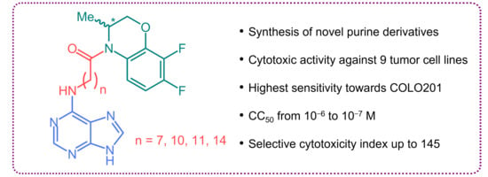
Synthesis and Cytotoxic Activity of the Derivatives of N-(Purin-6-yl)aminopolymethylene Carboxylic Acids and Related Compounds

by
Victor P. Krasnov
1,*,
Olga A. Vozdvizhenskaya
1,
Maria A. Baryshnikova
2,
Alexandra G. Pershina
3,4,
Vera V. Musiyak
1,
Tatyana V. Matveeva
1,
Kseniya V. Nevskaya
3,
Olga Y. Brikunova
3,
Dmitry A. Gruzdev
1 and
Galina L. Levit
1
1
Postovsky Institute of Organic Synthesis, Russian Academy of Sciences (Ural Branch), 620108 Ekaterinburg, Russia
2
Blokhin National Medical Research Center of Oncology, Ministry of Health of the Russian Federation, 115522 Moscow, Russia
3
Center of Bioscience and Bioengineering, Siberian State Medical University, 634050 Tomsk, Russia
4
Research School of Chemical and Biomedical Engineering, National Research Tomsk Polytechnic University, 634050 Tomsk, Russia
*
Author to whom correspondence should be addressed.
Molecules 2023, 28(4), 1853; https://doi.org/10.3390/molecules28041853
Submission received: 12 January 2023 / Revised: 6 February 2023 / Accepted: 13 February 2023 / Published: 15 February 2023
(This article belongs to the Special Issue Synthesis of Bioactive Compounds)

Abstract

:
Testing a number of N-[omega-(purin-6-yl)aminoalkanoyl] derivatives of 7,8-difluoro-3,4-dihydro-3-methyl-2H-[1,4]benzoxazine in a panel of nine tumor cell lines has shown that the studied compounds exhibit high cytotoxic activity, especially against 4T1 murine mammary carcinoma, COLO201 human colorectal adenocarcinoma, SNU-1 human gastric carcinoma, and HepG2 human hepatocellular carcinoma cells. Synthesis and study of structural analogs of these compounds made it possible to find that the presence of both a difluorobenzoxazine fragment and a purine residue bound via a linker of a certain length is crucial for the manifestation of the cytotoxic activity of this group of compounds. The study of the effect of the most promising compound on the cell cycle of the human tumor cell lines, the most sensitive and least sensitive to cytotoxic action (MDA-MB-231 breast adenocarcinoma and COLO201 colorectal adenocarcinoma, respectively), allows us to conclude that this compound is an inhibitor of DNA biosynthesis. The found group of purine conjugates may be of interest in the design of new antitumor agents.

Graphical Abstract

1. Introduction

The prevalence of cancer in the world is enormous. Despite advances in the field of tumor therapy, the number of reported cases of cancer is increasing every year. According to the International Agency for Research on Cancer, approximately 19.3 million new cases of cancer have been identified in 2020 [1]. The design of new agents for cancer treatment, primarily of the most lethal forms, is an urgent task. Preparation of new compounds that are highly toxic to tumor cells and are well tolerated at therapeutic doses can lay the foundation for new cancer treatment regimens and contribute to the reduction in cancer mortality.
Purine-containing compounds, such as nucleic acids, ATP, ADP, and NAD, play a very important role in cell metabolism and vital activity. Many compounds with antineoplastic activity have been found among purine derivatives. Purine-based compounds such as mercaptopurine, cladribine, clofarabine, and fludarabine (Figure 1) are among the clinically used anticancer agents.
In recent decades, efficient cytostatics with different mechanisms of action have been found among purine derivatives: inhibitors of cyclin-dependent kinases [2,3,4,5], apoptosis inducers [6,7,8,9,10] and some others [11,12,13,14]. Purine conjugates containing fragments of polymethylene carboxylic acids deserve special attention. It has been reported that these compounds are kinase inhibitors [15,16,17], histone deacetylase inhibitors with antiproliferative activity [18], and also bivalent modulators of A1 adenosine receptors (A1ARs) and β2-adrenergic receptors (β2ARs) [19,20] (Figure 2). At the same time, it has been reported that the introduction of an adenosine residue leads to a significant decrease in the toxicity of the ω-aminopolymethylenecarbonyl derivatives of batracylin [21].
In recent years, we have focused on the preparation of new purine and 2-aminopurine derivatives and the search for highly bioactive compounds in this series [22,23,24,25,26,27,28,29,30]. In particular, we have synthesized a number of purine conjugates with various N-heterocycles attached via a linker, the omega-amino acid residue, –NH(CH2)nCO–, to position 6 of the purine nucleus [22,23,24,25,26,27,28,29]. The results of biological testing have shown that conjugates of purine with racemic 7,8-difluoro-3,4-dihydro-3-methyl-2H-[1,4]benzoxazine (Figure 3, compounds 1a–f) attached via 6-aminohexanoyl (n = 5, compound 1b) or 8-aminooctanoyl (n = 7, compound 1c) residues, as well as (S)-enantiomer of compound 1b, exhibit high antiviral activity in vitro against herpes simplex virus type 1 (HSV-1), including acyclovir-resistant HSV-1 strain [26,27,28].
It has also been found that some of the synthesized compounds exhibit high cytotoxicity. Thus, the concentration of compound 1d (n = 10), which caused the death of 50% Vero E6 cells, was less than 30 µM [28]. In the framework of this work, in order to elucidate the structure–activity relationship, we have synthesized and studied the cytotoxic activity of purine conjugates containing fragments of omega-amino acids with different lengths of the polymethylene chain as linkers, and related compounds against tumor cell lines.

2. Results and Discussion

2.1. Chemistry

Synthesis of the studied compounds 1a–c, (S)-1b [26], and 1d–f [28] was described earlier. To reveal the structure–activity relationship, we synthesized a number of related compounds. Thus, N-(purin-6-yl)amino acids 2a–c were synthesized in moderate yields by analogy with a literature method [31], reacting the corresponding omega-amino acids with 6-chloropurine in an aqueous Na2CO3 solution under reflux (Scheme 1).
A major disadvantage of compound 1d, which limits its further study, is its low solubility in aqueous media. In order to increase the solubility and, possibly, to improve the profile of the cytotoxic action, we synthesized compound 7 containing a 2-hydroxyethoxymethyl fragment in position N(9) of purine (Scheme 2). Compounds 6 and 7 were synthesized starting from N-phthaloyl derivative 3 [28]. At the first step, compound 3 was treated with hydrazine hydrate in refluxing EtOH to afford to the corresponding amine 4, which (without isolation) was introduced into the reaction of nucleophilic substitution of chlorine in 9-[(2-acetoxyethoxy)methyl]-6-chloropurine (5) [32], thus resulting in compound 6. The subsequent alkaline hydrolysis in 1N NaOH gave the target compound 7 in good yield.
For comparative studies, we have obtained two compounds without purine residue, but the polymethylenecarboxylic acid amide fragment is retained (Scheme 3). As a result of the interaction of sebacoyl dichloride (8) with enantiomerically pure (S)-7,8-difluoro-3,4-dihydro-3-methyl-2H-[1,4]benzoxazine [33] in dichloromethane at room temperature in the presence of N,N-diethylaniline (PhNEt2) as a HCl acceptor, diamide (S)-9 was obtained; the reaction with racemic 7,8-difluoro-3,4-dihydro-3-methyl-2H-[1,4]benzoxazine gave a mixture of three diastereomers 9. Using chiral HPLC, the (R*,S*)–(S,S)–(R,R) diastereomeric ratio has been found to be approximately 2:1:1 (see the Supplementary Materials, Figures S19 and S20).

2.2. Cytotoxicity Assessment

The cytotoxicity of the test compounds was studied using the MTT assay [34] against a number of cell lines: CT-26 murine colon carcinoma, 4T1 murine mammary carcinoma, MDA-MB-231 human breast adenocarcinoma, COLO201 human colorectal adenocarcinoma, HepG2 human hepatocellular carcinoma, A549 human non-small-cell lung carcinoma, SK-BR-3 human breast adenocarcinoma, SNU-1 human gastric carcinoma, Jurkat human acute T-lymphoblastic leukemia, and WI-38 human lung fibroblasts (normal cells). Doxorubicin (Dox) was used as a reference drug. Cells were incubated with test compounds for 72 h.
Since primary data on cytotoxicity were obtained for compounds 1c–f (Figure 3) [28], we first carried out a detailed study of the cytotoxic activity of these compounds against various cells and determined their 50% cytotoxic concentration (CC50) and selective cytotoxicity index (SCI) (Table 1). Selective cytotoxicity index (SCI) (see, for example [35,36]) generally refers to the ability of a test compound to preferentially kill cancer cells, causing less or insignificant damage to normal cells (in our case, WI-38 human lung fibroblasts).
The COLO201 human colorectal adenocarcinoma and 4T1 murine mammary carcinoma cell lines were found to be more sensitive to compounds 1c–f (in some cases, CC50 < 1 µM), while the cell lines of SK-BR-3 and MDA-MB-231 human breast adenocarcinomas, as well as A549 human non-small-cell lung carcinoma, were the most resistant to the cytotoxic effects of the studied compounds (CC50 > 10 µM).
It should be emphasized that normal cells (WI-38 fibroblasts) are less sensitive to the highest of the studied concentrations (1 × 10⁻⁴ M) of compounds 1c–f than tumor cells (Table 2), which allows us to talk about the selectivity of action of these compounds. Compound 1d exhibits a high cytotoxic activity against a larger number of studied cell lines compared to compounds 1c,e,f, which makes it a promising candidate for further studies of antitumor activity.
The cytotoxicity of compounds 2a–c, 6, 7, and 9 was also studied using the MTT assay against a number of tumor cell lines: A549, SK-BR-3, SNU-1, Jurkat, as well as normal fibroblasts WI-38 (Table 3).
Compound 1a, in which purine and benzoxazine residues are separated by a single methylene group, was found to be non-cytotoxic (Table 3). Compound 1b, the linker of which contains five methylene groups, showed cytotoxic activity, but in general, it was inferior to compound 1d. It turned out that SK-BR-3 breast cancer cells were resistant to compounds 1b and (S)-1b (CC50 > 1 × 10⁻⁴), in contrast to compound 1d. The cytotoxic activity of the enantiomer (S)-1b practically did not differ from that of racemate 1b (except for the A549 cell line).
Among purine conjugates with omega-amino acids, compounds 2a,b were cytotoxic only against the Jurkat cell line (Table 3). Compound 2c showed no cytotoxic activity. Sebacates (S)-9 and 9 that do not contain a purine fragment, did not exhibit cytotoxic activity.
Apparently, the presence of both a purine fragment and difluorobenzoxazine connected via a linker of a certain length is important for the manifestation of cytotoxic activity.
Compounds 6 and 7 containing 2-acetoxy- and 2-hydroxy-ethoxymethyl fragments, respectively, exhibited approximately the same cytotoxicity as the most active compound 1d. It was not possible to achieve a significant increase in the solubility of conjugate 7 compared to compound 1d. At the same time, the preservation of the activity of compound 7 compared to 1d indicates that the modification at position 9 of purine does not lead to a loss of cytotoxic activity, so further introduction of hydrophilic groups, for example, sugar residues, can result in water-soluble conjugates.

2.3. Cell Cycle Analysis

To study the potential cytostatic effect of compound 1d, we chose the method of cell cycle analysis using flow cytometry by DNA staining with propidium iodide [37].
Based on experiments to determine the cytotoxic properties of compound 1d, the following two cell lines were selected for study: MDA-MB-231, which showed high resistance to the action of compound 1d, and COLO201, which showed the highest sensitivity among all tested lines. To analyze possible effects on the cell cycle, we tested three concentrations of compound 1d, which were close to CC50 calculated on the basis of viability assessment data (MTT assay). Figure 4 shows the phase distribution of the cell cycle of COLO201 and MDA-MB231 cells after incubation with compound 1d for 24 h.
According to the data obtained, compound 1d at concentrations close to CC50 after incubation for 24 h, had a dose-dependent cytostatic effect on COLO201 and MDA-MB-231 cell lines: a decrease in the proportion of cells in the S phase was observed (Figure 4A,B). In the case of COLO201, cell cycle arrest occurred in the G2/M phase; however, the cell proliferation index was in the range 35–37% and not changed compared to the control (see the Supplementary Materials, Table S2). For the MDA-MB-231 cell line, a decrease in the proliferation index (see the Supplementary Materials, Table S3) and arrest of the cell cycle in the G0/G1 phase were observed over the entire range of concentrations studied; when the concentration of 1d was increased to 30 µM, an increase in the proportion of cells in the G2/M phase was observed (Figure 4B).
When analyzing the possible mechanism of antitumor activity, it can be concluded that compound 1d at a dose lower CC50 blocks DNA synthesis in cells. This leads to the accumulation of cells in the G1 phase. An increase in the concentration of the substance above CC50 causes a pronounced death of cells in the G1 phase, which manifests itself in an increase in the relative proportion of cells in the G2/M phase with a progressive reduction in cells in the S phase. The delay in the G2/M phase is most likely due to the lengthening of the DNA repair process. It is important to note that the delay of cells in the G2/M phase leads to an increase in the calculated value of the cell proliferation index; thus, despite a pronounced cytostatic effect, no change in this indicator was recorded for COLO201 cells. Thus, compound 1d can be considered as a blocker of DNA synthesis.

3. Materials and Methods

3.1. Chemistry

3.1.1. Chemistry General Section

Compounds 1a–c, (S)-1b [26], 1d–f, 3 [28], and 5 [32] were obtained as previously described. Other reagents are commercially available and were purchased from Alfa Aesar (Heysham, UK). Melting points were obtained on a SMP3 apparatus (Barloworld Scientific, Staffordshire, UK) and are uncorrected. Optical rotations were measured on a Perkin Elmer M341 polarimeter (Perkin Elmer, Waltham, MA, USA). The reactions were monitored by thin layer chromatography (TLC) using silica gel precoated Sorbfil plates (Imid, Krasnodar, Russia); compounds were visualized by UV irradiation at 254 nm and iodine vapors. Flash column chromatography was performed using Silica gel 60 (230–450 mesh) (Alfa Aesar, Heysham, UK). The 1H, 19F, and 13C NMR spectra were recorded on a Bruker AVANCE 500 spectrometer (Bruker, Karlsruhe, Germany). Chemical shifts are given in ppm and are referenced to TMS (or DSS) and hexafluorobenzene as internal standards and multiplicities are reported as s (singlet), d (doublet), t (triplet), and m (multiplet). The 1H and 19F NMR spectra of compounds 6 and 7 were recorded in DMSO-d6 at 100 °C; the 1H and 13C NMR spectra of compounds 2b,c were recorded in a D2O–NaOD mixture at ambient temperature. For NMR spectra of the compounds obtained, see the Supplementary Materials, Figures S1–S18. CHN-Elemental analysis was performed using Perkin Elmer 2400 II analyzer (Perkin Elmer, Waltham, MA, USA). High resolution mass spectra were obtained on a Bruker ma**s Impact HD mass spectrometer (Bruker, Karlsruhe, Germany), electrospray ionization (ESI) with direct sample inlet (4 L/min flow rate). Analytical chiral HPLC of compounds (S)-9 and 9 was performed on an Agilent 1100 instrument (Agilent Technologies, Santa Clara, CA, USA) using a (S,S)-Whelk-O1 column (250 × 4.6 mm, 5 µm) (Phenomenex, Torrance, CA, USA); flow rate 0.8 mL/min, detection at 280 nm. For HPLC data for compounds (S)-9 and 9, see the Supplementary Materials, Figures S19 and S20.

3.1.2. Synthesis

General Procedure for the Synthesis of N-(Purin-6-yl)amino Carboxylic Acids 2a–c

6-Chloropurine (2.00 g, 12.9 mmol) and Na2CO3 (1.37 g, 12.9 mmol) were added to a suspension of appropriate ω-amino acid (25.9 mmol) in water (40 mL). The reaction mixture was refluxed for 3 h; then 4 N HCl (2 mL) was added, the precipitate formed was filtered off and recrystallized from ethanol (in the case of compounds 2a and 2b) or washed with hot ethanol (in the case of compound 2c).
N-(Purin-6-yl)-11-aminoundecanoic Acid (2a). Yield 2.10 g (51%). Colorless powder m.p. 172–174 °C (EtOH). 1H NMR (500 MHz, DMSO-d6) δ (ppm): 1.24 (br. s, 8H, 4×CH2), 1.28–1.37 (m, 4H, 2×CH2), 1.43–1.52 (m, 2H, CH2), 1.59–1.64 (m, 2H, CH2), 2.18 (dd, J = 7.4, 7.4 Hz, 2H, CH2CO), 3.53 (br. s, 2H, CH2NH), 8.33 (s, 1H, H-8 purine), 8.41 (br. s, 1H, H-2 purine), 8.82 (br. s, 1H, NH), 12,00 (br. s, 1H, NH purine), 13.65 (br. s, 1H, COOH). 13C NMR (125 MHz, DMSO-d6) δ (ppm): 24.4, 26.2, 28.5, 28.6 (3C), 28.7, 28.8, 33.6, 40.5 (br. s), 114.5 (br. s), 141.3, 148.5, 148.8, 152.1, 174.4. HRMS (ESI): m/z [M+H]+ calcd for [C16H26N5O2]+: 320.2082, found: 320.2080.
N-(Purin-6-yl)-12-aminododecanoic Acid (2b). Yield 2.36 g (55%). Colorless powder m.p. 190–191 °C (EtOH). 1H NMR (500 MHz, D2O + NaOD) δ (ppm): 1.03 (br. s, 6H, 3×CH2), 1.07–1.17 (m, 6H, 3×CH2), 1.19–1.25 (m, 2H, CH2), 1.45–1.51 (m, 2H, CH2), 1.51–1.57 (m, 2H, CH2), 2.16 (dd, J = 7.7, 7.6 Hz, 2H, CH2CO), 3.49 (dd, J = 6.5, 6.4 Hz, 2H, CH2NH), 7.93 (s, 1H, H-8 purine), 8.15 (s, 1H, H-2 purine). 13C NMR (125 MHz, D2O + NaOD) δ (ppm): 28.6, 28.7, 31.2, 31.26, 31.31, 31.33 (2C), 31.5, 31.6, 40.4, 43.5, 122.7, 152.3, 154.2, 156.7, 160.7, 186.7. HRMS (ESI): m/z [M+H]+ calcd for [C17H28N5O2]+: 334.2239, found: 334.2244.
N-(Purin-6-yl)-15-aminopentadecanoic Acid (2c). Yield 2.72 g (56%). Colorless powder m.p. 198–199 °C. 1H NMR (500 MHz, D2O + NaOD) δ (ppm): 1.10 (br. s, 12H, 6×CH2), 1.14 (br. s, 6H, 3×CH2), 1.22–1.29 (m, 2H, CH2), 1.45–1.51 (m, 2H, CH2), 1.52–1.58 (m, 2H, CH2), 2.14 (dd, J = 7.9, 7.7 Hz, 2H, CH2CO), 3.47 (dd, J = 6.5, 6.5 Hz, 2H, CH2NH), 7.89 (s, 1H, H-8 purine), 8.13 (s, 1H, H-2 purine). 13C NMR (125 MHz, D2O + NaOD) δ (ppm): 28.7, 29.0, 31.6, 31.7, 31.8, 31.9 (6C), 40.3, 43.4, 122.6, 152.0, 152.1, 153.7, 156.4, 160.5, 186.1. HRMS (ESI): m/z [M+H]+ calcd for [C20H34N5O2]+: 376.2708, found: 376,2711.
{11-[9-(2-Acetoxyethoxy)methylpurin-6-yl]aminoundecanoyl]-7,8-difluoro-3,4-dihydro-3-methyl-2H-[1,4]benzoxazine (6). Hydrazine monohydrate (0.22 mL, 4.52 mmol) was added to a solution of compound 3 (1.25 g, 2.51 mmol) in EtOH (25 mL). The reaction mixture was refluxed for 2 h, and then evaporated to dryness. The residue was treated with Et2O (15 mL) and kept at −16 °C overnight; the resulting precipitate was filtered off; filtrate was evaporated to dryness. The residue was re-dissolved in n-BuOH (10 mL); the formed solution was added to a solution of compound 5 (0.27 g, 1.00 mmol) and NEt3 (0.42 mL, 3.00 mmol) in n-BuOH (10 mL). The reaction mixture was refluxed for 6 h, cooled to room temperature, successively washed with 1N HCl (3 × 10 mL), a saturated aqueous NaCl solution (3 × 15 mL), and water (10 mL), and evaporated to dryness under reduced pressure. The residue was purified by flash column chromatography on silica gel (chloroform–EtOH, 98:2 as eluent) to afford 0.35 g (58%) of compound 6 as a colorless oil. 1H NMR (500 MHz, DMSO-d6, 100 °C) δ (ppm): 1.12 (d, J = 6.8 Hz, 3H, Me), 1.26 (br. s, 8H, 4×CH2), 1.29–1.36 (m, 4H, 2×CH2), 1.55–1.65 (m, 4H, 2×CH2), 1.91 (s, 3H, CH3CO), 2.43–2.49 (m, 1H, CH, partially overlapped with DMSO signal), 2.54–2.60 (m, 1H, CH), 3.56–3.60 (m, 2H, CH2), 3.74 (dd, J = 5.0, 4.7 Hz, 2H, OCH2), 4.08 (dd, J = 6.0, 4.7 Hz, 2H, CH2OAc), 4.13 (dd, J = 11.0, 2.9 Hz, 1H, H-2A benzoxazine), 4.33 (dd, J = 11.0, 1.5 Hz, 1H, H-2B benzoxazine), 4.71–4.76 (m, 1H, H-3 benzoxazine), 5.56 (s, 2H, N–CH2–O), 6.85 (td, J = 9.8, 8.3 Hz, 1H, H-6 benzoxazine), 7.20 (m, 1H, NH), 7.55 (ddd, J = 8.3, 5.6, 2.6 Hz, 1H, H-5 benzoxazine), 8.14 (br. s, 1H, H-8 purine), 8.19 (s, 1H, H-2 purine). 19F NMR (470 MHz, DMSO-d6, 100 °C) δ (ppm): 1.98 (ddd, J = 20.5, 8.0, 2.0 Hz, 1F, F-8), 20.04–20.14 (m, 1F, F-7). 13C NMR (125 MHz, DMSO-d6) δ (ppm): 15.1, 20.4 (2C), 24.6, 26.3, 28.5, 28.71, 28.73, 28.8, 28.9, 29.0 (br. s), 33.4, 44.8 (br. s), 62.7, 66.8, 69.8, 71.9, 106.7 (d, J = 18.0 Hz), 118.8, 119.3, 121.8, 135.7 (d, J = 8.5 Hz), 138.9 (dd, J = 243.0, 15.5 Hz), 140.7, 146.4 (d, J = 244.0 Hz), 148.8 (br. s), 152.9, 154.5, 170.1, 171.0. Calcd (%) for C30H40F2N6O5: C 59.79, H 6.69, F 6.30, N 13.94. Found (%): C 60.02, H 7.00, F 6.55, N 13.64.
4-{11-[9-(2-Hydroxyethoxy)methylpurin-6-yl]aminoundecanoyl}-7,8-difluoro-3,4-dihydro-3-methyl-2H-[1,4]benzoxazine (7). The 1N NaOH (1.8 mL) was added to a solution of compound 6 (0.21 g, 0.35 mmol) in EtOH (4 mL). The reaction mixture was stirred at room temperature for 1 day, neutralized with 4 N HCl to pH = 6, and evaporated to dryness. The residue was purified by flash chromatography on silica gel (eluent CHCl3–EtOH, 95:5). Yield 0.16 g (82%), colorless oil. 1H NMR (500 MHz, DMSO-d6, 100 °C) δ (ppm): 1.12 (d, J = 6.8 Hz, 3H, Me), 1.26 (br. s, 8H, 4×CH2), 1.29–1.36 (m, 4H, 2×CH2), 1.55–1.65 (m, 4H, 2×CH2), 2.43–2.49 (m, 1H, CH, partially overlapped with DMSO signal), 2.54–2.60 (m, 1H, CH), 3.48–3.50 (m, 2H, CH2), 3.55–3.60 (m, 4H, 2×CH2), 4.13 (dd, J = 10.9, 2.9 Hz, 1H, H-2A benzoxazine), 4.25 (br. s, 1H, OH), 4.33 (dd, J = 10.9, 1.4 Hz, 1H, H-2B benzoxazine), 4.71–4.76 (m, 1H, H-3 benzoxazine), 5.55 (s, 2H, N–CH2–O), 6.84 (td, J = 9.9, 8.2 Hz, 1H, H-6 benzoxazine), 7.17 (br. t, J = 5.6 Hz, 1H, NHCH2), 7.55 (ddd, J = 7.9, 5.4, 2.4 Hz, 1H, H-5 benzoxazine), 8.13 (br. s, 1H, H-8 purine), 8.19 (s, 1H, H-2 purine). 19F NMR (470 MHz, DMSO-d6, 100 °C) δ (ppm): 1.98 (ddd, J = 21.0, 8.0, 2.0 Hz, 1F, F-8), 20.06–20.13 (m, 1F, F-7). 13C NMR (125 MHz, DMSO-d6) δ (ppm): 15.1, 24.6, 26.3, 28.5, 28.71, 28.73, 28.8, 28.9, 29.0 (br. s), 45.0 (br. s), 59.8 (2C), 69.8, 70.7 (2C), 72.1, 106.8 (d, J = 18.0 Hz), 118.8 (br. s), 119.3, 121.8, 135.7 (dd, J = 10.0, 2.5 Hz), 138.9 (dd, J = 243.5, 15.5 Hz), 140.7, 146.5 (d, J = 243.5 Hz), 148.9 (br. s), 152.8, 154.5, 171.0. HRMS (ESI): m/z [M+H]+ calcd for [C28H39F2N6O4]+: 561.2995, found: 561.2999.
General Procedure for the Synthesis of Sebacates 9. Thionyl chloride (3.6 mL, 49.62 mmol) was added to sebacic acid (2.00 g, 9.89 mmol); the reaction mixture was refluxed for 1 h, then evaporated to dryness. The residue was dissolved in CH2Cl2 (25 mL); N,N-diethylaniline (3.2 mL, 20.0 mmol) and (S)-enantiomer or racemic 7,8-difluoro-3,4-dihydro-3-methyl-2H-[1,4]benzoxazine (3.60 g, 19.4 mmol) were added to the resulting solution. The reaction mixture was stirred at room temperature for 48 h, then successively washed with 4N HCl (3 × 10 mL), saturated NaCl solution (5 × 10 mL), 5% NaHCO3 solution (3 × 10 mL), and water (3 × 10 mL), dried over MgSO4, and evaporated to dryness under reduced pressure. The residue was recrystallized from a EtOH–water (2:1) mixture.
1,10-bis((S)-7,8-Difluoro-3-methyl-2H-benzo[b][1,4]oxazin-4(3H)-yl)decan-1,10-dione ((S)-9). Yield 2.87 g (54%), yellowish powder m.p. 41–42 °C. HPLC ((S,S)-Whelk-O1, MeOH–H2O, 85:15): t 17.3 min. 1H NMR (500 MHz, DMSO-d6) δ (ppm): 1.11 (d, J = 6.2 Hz, 6H, 2×Me), 1.27 (br. s, 8H, 4×CH2), 1.51–1.58 (m, 4H, 2×CH2), 2.42–2.48 (m, 2H, CH2), 2.60–2.66 (m, 2H, CH2), 4.14 (br. d, J = 10.4 Hz, 2H, 2×H-2A benzoxazine), 4.36 (d, J = 10.8 Hz, 2H, 2×H-2B benzoxazine), 4.71 (br. s, 2H, 2×H-3 benzoxazine), 6.92 (td, J = 9.4, 9.0 Hz, 2H, 2×H-6 benzoxazine), 7.65 (br. s, 2H, 2×H-5 benzoxazine). 19F NMR (470 MHz, DMSO-d6, 100 °C) δ (ppm): 1.86 (br. s, F-8), 19.56 (br. s, F-7). 13C NMR (125 MHz, DMSO-d6) δ (ppm): 15.1, 24.6, 28.5, 28.7, 33.4, 44.8 (br. s), 69.8, 106.8 (d, J = 18.0 Hz), 119.3, 121.8, 135.7 (dd, J = 9.5, 2.5 Hz), 138.9 (dd, J = 243.5, 15.5 Hz), 146.4 (d, J = 243.5 Hz), 171.0. Calcd (%) for C28H32F4N2O4: C 62.68, H 6.01, F 14.16, N 5.22. Found (%): C 62.47, H 6.15, F 14.16, N 5.44.
1,10-bis(-7,8-Difluoro-3-methyl-2H-benzo[b][1,4]oxazin-4(3H)-yl)decan-1,10-dione (mixture 9). Yield 2.61 g (49%), colorless powder m.p. 103–104 °C (EtOH-H2O). HPLC ((S,S)-Whelk-O1, MeOH–H2O, 85:15): tS,S 17.6 min (27%), tR*,S* 24.2 min (47%), tR,R 34.0 min (26%). 1H NMR (500 MHz, DMSO-d6) δ (ppm): 1.11 (d, J = 6.1 Hz, 6H, 2×Me), 1.27 (br. s, 8H, 4×CH2), 1.49–1.60 (m, 4H, 2×CH2), 2.42–2.48 (m, 2H, CH2), 2.60–2.66 (m, 2H, CH2), 4.14 (br. d, J = 10.0 Hz, 2H, 2×H-2A benzoxazine), 4.36 (d, J = 10.8 Hz, 2H, 2×H-2B benzoxazine), 4.71 (br. s, 2H, 2×H-3 benzoxazine), 6.92 (td, J = 9.6, 8.9 Hz, 2H, 2×H-6 benzoxazine), 7.66 (br. s, 2H, 2×H-5 benzoxazine). 19F NMR (470 MHz, DMSO-d6, 100 °C) δ (ppm): 1.84 (br. s, F-8), 19.40 (br. s, F-7). 13C NMR (125 MHz, DMSO-d6) δ (ppm): 15.1, 24.6, 28.5, 28.6, 33.4, 44.9 (br. s), 69.8, 106.7 (d, J = 18.0 Hz), 119.2, 121.8, 135.7 (dd, J = 9.5, 2.5 Hz), 138.9 (dd, J = 243.5, 15.5 Hz), 146.4 (d, J = 242.0 Hz), 171.0. Calcd (%) for C28H32F4N2O4: C 62.68, H 6.01, F 14.16, N 5.22. Found (%): C 62.76, H 6.15, F 14.03, N 5.25.

3.2. Cytotoxicity Assessment

3.2.1. Materials

The following materials were used: dimethyl sulfoxide (DMSO, PanEco, Moscow, Russia), DMEM/F12 (Gibco, Grand Island, NY, USA), RPMI-1640 (Gibco, Grand Island, NY, USA), 100 × Glutamax (Gibco, Grand Island, NY, USA), fetal bovine serum (FBS, Gibco, Grand Island, NY, USA), HEPES (Sigma-Aldrich, St. Louis, MO, USA), PBS (Sigma-Aldrich, St. Louis, MO, USA), propidium iodide (Sigma-Aldrich, St. Louis, MO, USA), RNAse A (Sigma-Aldrich, St. Louis, MO, USA), Triton X-100 (PanReac AppliChem, Barcelona, Spain), 100 × Penicillin-Streptomycin (PanEco, Moscow, Russia), and L-glutamine (PanEco, Moscow, Russia). TrypLE (Gibco, Grand Island, NY, USA) was used for dissociating adherent cells from the surface of plastic flasks.

3.2.2. Cell Lines

The following cell lines were used: CT-26 murine colon carcinoma (ATCC), 4T1 murine mammary carcinoma (ATCC), MDA-MB-231 human breast adenocarcinoma (ECACC), COLO201 human colorectal adenocarcinoma (ATCC), HepG2 human hepatocellular carcinoma (State Research Center of Virology and Biotechnology “Vector”, Koltsovo, Russia); A549 human non-small-cell lung carcinoma, SK-BR-3 human breast adenocarcinoma, SNU-1 human gastric carcinoma, Jurkat human acute T-lymphoblastic leukemia, and WI-38 human lung fibroblasts (normal cells) obtained from the Bioresource Collection of Cell Lines and Primary Tumors of the Blokhin National Medical Research Center of Oncology (Moscow, Russia). Cells were counted on a Countess® II FL automatic cell counter (Thermo Fisher Scientific, Singapore).

3.2.3. MTT Cytotoxicity Assay

Cells were seeded in flat-bottomed 96-well plates at 10,000 cells per well (for A549, SNU-1, WI-38, CT-26, MDA-MB-231, 4T1, and HepG2 cells) or 20,000 cells per well (for SK-BR-3, Jurkat, and COLO201 cells) and cultivated in complete culture mediums (for composition of each medium, see the Supplementary Materials, Table S1) in a CO2 incubator at 37 °C. At the first stage of this study, the test compounds dissolved in DMSO were added to the wells with cells at final concentrations from 1 × 10⁻⁴ to 1 × 10⁻¹⁰ mol/L (A549, SK-BR-3, SNU-1, Jurkat, and WI-38) or 1 × 10⁻³ to 1 × 10⁻⁸ mol/L (CT-26, MDA-MB-231, 4T1, COLO201, and HepG2); DMSO concentration in the wells was finally 1%. A complete cultural medium and DMSO were added to the control wells at the same concentration as in the experimental wells; 0.1% Triton X-100 solution (10 µL) was added to the corresponding wells as a non-specific positive control. After adding the test compounds, the cells were incubated for 72 h in an atmosphere containing 5% CO2. After that, the nutrient medium was removed from each well (in the case of SK-BR-3, Jurkat, and COLO201, plates were pre-centrifuged at 1500 rpm for 6 min). A solution of the commercial anticancer agent Doxorubicin (Dox, Sigma-Aldrich, St. Louis, MO, USA) in DMSO was used as a reference drug. Then, 150 µL of a complete medium with the MTT reagent (PanEco, Moscow, Russia) at a concentration of 0.5 mg/mL was added and incubated for 2 h. Formazan crystals were dissolved in DMSO (200 µL) and the absorbance in the wells was measured at 540 nm and a reference wavelength of 650 nm (Sunrise, Tecan, Groding, Austria). Experiments were performed in three parallel runs. Cell viability after incubation in the presence of test compounds was calculated in relation to cell viability in the control wells according to the Formula (1):
Cell viability (%) = (Aexp/Ac) × 100,
where Aexp is the optical density of solutions in the wells containing test compounds; Ac is the optical density of solutions in control wells without test compounds. For detailed information on cell viability of various cell lines, see the Supplementary Materials, Figures S21–S26.
At the second stage of this study, in order to more accurately calculate the CC50 (the half maximal inhibitory concentration), the cytotoxic effect of test compounds was evaluated at concentrations close to CC50 found at the first stage. The study of the cytotoxic activity of each substance towards the cells of each line was carried out in triplicate; the mean value was used to calculate CC50. The CC50 values were determined by non-linear regression analysis using Graph-Pad Prism7.

3.2.4. Cell Cycle Analysis

Cell cycle analysis was performed by flow cytometry by staining DNA with propidium iodide (PI). For this, cells were seeded into 6-well plates: 2 × 10⁵ COLO201 cells per well and 1.2 × 10⁵ MDA-MB-231 cells per well. The next day, a solution of compound 1d in DMSO at a concentration of 1 × 10⁻² mol/L was prepared, and then a series of sequential dilutions was obtained; for each cell line, compound 1d was tested at three concentrations close to CC50. The final concentration of DMSO in the culture medium was 0.1%; an equivalent volume was added to the control cells. The plates were incubated for 24 h in a CO2 incubator. Then, cells were detached from the plastic surface using TrypLE, washed with PBS, and fixed with 70% ethanol. Next, the cells were incubated for 1 h in the dark at 4 °C, then sedimented by centrifugation at 184× g for 10 min, and washed twice with PBS. PBS (0.3 mL) containing 0.1% Triton X-100, 1 µg/mL PI, and 0.2 mg/mL RNAse A was added to the pellet, incubated for 30 min at room temperature in the dark, and analyzed on a CytoFlex flow cytometer (Beckman Coulter, Brea, CA, USA).
The cell population in the test sample was found and gated on forward versus side scatter (FSC-A vs. SSC-A). Next, doublets were discriminated and removed via PI signal height versus PI signal area (PI-H vs. PI-A). For cell cycle phases analysis, PI-A was plotted against number of events (number of cells) (PI-A vs. Count).
The flow cytometry data were processed using the Kaluza Analysis 2.1 software (Beckman Coulter, Brea, CA, USA). Percentages of cell populations distributed in the various phases of the cell cycle (sub-G1, G0/G1, S, and G2M) were calculated. The result was considered significant if the coefficient of variation (CV) was less than 6.
Representative diagrams of the COLO201 and MDA-MB-231 cell distribution by cell cycle phases are presented in the Supplementary Materials (Figures S27 and S28).

3.2.5. Statistical Analysis

Statistical data processing was carried out using GraphPad Prism 5.0, GraphPad Prism 7 (GraphPad Software, San Diego, CA, USA), and Microsoft Office Excel software packages. Descriptive statistics were used for all data. The normality of data distribution was tested using the Shapiro–Wilks test. Data were presented as the mean ± standard deviation (M ± SD). To identify the significance of differences in the case of multiple comparisons, one-way analysis of variance (one-way ANOVA) was used for group comparison, then the Tukey’s test for pairwise comparisons between groups. Differences were considered significant at p ˂ 0.05.

4. Conclusions

In summary, the high cytotoxic activity of purinyl derivatives of omega-aminoalkanoyl-3,4-dihydro-3-methyl-7,8-difluoro-2H-[1,4]benzoxazine, especially compound 1d, against A549, SK-BR-3, SNU-1, Jurkat, CT-26, MDA-MB-231, 4T1, COLO201, and HepG2 tumor cells has been found. Among the cell lines studied, 4T1 murine breast carcinoma, COLO201 human colorectal adenocarcinoma, SNU-1 gastric cancer, and HepG2 human hepatocellular carcinoma cells showed the highest sensitivity. The cytotoxicity against these tumor cells in experiments exceeded the cytotoxicity against normal cells (human lung fibroblasts WI-38); selective cytotoxicity index reached 145.
We have obtained structural analogs of the most active compounds; the study of their cytotoxicity made it possible to find that the presence of both a purine fragment and difluorobenzoxazine bound via a polymethylene linker of a certain length is important for the manifestation of cytotoxic activity of this group of compounds. The introduction of a 2-hydroxyethoxymethyl group into the molecule of compound 1d resulted in a compound that was not inferior to it in terms of cytotoxic activity.
The study of the effect of the most promising compound 1d on the cell cycle of two types of human tumor cell lines, the most highly sensitive and the least sensitive to cytotoxic action, allows us to conclude that this compound is a blocker of DNA biosynthesis. Thus, we have found a group of potential antitumor agents, purine conjugates with high cytotoxicity against tumor cell lines.

Supplementary Materials

The following supporting information can be downloaded at: https://mdpi.longhoe.net/article/10.3390/molecules28041853/s1. Figures S1–S18: 1H, 19F, and 13C NMR spectra of compounds 2a–c, 6, 7, (S)-9, and 9. Figures S19 and S20: HPLC data for compounds (S)-9 and 9. Table S1: Cell lines and composition of culture medium. Figure S21: Viability of WI-38, A549, SK-BR-3, SNU-1, Jurkat, CT-26, MDA-MB-231, 4T1, COLO201, HepG2 cells vs. logarithmic concentration of compound 1c after a 72-h incubation. Figure S22: Viability of WI-38, A549, SK-BR-3, SNU-1, Jurkat, CT-26, MDA-MB-231, 4T1, COLO201, HepG2 cells vs. logarithmic concentration of compound 1d after a 72-h incubation. Figure S23: Viability of WI-38, A549, SK-BR-3, SNU-1, Jurkat, CT-26, MDA-MB-231, 4T1, COLO201, HepG2 cells vs. logarithmic concentration of compound 1e after a 72-h incubation. Figure S24: Viability of WI-38, A549, SK-BR-3, SNU-1, Jurkat, CT-26, MDA-MB-231, 4T1, COLO201, HepG2 cells vs. logarithmic concentration of compound 1f after a 72-h incubation. Figure S25. Viability of WI-38, A549, SK-BR-3, SNU-1, and Jurkat cells vs. logarithmic concentration of compound 6 after a 72-h incubation. Figure S26: Viability of WI-38, A549, SK-BR-3, SNU-1, and Jurkat cells vs. logarithmic concentration of compound 7 after a 72-h incubation. Figure S27: Diagrams of the COLO201 cell distribution by cell cycle phases. Figure S28: Diagrams of the MDA-MB-231 cell distribution by cell cycle phases. Table S2: Effect of compound 1d on the cell cycle of the COLO201 cells. Table S3: Effect of compound 1d on the cell cycle of the MDA-MB-231 cells.

Author Contributions

Conceptualization and methodology, V.P.K.; investigation, O.A.V., M.A.B., A.G.P., V.V.M., T.V.M., K.V.N. and O.Y.B.; writing—original draft preparation, V.P.K. and D.A.G.; writing—review and editing, V.P.K. and G.L.L.; supervision, V.P.K. All authors have read and agreed to the published version of the manuscript.

Funding

This research was funded by the Russian Science Foundation, Grant No. 19-13-00231-P.

Institutional Review Board Statement

Not applicable.

Informed Consent Statement

Not applicable.

Data Availability Statement

Not applicable.

Acknowledgments

Equipment of the Centre for Joint Use “Spectroscopy and Analysis of Organic Compounds” at the Postovsky Institute of Organic Synthesis was used.

Conflicts of Interest

The authors declare no conflict of interest.

Sample Availability

Not applicable.

References

  1. Sung, H.; Ferlay, J.; Siegel, R.L.; Laversanne, M.; Soerjomataram, I.; Jemal, A.; Bray, F. Global Cancer Statistics 2020: GLOBOCAN Estimates of Incidence and Mortality Worldwide for 36 Cancers in 185 Countries. CA Cancer J. Clin. 2021, 71, 209–249. [Google Scholar] [CrossRef] [PubMed]
  2. Chang, Y.-T.; Gray, N.S.; Rosania, G.R.; Sutherlin, D.P.; Kwon, S.; Norman, T.C.; Sarohia, R.; Leost, M.; Meijer, L.; Schultz, P.G. Synthesis and application of functionally diverse 2,6,9-trisubstituted purine libraries as CDK inhibitors. Chem. Biol. 1999, 6, 361–375. [Google Scholar] [CrossRef] [PubMed] [Green Version]
  3. Meijer, L.; Raymond, E. Roscovitine and Other Purines as Kinase Inhibitors. From Starfish Oocytes to Clinical Trials. Acc. Chem. Res. 2003, 36, 417–425. [Google Scholar] [CrossRef] [PubMed]
  4. Gucký, T.; Jorda, R.; Zatloukal, M.; Bazgier, V.; Berka, K.; Řezníčková, E.; Béres, T.; Strnad, M.; Kryštof, V. A Novel Series of Highly Potent 2,6,9-Trisubstituted Purine Cyclin-Dependent Kinase Inhibitors. J. Med. Chem. 2013, 56, 6234–6247. [Google Scholar] [CrossRef]
  5. Khalil, H.S.; Mitev, V.; Vlaykova, T.; Cavicchi, L.; Zhelev, N. Discovery and development of Seliciclib. How systems biology approaches can lead to better drug performance. J. Biotechnol. 2015, 202, 40–49. [Google Scholar] [CrossRef] [Green Version]
  6. Díaz-Gavilán, M.; Conejo-García, A.; Cruz-López, O.; Núñez, M.C.; Choquesillo-Lazarte, D.; González-Pérez, J.M.; Rodríguez-Serrano, F.; Marchal, J.A.; Aránega, A.; Gallo, M.A.; et al. Synthesis and Anticancer Activity of (R,S)-9-(2,3-Dihydro-1,4-Benzoxathiin-3-ylmethyl)-9H-Purines. ChemMedChem 2008, 3, 127–135. [Google Scholar] [CrossRef]
  7. Conejo-García, A.; García-Rubiño, M.E.; Marchal, J.A.; Núñez, M.C.; Ramírez, A.; Cimino, S.; García, M.Á.; Aránega, A.; Gallo, M.A.; Campos, J.M. Synthesis and anticancer activity of (RS)-9-(2,3-dihydro-1,4-benzoxaheteroin-2-ylmethyl)-9H-purines. Eur. J. Med. Chem. 2011, 46, 3795–3801. [Google Scholar] [CrossRef]
  8. Caba, O.; Díaz-Gavilán, M.; Rodríguez-Serrano, F.; Boulaiz, H.; Aránega, A.; Gallo, M.A.; Marchal, J.A.; Campos, J.M. Anticancer activity and cDNA microarray studies of a (RS)-1,2,3,5-tetrahydro-4,1-benzoxazepine-3-yl]-6-chloro-9H-purine, and an acyclic (RS)-O,N-acetalic 6-chloro-7H-purine. Eur. J. Med. Chem. 2011, 46, 3802–3809. [Google Scholar] [CrossRef]
  9. Knies, C.; Hammerbacher, K.; Bonaterra, G.A.; Kinscherf, R.; Rosemeyer, H. Nucleolipids of Canonical Purine β-D-Ribo-Nucleosides: Synthesis and Cytostatic/Cytotoxic Activities Toward Human and Rat Glioblastoma Cells. ChemistryOpen 2016, 5, 129–141. [Google Scholar] [CrossRef] [Green Version]
  10. Salas, C.O.; Zarate, A.M.; Kryštof, V.; Mella, J.; Faundez, M.; Brea, J.; Loza, M.I.; Brito, I.; Hendrychová, D.; Jorda, R.; et al. Promising 2,6,9-Trisubstituted Purine Derivatives for Anticancer Compounds: Synthesis, 3D-QSAR, and Preliminary Biological Assays. Int. J. Mol. Sci. 2020, 21, 161. [Google Scholar] [CrossRef]
  11. Parker, W.B. Enzymology of Purine and Pyrimidine Antimetabolites Used in the Treatment of Cancer. Chem. Rev. 2009, 109, 2880–2893. [Google Scholar] [CrossRef] [Green Version]
  12. Kumar, D.V.; Hoarau, C.; Bursavich, M.; Slattum, P.; Gerrish, D.; Yager, K.; Saunders, M.; Shenderovich, M.; Roth, B.L.; McKinnon, R.; et al. Lead optimization of purine based orally bioavailable Mps1 (TTK) inhibitors. Bioorg. Med. Chem. Lett. 2012, 22, 4377–4385. [Google Scholar] [CrossRef]
  13. De las Infantas, M.J.P.; Torres-Rusillo, S.; Unciti-Broceta, J.D.; Fernandez-Rubio, P.; Luque-Gonzalez, M.A.; Gallo, M.A.; Unciti-Broceta, A.; Molina, I.J.; Diaz-Mochon, J.J. Synthesis of 6,8,9 poly-substituted purine analogue libraries as pro-apoptotic inducers of human leukemic lymphocytes and DAPK-1 inhibitors. Org. Biomol. Chem. 2015, 13, 5224–5234. [Google Scholar] [CrossRef] [Green Version]
  14. Bosco, B.; Defant, A.; Messina, A.; Incitti, T.; Sighel, D.; Bozza, A.; Ciribilli, Y.; Inga, A.; Casarosa, S.; Mancini, I. Synthesis of 2,6-Diamino-Substituted Purine Derivatives and Evaluation of Cell Cycle Arrest in Breast and Colorectal Cancer Cells. Molecules 2018, 23, 1996. [Google Scholar] [CrossRef] [Green Version]
  15. Cancilla, M.T.; He, M.M.; Viswanathan, N.; Simmons, R.L.; Taylor, M.; Fung, A.D.; Cao, K.; Erlanson, D.A. Discovery of an Aurora kinase inhibitor through site-specific dynamic combinatorial chemistry. Bioorg. Med. Chem. Lett. 2008, 18, 3978–3981. [Google Scholar] [CrossRef]
  16. Enkvist, E.; Lavogina, D.; Raidaru, G.; Vaasa, A.; Viil, I.; Lust, M.; Viht, K.; Uri, A. Conjugation of Adenosine and Hexa-(D-arginine) Leads to a Nanomolar Bisubstrate-Analog Inhibitor of Basophilic Protein Kinases. J. Med. Chem. 2009, 52, 308–321. [Google Scholar] [CrossRef]
  17. Rubio-Ruíz, B.; Conejo-García, A.; Ríos-Marco, P.; Carrasco-Jiménez, M.P.; Segovia, J.; Marco, C.; Gallo, M.A.; Espinosa, A.; Entrena, A. Design, synthesis, theoretical calculations and biological evaluation of new non-symmetrical choline kinase inhibitors. Eur. J. Med. Chem. 2012, 50, 154–162. [Google Scholar] [CrossRef]
  18. Liu, R.; Wang, J.; Tang, W.; Fang, H. Design and synthesis of a new generation of substituted purine hydroxamate analogs as histone deacetylase inhibitors. Bioorg. Med. Chem. 2016, 24, 1446–1454. [Google Scholar] [CrossRef]
  19. Karellas, P.; McNaughton, M.; Bake, S.P.; Scammells, P.J. Synthesis of Bivalent β2-Adrenergic and Adenosine A1 Receptor Ligands. J. Med. Chem. 2008, 51, 6128–6137. [Google Scholar] [CrossRef]
  20. Barlow, N.; Baker, S.P.; Scammells, P.J. Effect of Linker Length and Composition on Heterobivalent Ligand-Mediated Receptor Cross-Talk between the A1 Adenosine and β2 Adrenergic Receptors. ChemMedChem 2013, 8, 2036–2046. [Google Scholar] [CrossRef]
  21. Januchta, W.; Serocki, M.; Dzierzbicka, K.; Cholewinski, G.; Gensicka, M.; Skladanowski, A. Synthesis and biological evaluation of novel analogues of batracylin with synthetic amino acids and adenosine: An unexpected effect on centromere segregation in tumor cells through a dual inhibition of topoisomerase IIα and Aurora B. RSC Adv. 2016, 6, 42794–42806. [Google Scholar] [CrossRef] [Green Version]
  22. Gruzdev, D.A.; Musiyak, V.V.; Chulakov, E.N.; Levit, G.L.; Krasnov, V.P. Synthesis of purine and 2-aminopurine conjugates bearing the fragments of heterocyclic amines at position 6. Chem. Heterocycl. Compd. 2015, 51, 738–744. [Google Scholar] [CrossRef]
  23. Krasnov, V.P.; Gruzdev, D.A.; Chulakov, E.N.; Vigorov, A.Y.; Musiyak, V.V.; Matveeva, T.V.; Tumashov, A.A.; Levit, G.L.; Charushin, V.N. Synthesis of novel purin-6-yl conjugates with heterocyclic amines linked via 6-aminohexanoyl fragment. Mendeleev Commun. 2015, 25, 412–414. [Google Scholar] [CrossRef]
  24. Eletskaya, B.Z.; Konstantinova, I.D.; Paramonov, A.S.; Esipov, R.S.; Gruzdev, D.A.; Vigorov, A.Y.; Levit, G.L.; Miroshnikov, A.I.; Krasnov, V.P.; Charushin, V.N. Chemoenzymatic arabinosylation of 2-aminopurines bearing the chiral fragment of 7,8-difluoro-3-methyl-3,4-dihydro-2H-[1,4]benzoxazines. Mendeleev Commun. 2016, 26, 6–8. [Google Scholar] [CrossRef]
  25. Eletskaya, B.Z.; Gruzdev, D.A.; Krasnov, V.P.; Levit, G.L.; Kostromina, M.A.; Paramonov, A.S.; Kayushin, A.L.; Muzyka, I.S.; Muravyova, T.I.; Esipov, R.S.; et al. Enzymatic synthesis of novel purine nucleosides bearing a chiral benzoxazine fragment. Chem. Biol. Drug Design. 2019, 93, 605–616. [Google Scholar] [CrossRef]
  26. Krasnov, V.P.; Musiyak, V.V.; Vozdvizhenskaya, O.A.; Galegov, G.A.; Andronova, V.L.; Gruzdev, D.A.; Chulakov, E.N.; Vigorov, A.Y.; Ezhikova, M.A.; Kodess, M.I.; et al. N-[omega-(Purin-6-yl)aminoalkanoyl] Derivatives of Chiral Heterocyclic Amines as Promising Anti-Herpesvirus Agents. Eur. J. Org. Chem. 2019, 2019, 4811–4821. [Google Scholar] [CrossRef]
  27. Krasnov, V.P.; Levit, G.L.; Musiyak, V.V.; Gruzdev, D.A.; Charushin, V.N. Fragment-based approach to novel bioactive purine derivatives. Pure Appl. Chem. 2020, 92, 1277–1295. [Google Scholar] [CrossRef]
  28. Vozdvizhenskaya, O.A.; Andronova, V.L.; Galegov, G.A.; Levit, G.L.; Krasnov, V.P.; Charushin, V.N. Synthesis and antiherpetic activity of novel purine conjugates with 7,8-difluoro-3-methyl-3,4-dihydro-2H-1,4-benzoxazine. Chem. Heterocycl. Compd. 2021, 57, 490–497. [Google Scholar] [CrossRef]
  29. Krasnov, V.P.; Zarubaev, V.V.; Gruzdev, D.A.; Vozdvizhenskaya, O.A.; Vakarov, S.A.; Musiyak, V.V.; Chulakov, E.N.; Volobueva, A.S.; Sinegubova, E.O.; Ezhikova, M.A.; et al. Novel purine–N-heterocycle conjugates: Synthesis and anti-influenza activity. Chem. Heterocycl. Compd. 2021, 57, 498–504. [Google Scholar] [CrossRef]
  30. Krasnov, V.P.; Musiyak, V.V.; Levit, G.L.; Gruzdev, D.A.; Andronova, V.L.; Galegov, G.A.; Orshanskaya, I.R.; Sinegubova, E.O.; Zarubaev, V.V.; Charushin, V.N. Synthesis of Pyrimidine Conjugates with 4-(6-Aminohexanoyl)-7,8-difluoro-3,4-dihydro-3-methyl-2H-[1,4]benzoxazine and Evaluation of Their Antiviral Activity. Molecules 2022, 27, 4236. [Google Scholar] [CrossRef]
  31. Ward, D.N.; Wade, J.; Walborg, E.F., Jr.; Osdene, T.S. The Synthesis of N-(6-Purinyl)amino Acids. Amino Acids with a Single Reactive Amino Group. J. Org. Chem. 1961, 26, 5000–5005. [Google Scholar] [CrossRef]
  32. Frydrych, J.; Poštová-Slavětínská, L.; Dračínský, M.; Janeba, Z. Efficient Synthesis of α-Branched Purine-Based Acyclic Nucleosides: Scopes and Limitations of the Method. Molecules 2020, 25, 4307. [Google Scholar] [CrossRef]
  33. Slepukhin, P.A.; Gruzdev, D.A.; Chulakov, E.N.; Levit, G.L.; Krasnov, V.P.; Charushin, V.N. Structures of the racemate and (S)-enantiomer of 7,8-difluoro-3-methyl-2,3-dihydro-4H-[1,4]benzoxazine. Russ. Chem. Bull. 2011, 60, 955–960. [Google Scholar] [CrossRef]
  34. Mosmann, T. Rapid colorimetric assay for cellular growth and survival: Application to proliferation and cytotoxicity assays. J. Immunol. Methods 1983, 65, 55–63. [Google Scholar] [CrossRef]
  35. Gutierrez, D.A.; DeJesus, R.E.; Contreras, L.; Rodriguez-Palomares, I.A.; Villanueva, P.J.; Balderrama, K.S.; Monterroza, L.; Larragoity, M.; Varela-Ramirez, A.; Aguilera, R.J. A new pyridazinone exhibits potent cytotoxicity on human cancer cells via apoptosis and poly-ubiquitinated protein accumulation. Cell Biol. Toxicol. 2019, 35, 503–519. [Google Scholar] [CrossRef]
  36. Mfotie Njoya, E.; Maza, H.L.D.; Mkounga, P.; Koert, U.; Nkengfack, A.E.; McGaw, L.J. Selective cytotoxic activity of isolated compounds from Globimetula dinklagei and Phragmanthera capitata (Loranthaceae). Z. Naturforsch. C 2020, 75, 135–144. [Google Scholar] [CrossRef]
  37. Rahmé, R. Assaying Cell Cycle Status Using Flow Cytometry. Methods Mol. Biol. 2021, 2267, 165–179. [Google Scholar] [CrossRef]
Figure 1. Purine-based anticancer agents.
Molecules 28 01853 g001
Figure 2. Selected literature examples of biologically active purine conjugates containing fragments of polymethylene carboxylic acids. Aurora A kinase inhibitor [15], histone deacetylase inhibitor [18], A1AR/β2AR heterobivalent ligand [20], cytotoxic batracylin analogue [21].
Molecules 28 01853 g002
Figure 3. Structures of the previously obtained purine conjugates with 7,8-difluoro-3,4-dihydro-3-methyl-2H-[1,4]benzoxazine.
Molecules 28 01853 g003
Scheme 1. Synthesis of N-(purin-6-yl)amino carboxylic acids 2a–c. (a) Na2CO3, H2O, Δ, 3 h.
Molecules 28 01853 sch001
Scheme 2. Synthesis of N(9)-substituted purine conjugates 6 and 7. (a) NH2NH2×H2O, EtOH, Δ, 2 h; (b) NEt3, n-BuOH, Δ, 6 h; (c) 1 N NaOH, EtOH, rt, 1 day.
Molecules 28 01853 sch002
Scheme 3. Synthesis of sebacates 9. (a) SOCl2, Δ, 1 h; (b) (S)-7,8-difluoro-3,4-dihydro-3-methyl-2H-[1,4]benzoxazine, PhNEt2, CH2Cl2, rt, 2 days; (c) (RS)-7,8-difluoro-3,4-dihydro-3-methyl-2H-[1,4]benzoxazine, PhNEt2, CH2Cl2, rt, 2 days.
Molecules 28 01853 sch003
Figure 4. Effect of compound 1d on the cell cycle of the (A) COLO201 and (B) MDA-MB231 cells. Data are presented as the mean ± SD (n = 3). * p ˂ 0.05 compared to control.
Molecules 28 01853 g004
Table 1. CC50 (µM) and SCI values for compounds 1c–f after incubation with cells for 72 h (MTT assay, n = 3).
Cell LineCompound
1c1d1e1f
CC50SCICC50SCICC50SCICC50SCI
WI-3850-21-71-35-
CT-26192.6191.13.4213.012
4T1232.2230.910.491452.315
MDA-MB-231441.2440.48125.9281.4
COLO2010.68730.68313.5201.327
HepG28.55.98.52.55.2149.73.6
A549431.2430.49750.95600.58
SK-BR-3750.67750.28850.83990.35
SNU-17.07.17.03.06.0126.05.8
Jurkat8.55.98.52.54.018321.1
SCI values were calculated dividing the CC50 of WI-38 normal cell line by the CC50 of the cancer cell line for each compound.
Table 2. Cell viability (%) of compounds 1c–e at a concentration of 1 × 10⁻⁴ M. Data are presented as the mean ± SD (n = 3).
CompoundCT-264T1MDA-MB-231COLO201HepG2A549SK-BR-3SNU-1JurkatWI-38
1c2 ± 12 ± 25 ± 52 ± 12 ± 17 ± 18 ± 06 ± 28 ± 221 ± 3
1d2 ± 12 ± 14 ± 21 ± 12 ± 16 ± 18 ± 25 ± 110 ± 421 ± 7
1e3 ± 22 ± 27 ± 52 ± 12 ± 16 ± 09 ± 25 ± 110 ± 422 ± 7
Table 3. CC50 (M) and SCI values for studied compounds 1a–d, 2a–c, 6, 7, and 9 after incubation with cells for 72 h (MTT assay, n = 3).
CompoundCell Lines
WI-38A549SK-BR-3SNU-1Jurkat
CC50CC50SCICC50SCICC50SCICC50SCI
1a>1.0 × 10⁻⁴>1.0 × 10–41.0>1.0 × 10–41.0>1.0 × 10–41.0>1.0 × 10–41.0
1b9.8 × 10–55.3 × 10–51.8>1.0 × 10–40.982.0 × 10–54.96.7 × 10–51.5
(S)-1b8.1 × 10–55.8 × 10–40.14>1.0 × 10–40.812.6 × 10–53.13.7 × 10–52.2
1d2.1 × 10–52.1 × 10–51.02.8 × 10–50.752.0 × 10–6101.0 × 10–52.1
2a>1.0 × 10⁻⁴>1.0 × 10–41.0>1.0 × 10–41.0>1.0 × 10–41.05.9 × 10–51.7
2b>1.0 × 10⁻⁴>1.0 × 10–41.0>1.0 × 10–41.0>1.0 × 10–41.04.9 × 10–52.0
2c>1.0 × 10⁻⁴>1.0 × 10–41.0>1.0 × 10–41.0>1.0 × 10–41.0>1.0 × 10–41.0
63.0 × 10–51.5 × 10–52.03.7 × 10–40.085.6 × 10–65.41.0 × 10–53.0
71.4 × 10–59.4 × 10–61.54.2 × 10–50.333.7 × 10–63.87.3 × 10–61.9
(S)-9>1.0 × 10⁻⁴>1.0 × 10–41.0>1.0 × 10–41.0>1.0 × 10–41.0>1.0 × 10–41.0
9>1.0 × 10⁻⁴>1.0 × 10–41.0>1.0 × 10–41.0>1.0 × 10–41.0>1.0 × 10–41.0
Dox2.3 × 10–72.2 × 10–71.01.5 × 10–60.151.4 × 10–71.61.2 × 10–71.9
SCI values were calculated dividing the CC50 of WI-38 normal cell line by the CC50 of the cancer cell line for each compound.
Disclaimer/Publisher’s Note: The statements, opinions and data contained in all publications are solely those of the individual author(s) and contributor(s) and not of MDPI and/or the editor(s). MDPI and/or the editor(s) disclaim responsibility for any injury to people or property resulting from any ideas, methods, instructions or products referred to in the content.

