Article

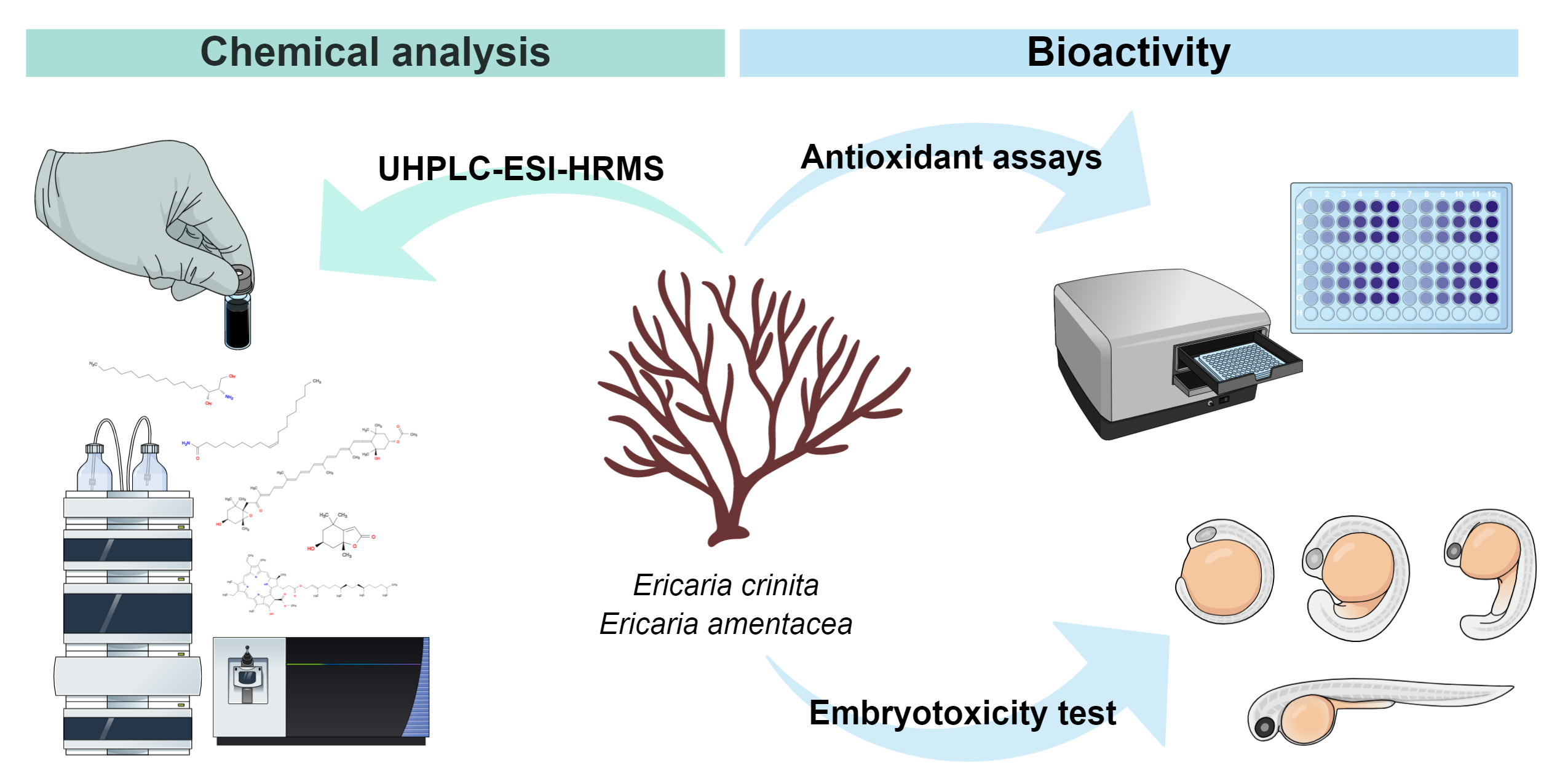
Bioprospecting of Less-Polar Fractions of Ericaria crinita and Ericaria amentacea: Developmental Toxicity and Antioxidant Activity

1
Department of Organic Chemistry, Faculty of Chemistry and Technology, University of Split, Ruđera Boškovića 35, 21000 Split, Croatia
2
Laboratory for Aquaculture Biotechnology, Division of Materials Chemistry, Ruđer Bošković Institute, Bijenička Cesta 54, 10000 Zagreb, Croatia
3
Department of Process Engineering, Faculty of Food Technology, Josip Juraj Strossmayer University of Osijek, Franje Kuhača 18, 31000 Osijek, Croatia
*
Author to whom correspondence should be addressed.
Mar. Drugs 2022, 20(1), 57; https://doi.org/10.3390/md20010057
Submission received: 20 December 2021 / Revised: 2 January 2022 / Accepted: 3 January 2022 / Published: 6 January 2022

Abstract

:
Ericaria crinita and Ericaria amentacea from the Adriatic Sea (Croatia) were investigated with respect to the presence of less-polar compounds for the first time after fractionation by solid-phase extraction (SPE). The composition of less-polar fractions of freeze-dried E. crinita (FdEc) and E. amentacea (FdEa) were analyzed by high-performance liquid chromatography–high-resolution mass spectrometry with electrospray ionization (UHPLC-ESI-HRMS). The major identified compounds were: amides of higher aliphatic acids (palmitoleamide, linoleamide, palmitamide, oleamide and erucamide) and related compounds, carotenoid (fucoxanthin), chlorophyll derivatives (pheophytin a and b and their derivatives) and higher terpenes (loliolide, isoamijiol with its oxidation product), β-stigmasterol and (3β,6α)-14-methylergosta-8,24(28)-diene-3,6-diol). The toxic effects observed on the less-polar fractions obtained from Ericaria species on zebrafish Danio rerio embryos could be associated with the high abundance of all five detected amides. The antioxidant activity of the fractions was evaluated by means of five independent assays, including the reduction of the radical cation (ABTS), the oxygen radical absorbance capacity (ORAC), ferric-reducing antioxidant power (FRAP), the 2,2-diphenyl-1-picryl-hydrazyl (DPPH) assay and the Folin–Ciocalteu method. A higher antioxidant activity of E. amentacea in comparison to that of the E. crinita fractions was found with IC50 concentrations of 0.072 and 1.177 mg/mL, respectively. The correlation between the activity and the chemical composition revealed that the synergistic effect of different compounds impacted their antioxidant response.

Graphical Abstract

1. Introduction

Cystoseira species are among the most important foundation species in the Mediterranean, forming complex habitats essential for biodiversity and ecosystem functioning [1,2]. Most of these species have been protected (Annex II of the Barcelona Convention, COM/2009/0585 FIN [3], Annex I of the Bern Convention [4] and the Habitat Directive [5]). In the Mediterranean Sea, about two-thirds of around 28 Cystoseira species are considered to be endemic [6].
Orellana et al. [7] concluded that the genus Cystoseira was polyphyletic and proposed the reinstatement of the genera for the species of the genus outside the clade of Cystoseira sensu stricto in order to achieve monophyly in the group classification. Molinari-Novoa et al. [8] reinvestigated the genera Ericaria Stackhouse and determined that Ericaria are the correct names for the “Cystoseira 2” and “Cystoseira 3” clades, as previously defined by Orellana et al. [7]. Consequently, Ericaria crinita (Duby) Molinari & Guiry 2020 is now accepted taxonomically instead of Cystoseira crinita (Duby) 1830, and Ericaria amentacea (C.Agardh) Molinari & Guiry 2020 is accepted as Cystoseira amentacea (C.Agardh) Bory.
Sterols and volatiles were investigated in E. crinita from the Black Sea [9]. Seven sterols were found with fucosterol as the main sterol, followed by 24-ethyl-cholest-5-en-3β-ol. The volatiles from the lipophilic extract were analyzed, and the major compounds were monoterpenes, from which dihydroactinidiolide was identified. The fatty acids (FAs) profile was investigated in E. crinita from Bulgarian Black Sea, and palmitic acid was found as the major compound, followed by linoleic acid and eicosapentaenoic acid [10]. The chemical composition of E. crinita from the Eastern Mediterranean [10] was also studied. Fourteen sterols have been identified with fucosterol as the main sterol, followed by 24-cholesterol and methyl-cholesta-5,24(28)-dien-3β-ol, while 24-ethyl-cholest-5-en-3β-ol was present only in traces and, among others, new sterol 24-nor-chol-5,22-dien-3β-ol was identified. The main FA was palmitic acid, followed by myristic and oleic acids. In the Mediterranean, the identified E. crinita [11] volatiles were dihydroactinidiolide, hexahydrofarnesyl acetone and 3,7-dimethylocta-1,6-diene-3-ol-2-aminobenzoate, along with the chlorinated compounds (ethanes substituted with chlorine and ethoxy groups). Nuclear magnetic resonance (NMR), mass spectrometry (MS), ultraviolet (UV) and infrared (IR) spectroscopy determined the meroterpenoids in E. crinita from the south coast of Sardinia [12]. Six new tetraprenyltoluquinol derivatives, two new triprenyltoluquinol derivatives, two new tetraprenyltoluquinone derivatives and four known tetraprenyltoluquinol derivatives were isolated and identified. The hydroquinones were found to have a powerful antioxidant activity, but none of the tested compounds showed antibacterial activity, while a moderate antitumor activity was found for several tested compounds. The phytochemical analysis of E. crinita collected from the French Riviera coast revealed the presence of new tetraprenyltoluquinols [13]. Diterpenes and sesquiterpenes from the Cystoseria genus were reviewed, including the data for E. crinita [14].
E. amentacea collected from the Sicilian coast has been studied [15,16,17,18,19]. Many tetraprenyltoluquinols have been isolated, either with a regular or irregular diterpenoid moiety. The regular tetraprenyltoluquinols that were found were: strictaketal, isocystoketal, isostrictaketal, isobalearone, (Z,E)-bifurcarenone, amentaepoxide and amentadione. The identified irregular or rearranged tetraprenyltoluquinols were: neobalearone and 2-epi-neobalearone. Two new cystoketal derivatives, demethoxy cystoketal chromane and cystoketal quinone, were isolated from E. amentacea collected from the French Riviera in addition to the sterols and previously described meroditerpenes [15]. Two new compounds were found: 4′-methoxy-(2E)-bifurcarenone and its chromene derivative. In a biological screening it was demonstrated that 4′-methoxy-(2E)-bifurcarenone inhibits cell division, since it stops the development of the fertilized eggs of the common sea-urchin Paracentrotus lividus [20]. DMSO and 50%-ethanolic extracts from E. amentacea from the Ligurian Sea (northwestern Mediterranean) were obtained, and their bioactive properties were investigated [21]. Meroditerpenes were found in both extracts with the structures containing the chromane or quinone groups, such as: cystoketal quinone, demethylcystoketal chromane and cystoketal chromane, and/or cystoketal. The extracts showed a very low grade of toxicity on RAW 264.7 macrophages and L929 fibroblasts and a plethora of antioxidant and anti-inflammatory effects [21]. The different efficacy of the two extracts in the various antioxidant and anti-inflammatory tests may be attributable to the quantitative, and not qualitative, difference of the present natural organic compounds. The FAs profile of E. amentacea lipid extract was characterized by high amounts of saturated FAs (SFAs), around 40.51% of the total FAs, and palmitic acid was the predominant one [22].
Although it is known that these two species contain a wide variety of secondary metabolites (terpenes and meroterpenes are described as the predominant constituents [15]), detailed compositions of their less-polar fractions have not been reported yet, and there are no reports of E. crinita and E. amentacea from the Adriatic Sea. We expect to identify pigments and other less-polar constituents after the fractionation by solid-phase extraction (SPE) and to evaluate their embryotoxic and antioxidant potential. Therefore, the main goals of the present research were to: (a) determine the composition of less-polar fractions of freeze-dried E. crinita (FdEc) and E. amentacea (FdEa) by high-performance liquid-chromatography-high-resolution mass spectrometry with electrospray ionization (UHPLC-ESI-HRMS) on two columns; (b) compare the obtained chemical profiles of FdEc and FdEa; (c) determine the fractions’ embryotoxic potential using zebrafish Danio rerio embryos; (d) determine the antioxidant potential of fractions obtained from Ericaria species by utilizing five in vitro assays (Folin-Ciocalteu, reduction of radical cation ABTS+, 2,2-diphenyl-1-picryl-hydrazyl (DPPH) assay, ferric-reducing antioxidant power (FRAP) and an oxygen radical absorbance capacity (ORAC) assay); and (e) discuss the observed antioxidant activity with respect to the determined composition of the fractions.

2. Results and Discussion

Although it is known that E. crinita and E. amentacea contain a wide variety of secondary metabolites [15], the detailed composition of their less-polar fractions has not been reported yet, and there are no reports on these algae from the Adriatic Sea. Therefore, FdEc and FdEa samples were fractionated by SPE (Section 3.3) to obtain less-polar fractions F3 and F4. The obtained fractions were analyzed by UHPLC-ESI(+)-HRMS. Two columns were used for the analysis: USP L7 (Acquity BEH C8) and USP L11 (Acquity CSH phenyl-hexyl). A slightly better separation as well as peak shapes were achieved on the Acquity CSH phenyl-hexyl column with charged surface hybrid particle technology. The major compounds (in the positive ion mode) were tentatively identified on the basis of their elemental compositions and tandem mass spectra. The major identified compounds were: amides of higher aliphatic acids, carotenoid, chlorophyll derivatives and higher terpenes.
The total ion chromatograms (TICs) of the fractions F3 and F4 are shown in Figure 1, and the extracted ion chromatograms (XICs) of the most abundant ions in the fractions F3 and F4 are shown in Figure 2.
Oleamide (amide of oleic acid, entry 5, Table 1 and Table 2) was the most abundant compound in F3 of FdEc and F4 of FdEa. It has been reported as a natural compound in different natural extracts [23,24] including the algae Rhizoclonium hieroglyphicum [25] and Prymnesium parvum carter [26]. Oleamide is an endogenous bioactive signaling molecule that acts in diverse cell types and consequently triggers different biological and pharmacological effects [23,24,27,28,29]. Palmitoleamide (entry 1, Table 1 and Table 2) and palmitamide (entry 3, Table 1 and Table 2) were also present. Palmitoleamide was isolated and characterized as one of the major toxic compound exuded from cells into the surrounding environment from both laboratory-grown and field samples of P. parvum [26]. Palmitoleamide and octadecanamide were both identified in the essential oil from the seagrass Zostera marina [30]. Erucamide (entry 7, Table 1 and Table 2) was also present in both algae with a higher abundance in F3, and previously it was identified in P. parvum [26]. Fatty acid amides were also identified from freshwater green alga Rhizoclonium hieroglyphicum [25]. Another group of present fatty acid derivatives were fatty acid glycerides: 2,3-dihydroxypropyl palmitate (entry 4, Table 1 and Table 2) and 2,3-dihydroxypropyl stearate (entry 6, Table 1 and Table 2), as well as 2-hydroxypropyl stearate (entry 8, Table 1 and Table 2). Mono- and diglycerides of stearic, palmitic, oleic and arachidonic acids were found to prevail in Fucus virsoides FA composition [31]. In addition, 1-hydroxy-3-(tetradecanoyloxy)-2-propanyl (9Z)-octadec-9-enoate (entry 9, Table 1 and Table 2) and hexadecasphinganine (entry 21, Table 1 and Table 2) were found in both fractions of FdEc and FdEa. Sphinganines are dihydro derivatives of the long chain amino alcohol sphingosine that is the core moiety of sphingolipids. It was previously found as the product of Pseudovibrio sp. W64 marine sponge-derived bacterium [32], and it showed biological activities including antiseptic, anti-inflammatory [33] and antimicrobial activity [34].
Among xanthophyll carotenoids, only fucoxanthin (entry 18, Table 1 and Table 2) was found in F3 and F4 of both algae, being the most abundant in F3 of FdEc followed by F3 of FdEa. Remarkably lower values were found in F4 fractions of both algae. It has been reported as the main carotenoid pigment in all brown algae [35] that exhibited different biological activities, i.e., antioxidant and anticancer [36,37]. It was found previously with higher abundance in the algae, e.g., Himanthalia elongata, Laminaria ochroleuca, Undaria pinnatifida [38], Fucus serratus, Fucus vesiculosus [39,40] or F. virsoides [31]. The differences among the fucoxanthin contents of different brown algae species can be associated with different environmental factors and species-inherent characteristics [36,41].
Chlorophyll was not detected in the fractions, but its derivatives devoid of magnesium atoms were found to be more abundant in F4 of both algae (Table 1 and Table 2). A similar finding was reported for F. virsoides [31]. The sub-group containing 55 carbon atoms (with the aliphatic side chain) was represented with five compounds (entries 10, 11, 12, 19 and 20, Table 1 and Table 2). In contrast to Codium adhaerens [42], the subgroup with 35 carbon atoms (without the aliphatic side chain) was not present. Pheopthyin b (entry 19, Table 1 and Table 2) was more abundant in E. crinita, particularly in F4 of FdEc, while pheophytin a (entry 20, Table 1 and Table 2) was more abundant in F4 of FdEa. Pheophytin a has been found previously in notable amounts in different macroalgae [43], like green algae Enteromorpha prolifera [36], or C. adhaerens [42], as well as brown algae Sargassum fulvellum [44] or F. virsoides [31]. The other three compounds of this subgroup were pheophytin a derivatives characterized by the presence of an additional double bond, carbonyl or hydroxyl group in their composition (entries 10, 11 and 12, Table 1 and Table 2). Pheophytin a and related compounds, among other biological activities [45,46], exhibited antioxidant activity for the autooxidation of lipids [47,48].
Terpenes and steroids comprised another group of identified compounds (Table 1 and Table 2). A monoterpene lactone, loliolide (entry 13, Table 1 and Table 2) was found in both algae fractions, being more abundant in F3 of FdEc and FdEa, probably since it is a more polar-containing lactone group and hydroxyl group. It was previously isolated from brown seaweed Sargassum ringgoldianum, subsp. coreanum [49]. It showed moderate antioxidant activity as well as positive dose-dependent protective effects against H2O2-induced cell damage [49]. Diterpene isoamijiol (with two free hydroxyl groups, entry 15, Table 1 and Table 2) and its oxidation product C20H30O2 (with one keto group, entry 14, Table 1 and Table 2) were more abundant in F3 of both algae, since they are more polar. Isoamijiol was isolated from the brown seaweed Dictyota linearis [50]. Triterpenes β-stigmasterol (entry 16, Table 1 and Table 2) and structurally related sterol (3β,6α)-14-methylergosta-8,24(28)-diene-3,6-diol (entry 17, Table 1 and Table 2) were present in F3 and F4 of both algae. Previously, they were found in C. adhaerens [42] and F. virsoides [31].
The structural formulas of the most important identified components are presented in Figure 3.

2.1. Toxicological Screening of Fractions Obtained from Ericaria Species

Zebrafish Embryotoxicity Test

The main goal of bioprospecting is the discovery of compound(s) that can be further developed for commercialization, but during the process, toxicological assays and the determination of safety should not be overlooked. Zebrafish has been utilized within this research as one of the most perspective vertebrate models used in different research areas, from genetics, developmental biology, environmental science and (eco)toxicology to drug screening [51]. The results of the conducted embryotoxicity test are presented as concentration-response curves (Figure 4). Among the tested samples, FdEa F3 fraction demonstrated the highest toxicity (50% lethal concentration (LC50) = 25.37 ± 1.11 µg/mL, and 50% effective concentration (EC50) = 23.12 ± 0.67 µg/mL), followed by FdEc F4 fraction, that revealed a negative effect within a 40–50 µg/mL concentration range, causing 25–45% of mortality and 43–72% of malformations in exposed specimens. Although no mortality was observed during the exposure to FdEa F4 fraction, higher concentrations induced developmental abnormalities, i.e., 75% affected larvae at 194 µg/mL and 25% at 97 µg/mL. The FdEc F3 fraction demonstrated no negative effect on zebrafish survival and development, but one should notice that, in comparison to FdEa F4 fraction, the lower concentration range (4–50 µg/mL) was tested. Morphological abnormalities observed on the tested fractions mainly included pericardial edema and scoliosis. The lethality of the negative control and solvent controls groups was less than 10%.
As can be observed from the results of UHPLC-ESI(+)-HRMS analysis, the tested fractions contained a high amount of fatty acid amides, specifically palmitoleamide, linoleamide, palmitamide, oleamide and erucamide, that might be responsible for the observed toxicity. Bertin et al. [26] demonstrated the presence of seven fatty acid amides (myristamide, palmitamide, oleamide, linoleamide, elaidamide, stearamide and erucamide) in laboratory-grown and field-sampled microalgae P. parvum collected during an ichthytoxic harmful algal bloom event, as well as in the culture media and field-collected water. The same authors utilized Sciaenops ocellatus and recorded high acute toxicity (100% mortality) upon 6 h of larval exposure to the mixture of seven detected amides (100 ppm) [26]. Such findings indicate that the fatty acid amides detected within this study might be excreted from Ericaria species upon the cell lysis, which could translate into elevated mortality and/or a developmental abnormality rate of the exposed zebrafish. Nevertheless, possible synergistic/antagonistic interactions between the detected fatty acid amides, as well as among other molecules in such a complex mixture, should not be excluded. For this reason, further toxicological studies on fatty acid amides, individually and in a mixture, are needed to determine their impact on aquatic vertebrates, and consequently on humans.

2.2. Screening of the Antioxidant Activity of Fractions Obtained from Ericaria Species

In the research, the evaluation of the antioxidant activity of two less-polar fractions F3 and F4 of FdEc and FdEa was performed using five different spectroscopic methods: ORAC, FRAP, DPPH, ABTS and Folin–Ciocalteu (F-C) assays.
As can be seen from Figure 5, by implementing different methods for the antioxidant activity determination, diverse results were obtained. ORAC assay measures a fluorescent signal from a probe that is quenched in the presence of reactive oxygen species (ROS). The results revealed a 10- and 5-fold higher activity for FdEc and FdEa F3, then F4 fractions, respectively (Figure 5a). Considering the polarity of these fractions, the obtained order could be ascribed to the highest fucoxanthin content in F3 (Table 1 and Table 2). Fucoxanthin is a major carotenoid in seaweeds with already proven antioxidant activity [52]. When compared to our previous research on the endemic brown alga F. virsoides [31], it can be concluded that lower values were obtained for both Ericaria species.
The FRAP assay was employed because it is based on single electron transfer (SET) and gives a better understanding of the antioxidant reaction mechanism. Interestingly, the results presented in Figure 5b showed an approximately 2-fold (p < 0.0001) higher activity for both F3 and F4 fractions of FdEc in comparison to FdEa. However, within the same species, discrepancies can be observed. In FdEc, a higher FRAP value (p < 0.0001) was observed for F3 fraction, probably due to the synergistic effect and higher content of found antioxidant compounds (loliolide, isoamijiol and its oxidation product, β-stigmasterol, (3β,6α)-14-methylergosta-8,24(28)-diene-3,6-diol, fucoxanthin, pheophytin a and b), while in FdEa the opposite result was obtained, i.e., higher activity (p < 0.0001) was observed for F4 fraction, indicating that one or more of the found compounds act through the SET mechanism. In this case, it is probably due to (3β,6α)-14-methylergosta-8,24(28)-diene-3,6-diol, the phytosterol abundantly found in F4 of FdEa, since phytosterols have a proven antioxidant activity [53].
The DPPH assay implies a dominant reaction through single electron transfer (SET), but can also react through the transfer of hydrogen atoms (HAT). The inhibition percentage using the DPPH assay for a tested concentration of 1 mg/mL for F3 of FdEa was around 70%, while for F4 this percentage was only 10%. When normalized per gram of the fraction, F3 (369.32 ± 5.85 mg AAE/g fraction) showed a significantly higher (p < 0.0001) antioxidant activity than F4 (16.70 ± 4.52 mg AAE/g fraction). For FdEc, no inhibitory percentage could be found using the DPPH assay (Figure 5c).
Further on, although the Folin–Ciocalteu assay is commonly known as a measure of total polyphenolic content, it represents here the rate of the overall antioxidant activity, since the extracts do not contain polyphenols (see Table 1 and Table 2). The activity using the Folin–Ciocalteau assay was only observed for F3 from both species, with a 70-fold higher (p < 0.0001) activity for FdEa (278.77 ± 2.67 mg GAE/g) than FdEc (4.12 ± 0.56 mg GAE/g).
The fifth method implemented in this research was the reduction of the radical cation by implementing an ABTS assay. Different concentrations of F3 and F4 for both algae were prepared ranging from 0.005 to 5 mg/mL to obtain IC50 curves (Figure 6). The IC50 values for both samples were calculated as shown in Table 3, with the corresponding confidence interval, slope and coefficient of determination (R2). As can be seen, the lowest IC50 value, i.e., the highest antioxidant activity was obtained for F3 of FdEa, which is 15 times lower than for F4 of FdEa and F3 of FdEc. This could be explained by the synergistic effect of all found antioxidant compounds with a significantly higher amount of pheophytin a. Interestingly, although the highest amount of pheophytin a was recorded in F4 of FdEc, its IC50 value could not be determined because the upper inhibition plateau could not be reached, i.e., for the highest tested concentrations, the inhibition percentage was around 40%. This discrepancy suggests that, although pheophytin a exhibits some antioxidant activity [54], other compounds like fucoxanthin, phytosterols and terpenes have a more significant role in oxidative damage prevention.
The correlation between the antioxidant activity assays for all samples and found antioxidant compounds was preformed using Pearson’s correlation coefficient [55]. The ORAC results showed a higher negative correlation to β-stigmasterol (r = −0.770), pheophytin a (r = −0.857) and pheophytin b (r = −0.755) and a positive correlation to fucoxanthin (r = 0.914). The FRAP results showed a higher positive correlation to loliolide (r = 0.719), isoamijiol and its oxidation product (r = 0.846 and r = 0.879) and (3β,6α)-14-methylergosta-8,24(28)-diene-3,6-diol (r = 0.823). The FRAP results showed a higher positive correlation to loliolide (r = 0.719), isoamijiol and its oxidation product (r = 0.846 and r = 0.879) and (3β,6α)-14-methylergosta-8,24(28)-diene-3,6-diol (r = 0.823). A moderate negative correlation was found between ABTS results and β-stigmasterol (r = −0.532), pheophytin a (r = −0.415) and pheophytin b (r = −0.514), while moderate positive correlation was observed with fucoxanthin (r = 0.556). However, the significance level was not sufficient enough for any of them and only marginally significant for ORAC results correlated to fucoxanthin (p = 0.08), suggesting that the antioxidant activity is the response of a mixture of compounds and their different antioxidant modes of action.

3. Materials and Methods

3.1. Chemicals

The standards of gallic acid (>97.5%), L-ascorbic acid (≥99%), DPPH (2,2-diphenyl-1-picrylhydrazyl), ABTS (diammonium salt of 2,2′-azino-bis(3-ethylbenzthiazolin-6-yl)sulfonic acid, >99.0%), TPTZ (2,4,6-tripyridyl-S-triazine, ≥98%), dichloro-dihydro-fluorescein diacetate (≥97%, DCF-DA), AAPH (2,2-azobis (2-methylpropionamidine) dihydrochloride, 97%) and 2′,7′-dichlorofluorescin diacetate were purchased from Sigma-Aldrich (St. Louis, MO, USA).
Dimethyl sulfoxide (DMSO, p.a.), methanol (p.a.), ethanol (p.a.), iron (III) chloride (FeCl3, p.a.), hydrochloric acid (HCl, p.a.), NaHCO3 (p.a.) and Folin–Ciocalteu reagent were obtained from Kemika (Zagreb, Croatia). Hydrogen peroxide (H2O2, 30%) was purchased from Alkaloid (Skopje, Macedonia) and potassium persulfate (>98%) from Scharlau, (Regensburg, Germany).
The solvents used for SPE were of HPLC grade and were obtained from J.T. Baker (New Jersey, PA, USA).
Acetonitrile with 0.1% (v/v) formic acid and water with 0.1% (v/v) formic acid, both hypergrade for HPLC-MS LiChrosolv®, were purchased from Supelco Co. (Bellefonte, PA, USA).

3.2. Macroalga Samples

Ericaria crinita (Duby) Molinari & Guiry 2020 and Ericaria amentacea (C.Agardh) Molinari & Guiry 2020 were collected by a single point collection from the Adriatic Sea. E. crinita was collected in November 2020 at the southwest coast of Novigrad Sea with the sampling geographical coordinates: 44°12′02″ N; 15°28′51″ E at the depth of 2 m. The sea temperature was 14 °C. E. amentacea was collected in April 2021 at the offshore side of Dugi otok, 1 km northwest of Brbišćica Bay, with the sampling geographical coordinates: 43°03′16″ N; 14°59′14″ E. The sea depth was 0.5 m, with the sea temperature at 16 °C. The samples were transferred to the laboratory and washed with water (5 times) and deionized water (2 times), and then the samples were cut in 5–10 mm slices. The slices were frozen in an ultra-low freezer (CoolSafe PRO, Labogene, Denmark) at −60 °C for 24 h. A high vacuum (0.13–0.55 hPa) at −30 °C and 20 °C as the primary and secondary drying temperatures was applied for freeze-drying for 24 h.

3.3. Fractionation by Solid-Phase Extraction (SPE)

The freeze-dried E. crinita (FdEc) and E. amentacea (FdEa) were extracted (10 mL/g solvent:solid ratio) three times with the solvent methanol:dichloromethane (MeOH/DCM, 1:1, v/v) in an ultrasound-bath (Elma, Elmasonic P 70 H, Singen, Germany; 37 kHz/50 W) for 5 min. The obtained extracts were evaporated by nitrogen (5.0, Messer, Croatia) and were further mixed with C18 powder (40–63 µm, Macherey-Nagel Polygoprep 60–50 C18, Fisher Scientific, MA, USA). The SPE cartridge was conditioned with MeOH and ultrapure water. The obtained dry extracts were then placed on an SPE cartridge (C18, particle size 40 µm, bed weight 1 g, column capacity 6 mL, Agilent Bond Elut, Waldbronn, Germany) and were eluted with different solvents to obtain the fractions F1 to F4 as in our previous paper [11]: F1 (with H2O), F2 (with H2O/MeOH (1:1, v/v)), F3 (with MeOH) and F4 (with MeOH/DCM (1:1, v/v)). Less-polar compounds were obtained in F3 and F4 fractions that were dried by SpeedVac (SPD1030, Thermo Scientific, Waltham, MA, USA) and were stored in the dark at 4 °C.

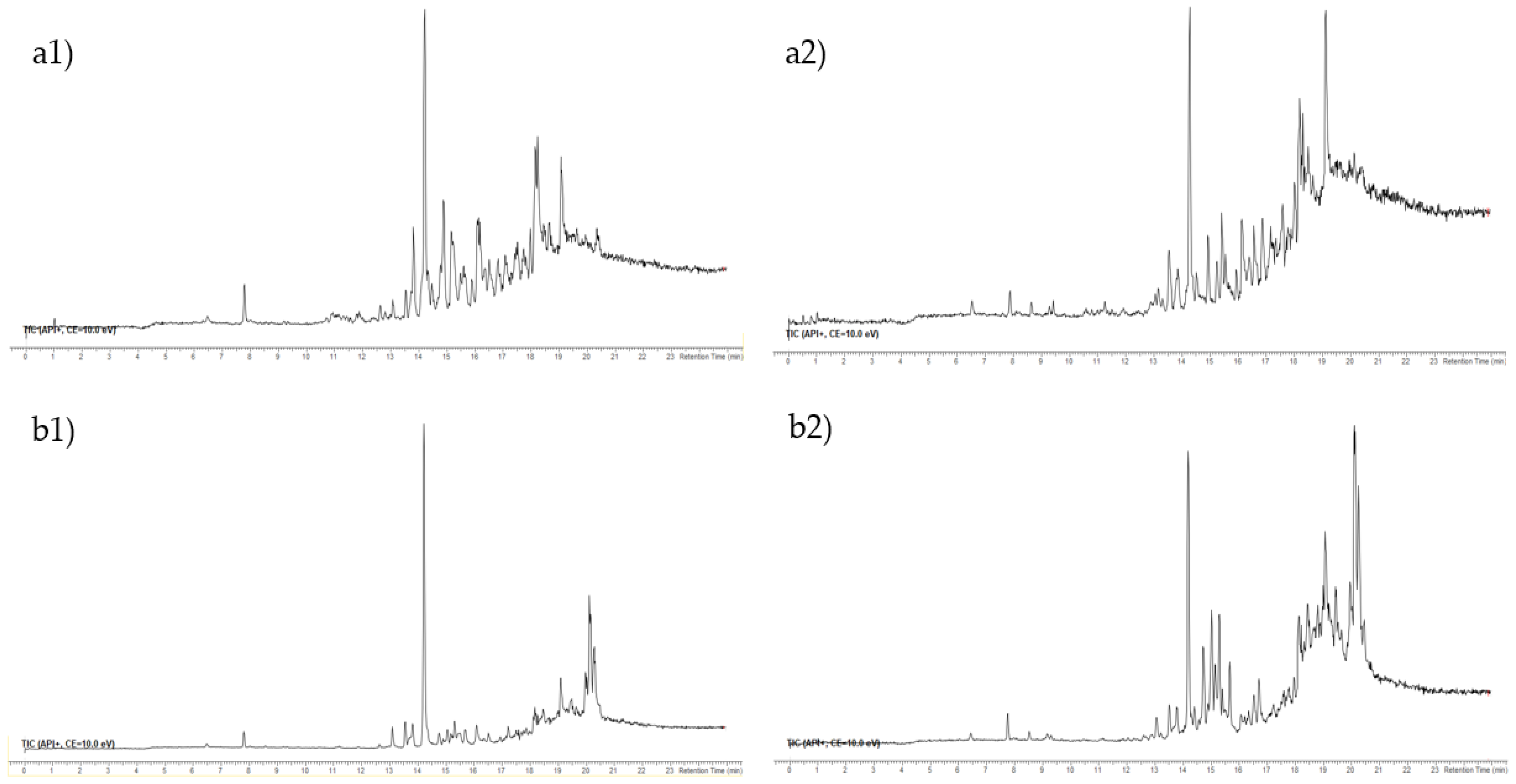
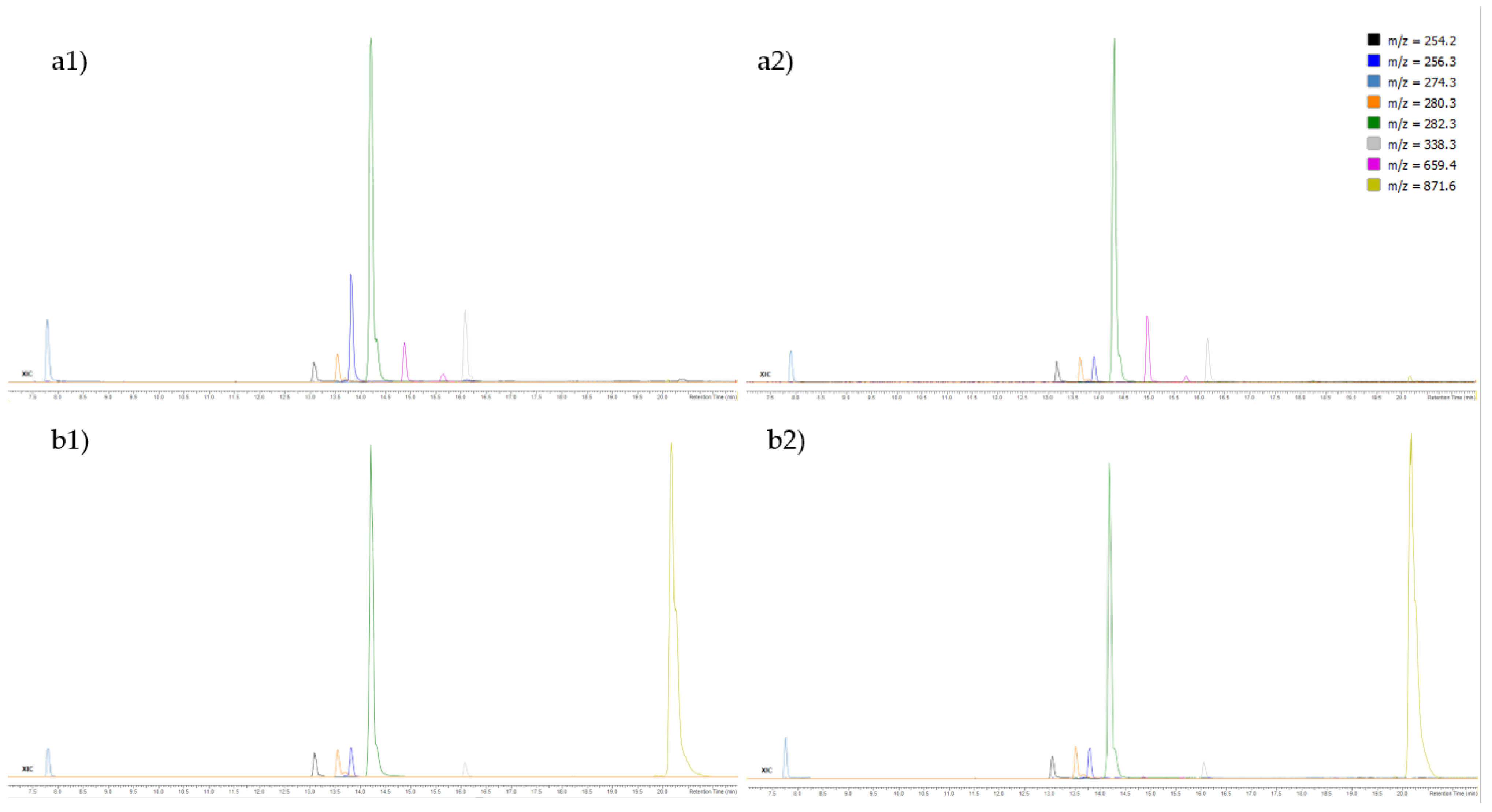
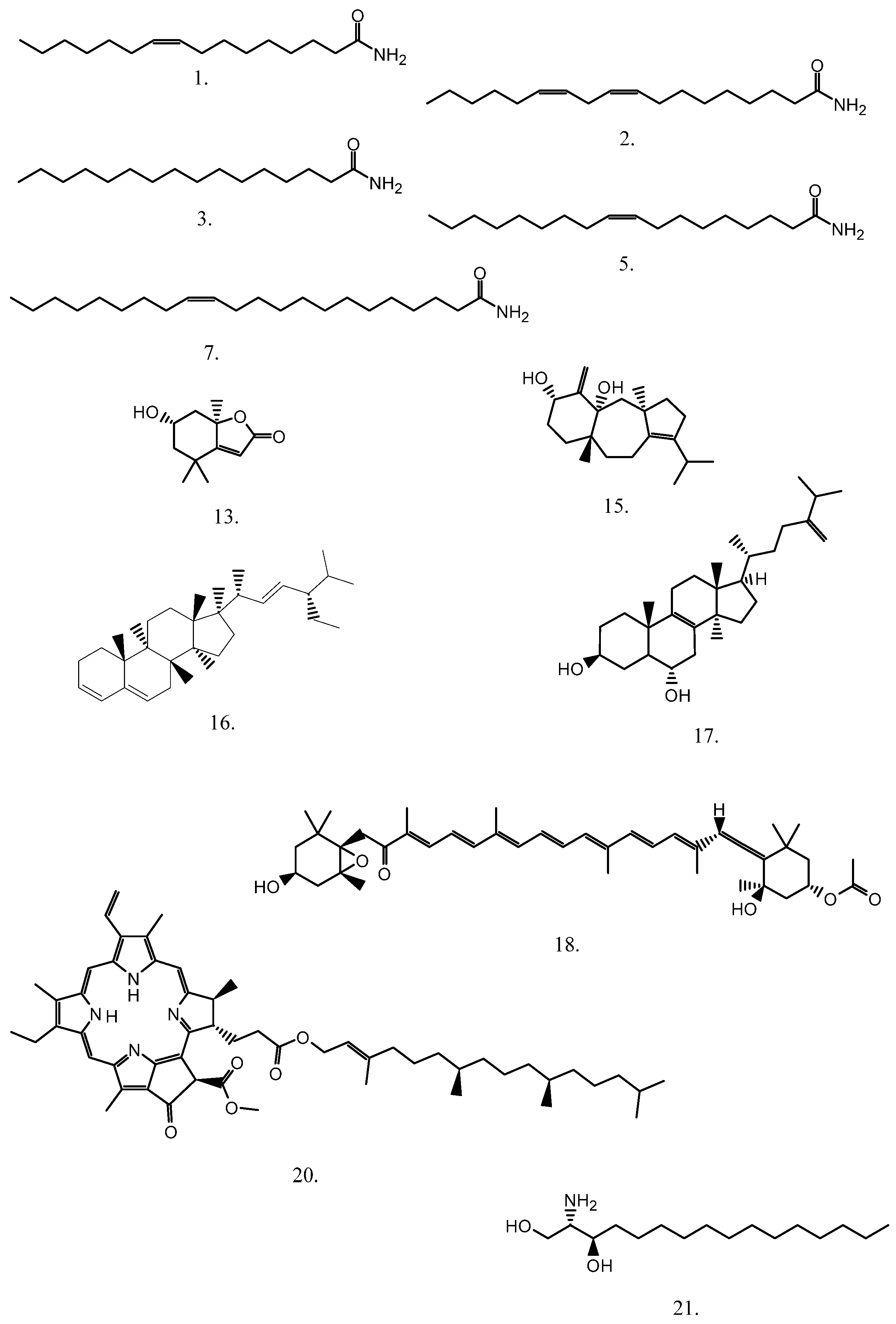
3.4. Ultra-High Performance Liquid Chromatography-High-Resolution Mass Spectrometry (UHPLC-ESI-HRMS) of F3 and F4 Fractions

The UHPLC-HRMS analyses were carried out on an ExionLC AD system (AB Sciex, Concord, ON, Canada) equipped with the ExionLC AD Autosampler, ExionLC AD Pump, ExionLC AD Degasser, ExionLC solvent delivery system, ExionLC AD Column oven and ExionLC Controller combined with a TripleTOF 6600+ (AB Sciex, Concord, ON, Canada) quadrupole-time-of-flight (Q-TOF) mass spectrometer with a Duospray ion source. The chromatographic separation of the compounds in F3 and F4 fractions was achieved using the analytical columns: Acquity UPLC BEH C8 2.1 × 100 mm, particle size 1.7 µm (Waters, Milford, MA, USA) and Acquity UPLC CSH Phenyl-Hexyl, 2.1 × 100 mm, particle size 1.7 µm (Waters, Milford, MA, USA). The mobile phases were water (A) and acetonitrile (B), both containing 0.1% formic acid with the flow rate set at 0.4 mL/min. After the isocratic condition (0.6 min) with 2% of B, the applied gradient elution program was: 0.6–18.5 min (B linear gradient to 100%), 18.5–25 min (100% B). The column temperature was set at 30 °C, and the injection volume was 4 µL.
Mass spectrometry detection was carried out in the positive electrospray ionization (ESI+). A collision-induced dissociation (CID) in information-dependent acquisition (IDA) mode of tandem (MS/MS) mass spectra was used for precursor ions with the signal intensities above a 200 cps threshold. The maximum number of precursor ions simultaneously subjected to CID was 15. The ion source parameters were: ESI capillary voltage 5.5 kV and source temperature 300 °C, nebulizing gas (air, gas 1) pressure 40 psi, heater gas (air, gas 2) pressure 15 psi, curtain gas (nitrogen) pressure 30 psi. The recording mass spectra parameters were: 80 V declustering potential, m/z range 100–1000 (MS) and 20–1000 (MS/MS), and accumulation time 100 ms. Nitrogen was the collision gas, with a collision energy of 40 eV and a spread of 20 eV. The mass scale calibrations (in the MS and MS/MS modes) were done prior to each run with the ESI Positive Calibration Solution 5600 (AB Sciex, Concord, ON, Canada).
The data were processed with ACD/Spectrus Processor 2021.1.0 (ACD/Labs, Toronto, ON, Canada). The compounds’ elemental compositions were determined based on the accurate masses of the corresponding protonated molecules, their isotopic distributions and the product ions m/z in MS/MS spectra. The identification of detected components was performed based on their elemental compositions, mass spectra and search in the ChemSpider database. The selection among the suggested hits was based on a matching with MS/MS data.

3.5. Zebrafish Embryotoxicity Test

Mature zebrafish Danio rerio (a wild-type strain obtained from European Zebrafish Resource Center, Karlsruhe Institute of Technology (KIT), Karlsruhe, Germany) were utilized for egg production, following the procedure [32].
A zebrafish embryotoxicity test was conducted in accordance with the OECD Test Guideline, with slight modifications already described in Babić et al. [56]. The embryos (n = 20, 4–64 blastomeres) were exposed to a wide range of concentrations (FdEa F3 (8–40 µg/L), FdEa F4 (12–194 µg/L), FdEc F3 and FdEc F4 (4–50 µg/L)). The final solvent concentration (MeOH in F3 fraction and DMSO in F4 fraction) did not exceed 1% [57], which was also the limiting factor during the determination of the concentration range. Artificial water was used as a negative control [58], while MeOH and DMSO (1%) were used as the solvent controls. The exposed zebrafish specimens were incubated at 27.5 ± 0.5 °C (Innova 42 incubator, New Brunswick, Canada). At 96 h of exposure to the tested fractions, mortality and developmental abnormalities were observed using an inverted microscope (Olympus CKX41).
Zebrafish maintenance and spawning were performed in aquaria units approved by the Croatian Ministry of Agriculture and according to the Directive 2010/63/EU. An embryotoxicity test was conducted on the non-protected embryonic/larval stages (up to 96 hpf), which do not require permission by animal welfare commissions [59].

3.6. Antioxidant Activity of Tested Fractions

In this study, five methods for the determination of antioxidant activity were employed, including oxygen radical absorbance capacity (ORAC), ferric-reducing antioxidant power (FRAP), a 2,2-diphenyl-1-picryl-hydrazyl (DPPH) assay, the Folin–Ciocalteu method and a reduction of the radical cation (ABTS). The measurements were carried out in triplicates in 96-well plates using a UV/Vis microplate reader (Infinite M200 PRO, TECAN, Switzerland), while all the results were expressed as mean ± standard deviation (n = 4).
The reduction of the radical cation assay (ABTS) was measured by spectroscopy at 734 nm [60] with slight modifications. The ABTS radical cation stock solution was prepared by mixing 7 mM ABTS and 2.45 mM potassium persulfate in the same ratio. The obtained mixture was allowed to react at room temperature in the dark for 17 h. The working solution of the ABTS radical cation was adjusted to an absorbance of 0.700 ± 0.02. The reaction mixture was prepared by adding the sample and an ABTS working solution, achieving the inhibition percentage between 10% and 100% (the blank was represented by the used solvent for each fraction). The results were expressed as milligram Trolox equivalents per gram of sample (mg TE/g). Moreover, an IC50 curve was constructed for both samples.
The oxygen radical absorbance capacity (ORAC) was determined as described by Huang and colleagues [61], with some modifications. A 25 μL diluted sample was added in black 96-well flat-bottom plates. Afterwards, 150 μL of a DCF-DA solution (1:1000, v/v in 25 mL 75 mM PBS) was added, followed by incubation at 37 °C for 30 min in the shaking incubator (New Brunswick, Innova 42). To start the reaction, 25 μL AAPH was added to the mixture, and the loss of fluorescence was monitored every 5 min for 180 min. The excitation wavelength was 485 nm and the emission wavelength was 528 nm, with an optimal fluorescence gain of 209. The results were expressed as μmol Trolox equivalents (TE)/g of the fraction.
For the FRAP assay [62], the FRAP regent must first be prepared by mixing the equal volumes of a 10 mM TPTZ solution in 40 mM HCl and an aqueous 20 mM FeCl3 solution. Then, the mixture was diluted five times in 0.25 M acetate buffer (pH 3.6) and heated to 37 °C. The 100 μL of the sample was mixed with 3.9 mL of the FRAP reagent, and the absorbance was determined at 593 nm after the incubation at 37 °C for 10 min. The results are expressed as mmol/g ferrous equivalents.
For the DPPH radical scavenging assay [63], the volume of 25 μL of the prepared sample was mixed with 200 μL of methanol and the prepared DPPH reagent in methanol (240 μg/mL). The reaction mixture was kept in the dark for 30 min, after which the absorbance of the solution was measured at 490 nm. The results were expressed as milligram ascorbic acid equivalents per gram of the sample (mg/gAAE).
The Folin–Ciocalteu method was used [64] with some adaptations. Briefly, 100 μL of the sample was mixed with 750 μL of a 10-fold diluted Folin–Ciocalteu reagent. After 5 min of incubation at room temperature, 750 μL of sodium bicarbonate solution (60 g/L) was added to the mixture and incubated in the dark at room temperature for 90 min. Absorbance was measured at 750 nm, and the results were expressed as mg gallic acid equivalent (GAE) per g of the sample.

3.7. Statistical Analysis

GraphPad Prism software version 8 was used for statistical analysis and graph presentation. p ≤ 0.05 was used as a cut-off value of statistical significance throughout the manuscript. Prior to LC50/EC50 determination, the obtained values were subjected to logarithmic transformation. A one-way analysis of variance (ANOVA) was performed to examine the significance between tested samples, as well as among treatments. The correlation between the antioxidant assay results and the chemical composition was tested through Pearson correlation analysis.

4. Conclusions

E. crinita and E. amentacea from the Adriatic Sea (Croatia) were investigated for the first time with respect to the presence of less-polar compounds. After fractionation by solid-phase extraction (SPE), the major identified compounds by UHPLC-ESI-HRMS were: amides of higher aliphatic acids (palmitoleamide, linoleamide, oleamide and erucamide) and related compounds, carotenoid (fucoxanthin), chlorophyll derivatives (pheophytin a and b and their derivatives) and higher terpenes (loliolide, isoamijiol and its oxidation products), β-stigmasterol and (3β,6α)-14-methylergosta-8,24(28)-diene-3,6-diol). Fucoxanthin was found to be the most abundant in F3 of FdEc and FdEa. Pheopthyin b was more abundant in F4 of FdEc, while pheophytin a was more abundant in F4 of FdEa. Loliolide and isoamijiol were found in both algae fractions, being more abundant in F3.
The results of toxicological testing obtained within this study emphasize the need to employ model organisms for determining the toxicological effect of bioactive molecules, all in order to provide answers on their safety for non-target organisms and, consequently, humans.
Higher antioxidant activities of FdEa in comparison to FdEc fractions were found by implementing all five assays (ABTS, ORAC, FRAP DPPH, Folin–Ciocalteu), with inhibitory concentrations of 0.072 and 1.177 mg/mL for the fractions, respectively. By correlating the results of the antioxidant analysis with the chemical composition, it was determined that the synergistic effect of different compounds, as well as their modes of action, impact the antioxidant response. The high antioxidant activity of E. amentacea suggests its potential as a source of natural antioxidants. The correlations between two Ericaria species show that the same species has a different composition and consequently diverse activities.

Author Contributions

Conceptualization, I.J., R.Č.-R. and S.J.; methodology, S.R., A.-M.C., L.Č. and S.B.; formal analysis, S.R., L.Č. and S.B.; investigation, S.R., L.Č., S.B. and I.J.; resources, R.Č.-R. and I.J.; data curation, S.R., S.B. and L.Č.; writing—original draft preparation, I.J., S.B. and L.Č.; writing—review and editing, I.J., S.J., R.Č.-R., S.R., L.Č. and S.B.; supervision, I.J., S.J. and R.Č.-R.; project administration, R.Č.-R.; funding acquisition, R.Č.-R. and I.J. All authors have read and agreed to the published version of the manuscript.

Funding

We would like to thank the Croatian Government and the European Union (European Regional Development Fund—the Competitiveness and Cohesion Operational Program—KK.01.1.1.01) for funding this research through the project Bioprospecting of the Adriatic Sea (KK.01.1.1.01.0002), granted to The Scientific Centre of Excellence for Marine Bioprospecting—BioProCro.

Institutional Review Board Statement

Animal housing and spawning were performed in aquaria units approved by the Croatian Ministry of Agriculture and according to the Directive 2010/63/EU. All experiments in this study were conducted on the non-protected embryonic stages (up to 96 hpf), which do not require permission by animal welfare commissions.

Informed Consent Statement

Not applicable.

Data Availability Statement

Data are contained within the article.

Acknowledgments

We would like to thank the Croatian Government and the European Union (European Regional Development Fund—the Competitiveness and Cohesion Operational Program—KK.01.1.1.01) for funding this research through the project Bioprospecting of the Adriatic Sea (KK.01.1.1.01.0002), granted to The Scientific Centre of Excellence for Marine Bioprospecting—BioProCro. We also thank Donat Petricioli for the sample collection and identification.

Conflicts of Interest

The authors declare no conflict of interest. The funders had no role in the design of the study; in the collection, analyses, or interpretation of data; in the writing of the manuscript, or in the decision to publish the results.

References

  1. Bulleri, F.; Benedetti-Cecchi, L.; Acunto, S.; Cinelli, F.; Hawkins, S.J. The influence of canopy algae on vertical patterns of distribution of low-shore assemblages on rocky coasts in the northwest Mediterranean. J. Exp. Mar. Bio. Ecol. 2002, 267, 89–106. [Google Scholar] [CrossRef]
  2. Cheminée, A.; Sala, E.; Pastor, J.; Bodilis, P.; Thiriet, P.; Mangialajo, L.; Cottalorda, J.-M.; Francoura, P. Nursery value of Cystoseira forests for Mediterranean rocky reef fishes. J. Exp. Mar. Bio. Ecol. 2013, 442, 70–79. [Google Scholar] [CrossRef]
  3. United Nations Environment Programme (UNEP). Convention for the Protection of the Marine Environment and the Coastal Region of the Mediterranean and Its Protocols. Mediterranean Action Plan-Barcelona Convention Secretariat. 2019, p. 153. Available online: https://wedocs.unep.org/bitstream/handle/20.500.11822/31970/bcp2019_web_eng.pdf (accessed on 5 December 2021).
  4. Council of Europe. Bern Convention/Convention de Berne: Convention on the Conservation of European Wildlife and Natural Habitats/Convention Relative à la Conservation Dela Vie Sauvage et du Milieu Naturel de l’Europe; Appendix/Annexe I, 19.IX.1979; Council of Europe: Strasbourg, France, 1979. [Google Scholar]
  5. Directive, H. Council Directive 92/43/EEC of 21 May 1992 on the conservation of natural habitats and of wild fauna and flora. Off. J. Eur. Union 1992, 206, 7–50. [Google Scholar]
  6. Guiry, M.D.; Guiry, G.M. AlgaeBase. World-Wide Electronic Publication, National University of Ireland, Galway. Available online: http://www.algaebase.org (accessed on 5 December 2021).
  7. Orellana, S.; Hernández, M.; Sansón, M. Diversity of Cystoseira sensu lato (Fucales, Phaeophyceae) in the eastern Atlantic and Mediterranean based on morphological and DNA evidence, including Carpodesmia gen. emend. and Treptacantha gen. emend. Eur. J. Phycol. 2019, 54, 447–465. [Google Scholar] [CrossRef]
  8. Molinari-Novoa, E.A.; Guiry, M.D. Reinstatement of the genera Gongolaria Boehmer and Ericaria Stackhouse (Sargassaceae, Phaeophyceae). Not. Algarum 2020, 171, 1–10. [Google Scholar]
  9. Milkova, T.; Talev, G.; Christov, R.; Dimitrova-Konaklieva, S.; Popov, S. Sterols and volatiles in Cystoseira barbata and Cystoseira crinita from the Black sea. Phytochemistry 1997, 45, 93–95. [Google Scholar] [CrossRef]
  10. Ivanova, V.; Stancheva, M.; Petrova, D. Fatty acid composition of black sea Ulva rigida and Cystoseira crinite. Bulg. J. Agric. Sci. 2013, 19, 42–47. [Google Scholar]
  11. Kamenarska, Z.; Yalçin, F.N.; Ersöz, T.; Çaliş, I.; Stefanov, K.; Popov, S. Chemical composition of Cystoseira crinita Bory from the eastern Mediterranean. Z. Naturforsch. 2002, 57, 584–590. [Google Scholar] [CrossRef]
  12. Fisch, K.M.; Böhm, V.; Wright, A.D.; König, G.M. Antioxidative meroterpenoids from the brown alga Cystoseira crinita. J. Nat. Prod. 2003, 66, 968–975. [Google Scholar] [CrossRef]
  13. Praud, A.; Valls, R.; Piovetti, L.; Banaigs, B.; Benaim, J.-Y. Meroditerpenes from the brown alga Cystoseira crinita off the French Mediterranean coast. Phytochemistry 1995, 40, 495–500. [Google Scholar] [CrossRef]
  14. Gouveia, V.; Seca, A.M.; Barreto, M.C.; Pinto, D.C. Di-and sesquiterpenoids from Cystoseira genus: Structure, intra-molecular transformations and biological activity. Mini Rev. Med. Chem. 2013, 13, 1150–1159. [Google Scholar] [CrossRef] [PubMed]
  15. De Sousa, C.B.; Gangadhar, K.N.; Macridachis, J.; Pavão, M.; Morais, T.R.; Campino, L.; Varela, J.; Lago, J.H.G. Cystoseira algae (Fucaceae): Update on their chemical entities and biological activities. Tetrahedron Asymmetry 2017, 28, 1486–1505. [Google Scholar] [CrossRef]
  16. Amico, V.; Cunsolo, F.; Piatteli, M.; Ruberto, G.; Mayol, L. Strictaketal, a new tetraprenyltoluquinol with a heterotetracyclic diterpene moiety from the brown alga Cystoseira stricta. J. Nat. Prod. 1987, 50, 449–454. [Google Scholar] [CrossRef]
  17. Amico, V.; Cunsolo, F.; Piatteli, M.; Ruberto, G. Prenylated O-methyltoluquinols from Cystoseira stricta. Phytochemistry 1987, 26, 1719–1722. [Google Scholar] [CrossRef]
  18. Amico, V.; Oriente, G.; Neri, P.; Piatteli, M.; Ruberto, G. Tetraprenyltoluquinols from the brown alga Cystoseira stricta. Phytochemistry 1987, 26, 1715–1718. [Google Scholar] [CrossRef]
  19. Amico, V.; Piatteli, M.; Cunsolo, F.; Neri, P.; Ruberto, G. Two epimeric, irregular diterpenoid toluquinols from the brown alga Cystoseira stricta. J. Nat. Prod. 1989, 52, 962–969. [Google Scholar] [CrossRef]
  20. Mesguiche, V.; Valls, R.; Piovetti, L.; Banaigs, B. Meroditerpenes from Cystoseira amentacea var. stricta collected off the mediterranean coasts. Phytochemistry 1997, 45, 1489–1494. [Google Scholar] [CrossRef]
  21. De La Fuente, G.; Fontana, M.; Asnaghi, V.; Chiantore, M.; Mirata, S.; Salis, A.; Damonte, G.; Scarfì, S. The Remarkable Antioxidant and anti-inflammatory potential of the extracts of the brown alga Cystoseira amentacea var. stricta. Mar. Drugs 2021, 19, 2. [Google Scholar] [CrossRef]
  22. Bouafif, C.; Messaoud, C.; Boussaid, M.; Langar, H. Fatty acid profile of Cystoseira C. Agardh (Phaeophyceae, Fucales) species from the Tunisian coast: Taxonomic and nutritional assessments. Cienc. Mar. 2018, 44, 169–183. [Google Scholar] [CrossRef] [Green Version]
  23. Cravatt, B.F.; Prospero-Garcia, O.; Siuzdak, G.; Gilula, N.B.; Henriksen, S.J.; Boger, D.L.; Lerner, R.A. Chemical characterization of a family of brain lipids that induce sleep. Science 1995, 268, 1506–1509. [Google Scholar] [CrossRef] [Green Version]
  24. Yang, W.-S.; Lee, S.R.; Jeong, Y.L.; Park, D.W.; Cho, Y.M.; Joo, H.M.; Kim, I.; Seu, Y.B.; Sohn, E.H.; Kang, S.C. Antiallergic activity of ethanol extracts of Arctium lappa L. undried roots and its active compound, oleamide, in regulating FcεRI-mediated and MAPK signaling in RBL-2H3 cells. J. Agric. Food Chem. 2016, 64, 3564–3573. [Google Scholar] [CrossRef]
  25. Dembitsky, V.M.; Shkrob, I.; Rozentsvet, O.A. Fatty acid amides from freshwater green alga Rhizoclonium hieroglyphicum. Phytochem. 2000, 54, 965–967. [Google Scholar] [CrossRef]
  26. Bertin, M.J.; Zimba, P.V.; Beauchesne, K.R.; Huncik, K.M.; Moeller, P.D.R. Identification of toxic fatty acid amides isolated from the harmful alga Prymnesium parvum carter. Harmful Algae 2012, 20, 111–116. [Google Scholar] [CrossRef]
  27. Rueda-Orozco, P.E.; Montes-Rodriguez, C.J.; Ruiz-Contreras, A.E.; Mendez-Diaz, M.; Prospero-Garcia, O. The effects of anandamide and oleamide on cognition depend on diurnal variations. Brain Res. 2017, 1672, 129–136. [Google Scholar] [CrossRef]
  28. Langstein, J.; Hofstäder, F.; Schwarz, H. cis-9,10-Octadecenoamide, an endogenous sleep-inducing CNS compound, inhibits lymphocyte proliferation. Res. Immunol. 1996, 147, 389–396. [Google Scholar] [CrossRef]
  29. Ranger, C.M.; Winter, R.E.; Rottinghaus, G.E.; Backus, E.A.; Johnson, D.W. Mass spectral characterization of fatty acid amides from alfalfa trichomes and theirdeterrence against the potato leafhopper. Phytochemistry 2005, 66, 529–541. [Google Scholar] [CrossRef] [PubMed]
  30. Kawasaki, W.; Matsui, K.; Akakabe, Y.; Itai, N.; Kajiwara, T. Volatiles from Zostera marina. Phytochemistry 1998, 47, 27–29. [Google Scholar] [CrossRef]
  31. Jerković, I.; Cikoš, A.-M.; Babić, S.; Čižmek, L.; Bojanić, K.; Aladić, K.; Ul’yanovskii, N.V.; Kosyakov, D.S.; Lebedev, A.T.; Čož-Rakovac, R.; et al. Bioprospecting of less-polar constituents from endemic brown macroalga Fucus virsoides J. Agardh from the Adriatic Sea and targeted antioxidant effects in vitro and in vivo (zebrafish model). Mar. Drugs 2021, 19, 235. [Google Scholar] [CrossRef] [PubMed]
  32. Choudhary, A.; Naughton, L.M.; Dobson, A.D.W.; Rai, D.K. High-performance liquid chromatography/electrospray ionisation mass spectrometric characterisation of metabolites produced by Pseudovibrio sp. W64, a marine sponge derived bacterium isolated from Irish waters. Rapid Commun. Mass Spectrom. 2018, 32, 1737–1745. [Google Scholar] [CrossRef] [PubMed]
  33. Aoki, M.; Aoki, H.; Ramanathan, R.; Hait, N.C.; Takabe, K. Sphingosine-1-phosphate signaling in immune cells and inflammation: Roles and therapeutic potential. Mediat. Inflamm. 2016, 2016, 8606878. [Google Scholar] [CrossRef] [Green Version]
  34. Fischer, C.L.; Drake, D.; Dawson, D.V.; Blanchette, D.R.; Brogden, K.A.; Wertz, P.W. Antibacterial activity of sphingoid bases and fatty acids against gram-positive bacteria and gram-negative bacteria. Antimicrob. Agents Chemother. 2012, 56, 1157–1161. [Google Scholar] [CrossRef] [PubMed] [Green Version]
  35. Terasaki, M.; Hirose, A.; Narayan, B.; Baba, Y.; Kawagoe, C.; Yasui, H.; Saga, N.; Hosokawa, M.; Miyashita, K. Evaluation of recoverable functional lipid components of several brown seaweeds (Phaeophyta) from japan with special reference to fucoxanthin and fucosterol contents. J. Phycol. 2009, 45, 974–980. [Google Scholar] [CrossRef] [PubMed]
  36. Maeda, H.; Fukuda, S.; Izumi, H.; Saga, N. Anti-oxidant and fucoxanthin contents of brown alga ishimozuku (Sphaerotrichia divaricata) from the west coast of Aomori Japan. Mar. Drugs 2018, 16, 255. [Google Scholar] [CrossRef] [PubMed] [Green Version]
  37. Méresse, S.; Fodil, M.; Fleury, F.; Chénais, B. Fucoxanthin, a marine-derived carotenoid from brown seaweeds and microalgae: A promising bioactive compound for cancer therapy. Int. J. Mol. Sci. 2020, 21, 9273. [Google Scholar] [CrossRef] [PubMed]
  38. Osório, C.; Machado, S.; Peixoto, J.; Bessada, S.; Pimentel, F.B.; Alves, R.C.; Oliveira, M.B.P.P. Pigments content (chlorophylls, fucoxanthin and phycobiliproteins) of different commercial dried algae. Separations 2020, 7, 33. [Google Scholar] [CrossRef]
  39. Haugan, J.A.; Liaaen-Jensen, S. Algal carotenoids 54. Carotenoids of brown algae (Phaeophyceae). Biochem. Syst. Ecol. 1994, 22, 31–41. [Google Scholar] [CrossRef]
  40. Haugan, J.A.; Liaaen-Jensen, S. Improved isolation procedure for fucoxanthin. Phytochemistry 1989, 28, 2797–2798. [Google Scholar] [CrossRef]
  41. Lalegerie, F.; Gager, L.; Stiger-Pouvreau, V.; Connan, S. The stressful life of red and brown seaweeds on the temperate intertidal zone: Effect of abiotic and biotic parameters on the physiology of macroalgae and content variability of particular metabolites. In Advances in Botanical Research; Elsevier BV: Amsterdam, The Netherlands, 2020; pp. 247–287. [Google Scholar]
  42. Radman, S.; Cikoš, A.-M.; Flanjak, I.; Babić, S.; Čižmek, L.; Šubarić, D.; Čož-Rakovac, R.; Jokić, S.; Jerković, I. Less polar compounds and targeted antioxidant potential (in vitro and in vivo) of Codium adhaerens C. Agardh 1822. Pharmaceuticals 2021, 14, 944. [Google Scholar] [CrossRef]
  43. Kuczynska, P.; Jemiola-Rzeminska, M.; Strzalka, K. Photosynthetic pigments in diatoms. Mar. Drugs 2015, 13, 5847–5881. [Google Scholar] [CrossRef]
  44. Ina, A.; Hayashi, K.-I.; Nozaki, H.; Kamei, Y. Pheophytin a, a low molecular weight compound found in the marine brown alga Sargassum fulvellum, promotes the differentiation of PC12 cells. Int. J. Dev. Neurosci. 2007, 25, 63–68. [Google Scholar] [CrossRef]
  45. Okai, Y.; Higashi-Okai, K. Potent anti-inflammatory activity of pheophytin a derived from edible green alga, Enteromorpha prolifera (Sujiao-nori). Int. J. Immunopharmacol. 1997, 19, 355–358. [Google Scholar] [CrossRef]
  46. Okai, Y.; Ishizaka, S.; Higashi-Okai, K.; Yamashita, U. Detection of immunomodulating activities in an extract of Japanese edible seaweed Laminaria japonica (Makonbu). J. Sci. Food Agric. 1996, 72, 455–460. [Google Scholar] [CrossRef]
  47. Cahyana, A.H.; Shuto, Y.; Kinoshita, Y. Pyropheophytin a as an antioxidative substance from the marine alga Arame (Eisenis bicyclis). Biosci. Biotechnol. Biochem. 1992, 56, 1533–1535. [Google Scholar] [CrossRef]
  48. Nishibori, S.; Namiki, K. Antioxidative substances in the green fractions of the lipid of Aonori (Enteromorpha sp.). J. Home Econ. 1998, 39, 1173–1178. [Google Scholar] [CrossRef]
  49. Yang, X.; Kang, M.-C.; Lee, K.-W.; Kang, S.-M.; Lee, W.-W.; Jeon, Y.-J. Antioxidant activity and cell protective effect of loliolide isolated from Sargassum ringgoldianum subsp. Coreanum. Algae 2011, 26, 201–208. [Google Scholar] [CrossRef]
  50. Ochi, M.; Watanabe, M.; Miura, I.; Taniguchi, M.; Tokoroyama, T. Amijiol, isoamijiol, and 14-deoxyamijiol, three new diterpe- noids from the brown seaweed Dictyota linearis. Chem. Lett. 1980, 9, 1229–1232. [Google Scholar] [CrossRef]
  51. Hung, M.W.; Zhang, Z.J.; Li, S.; Lei, B.; Yuan, S.; Cui, G.Z.; Hoi, P.; Chain, K.; Lee, S.M.Y. From omics to drug metabolism and high content screen of natural product in zebrafish: A new model for discovery of neuroactive compound. Evid. Based Complementary Altern. Med. 2012, 2012, 605303. [Google Scholar] [CrossRef] [PubMed]
  52. **a, S.; Wang, K.; Wan, L.; Li, A.; Hu, Q.; Zhang, C. Production, characterization, and antioxidant activity of fucoxanthin from the marine diatom Odontella aurita. Mar. Drugs 2013, 11, 2667–2681. [Google Scholar] [CrossRef]
  53. Yoshida, Y.; Niki, E. Antioxidant effects of phytosterol and its components. J. Nutr. Sci. Vitaminol. 2003, 49, 277–280. [Google Scholar] [CrossRef] [PubMed]
  54. Pangestuti, R.; Kim, S.-K. Biological activities and health benefit effects of natural pigments derived from marine algae. J. Funct. Foods 2011, 3, 255–266. [Google Scholar] [CrossRef]
  55. Utakod, N.; Laosripaiboon, W.; Chunhachart, O.; Issakul, K. The efficiency and the correlation between testing methods on antimicrobial and antioxidant activities of selected medicinal essential oils. Int. Food Res. J. 2017, 24, 2616–2624. [Google Scholar]
  56. Babić, S.; Čižmek, L.; Maršavelski, A.; Malev, O.; Pflieger, M.; Strunjak-Perović, I.; Topić Popović, N.; Čož-Rakovac, R.; Trebše, P. Utilization of the zebrafish model to unravel the harmful effects of biomass burning during Amazonian wildfires. Sci. Rep. 2021, 11, 2527. [Google Scholar] [CrossRef] [PubMed]
  57. Maes, J.; Verlooy, L.; Buenafe, O.E.; De Witte, P.A.; Esguerra, C.V.; Crawford, A.D. Evaluation of 14 organic solvents and carriers for screening applications in zebrafish embryos and larvae. PLoS ONE 2012, 7, e43850. [Google Scholar] [CrossRef]
  58. ISO. Water Quality—Determination of the Acute Lethal Toxicity of Substances to a Freshwater Fish (Brachydanio Rerio Hamilton-Buchanan (Teleostei, Cyprinidae)); Parts 1–3; International Organization for Standardization: Geneva, Switzerland, 1996. [Google Scholar]
  59. Council Directive 2010/63/EU on the protection of animals used for scientific purposes. Off. J. Eur. Union 2010, 276, 33–79. [CrossRef]
  60. Re, R.; Pellegrini, N.; Proteggente, A.; Pannala, A.; Yang, M.; Rice-Evans, C. Antioxidant activity applying an improved ABTS radical cation decolorization assay. Free Radic. Biol. Med. 1999, 26, 1231–1237. [Google Scholar] [CrossRef]
  61. Huang, D.; Ou, B.; Hampsch-Woodill, M.; Flanagan, J.A.; Prior, R.L. High-throughput assay of oxygen radical absorbance capacity (ORAC) using a multichannel liquid handling system coupled with a microplate fluorescence reader in 96-well format. J. Agric. Food Chem. 2002, 50, 4437–4444. [Google Scholar] [CrossRef]
  62. Benzie, I.F.F.; Strain, J.J. The ferric reducing ability of plasma (FRAP) as a measure of “antioxidant power”: The FRAP assay. Anal. Biochem. 1996, 239, 70–76. [Google Scholar] [CrossRef] [Green Version]
  63. Brand-Williams, W.; Cuvelier, M.; Berset, C. Use of a free radical method to evaluate antioxidant activity. LWT 1995, 28, 25–30. [Google Scholar] [CrossRef]
  64. Singleton, V.L.; Rossi, J.A. Colorimetry of total phenolics with phosphomolybdic-phosphotungstic acid reagents. Am. J. Enol. Vitic. 1965, 16, 144–158. [Google Scholar]
Figure 1. Total ion chromatogram (TIC) (a) F3 and (b) F4 of (1) E. crinita and (2) E. amentacea on Acquity CSH phenyl-hexyl column.
Marinedrugs 20 00057 g001
Figure 2. Extracted ion chromatograms (XIC) zoom of the most abundant ions in the fractions (a) F3 and (b) F4 of (1) E. crinita and (2) E. amentacea on Acquity CSH phenyl-hexyl column.
Marinedrugs 20 00057 g002
Figure 3. Structural formulas of the most important components identified by HPLC-ESI(+)-HRMS labeled by the numbers depicted in Table 1 and Table 2.
Marinedrugs 20 00057 g003
Figure 4. Dose-response curves for the mortality and malformation of zebrafish Danio rerio at 96 h of exposure to F3 and F4 fractions of macroalgae (a) E. crinita (FdEc) and (b) E. amentacea (FdEa).
Marinedrugs 20 00057 g004
Figure 5. Radical scavenging effect of less-polar fractions from two Ericaria macroalgae using (a) oxygen radical absorbance capacity (ORAC), (b) ferric-reducing antioxidant power (FRAP), (c) 2,2-diphenyl-1-picryl-hydrazyl (DPPH) and (d) Folin–Ciocalteu in vitro assays (mean ± SD; n = 4). An asterisk indicates a significant difference between two Ericaria samples for F3 and F4 (*** p < 0.001; **** p < 0.0001). ND-none determined.
Marinedrugs 20 00057 g005
Figure 6. Concentration-inhibition response curves for (a) FdEc and (b) FdEa F3 and F4 used for the calculation of their antioxidant activity by implementing the ABTS assay.
Marinedrugs 20 00057 g006
Table 1. Major identified compounds in F3 and F4 fractions of E. crinita and their tentative identification by UHPLC-ESI(+)-HRMS.
No.CompoundElemental Composition[M + H]+Error **
(ppm)
AB
RT (min)Area (Counts)RT (min)Area (Counts)
F3F4F3F4
1.PalmitoleamideC16H31NO254.247840.513.609110811632080113.08513092023128998
2.LinoleamideC18H33NO280.263490.714.0861390454316636013.54615742983629020
3.PalmitamideC16H33NO256.263490.914.4605498614383404813.81863964273991527
4.2,3-Dihydroxypropyl palmitateC19H38O4331.284294.014.8172389831679714.12929578521659
5.OleamideC18H35NO282.279140.514.868252848265422897414.2092609620853855452
6.2,3-Dihydroxypropyl stearateC21H42O4359.315594.015.9743309175897715.16240978470222
7.ErucamideC22H43NO338.341740.116.9815433654143826416.08446821271767616
8.2-Hydroxypropyl stearateC21H42O3343.320671.517.26938981121416.254463731025
9.(2S)-1-Hydroxy-3-(tetradecanoyloxy)-2-propanyl (9Z)-9-octadecenoateC35H66O5567.498301.319.2312858661054717.97634885914426
10.3-Phorbinepropanoic acid, 3,4-didehydro-9-ethenyl-14-ethyl-24,25-dihydro-21-(methoxycarbonyl)-4,8,13,18-tetramethyl-20-oxo-, (2E)-3,7,11,15-tetramethyl-2-hexadecen-1-yl esterC55H72N4O5869.557551.120.3734307111089719.06653501005380
11.Methyl (3R,10Z,14Z,20Z,22S,23S)-12-ethyl-3-hydroxy-13,18,22,27-tetramethyl-5-oxo-23-(3-oxo-3-{[(2E,7R,11R)-3,7,11,15-tetramethyl-2-hexadecen-1-yl]oxy}propyl)-17-vinyl-4-oxa-8,24,25,26-tetraazahexacyclo [19.2.1.16,9.111,14.116,19.02,7]heptacosa-1(24),2(7),6(27),8,10,12,14,16,18,20-decaene-3-carboxylateC55H74N4O7903.563031.820.44145606294071019.987382742268701
12.3-Phorbinepropanoic acid, 9-acetyl-14-ethylidene-13,14-dihydro-21-(methoxycarbonyl)-4,8,13,18-tetramethyl-20-oxo-, 3,7,11,15-tetramethyl-2-hexadecen-1-yl esterC55H74N4O6887.568111.920.5943325321127710319.95340103113903240
13.LoliolideC11H16O3197.117221.26.2258815046036.323867655693
14.Isoamijiol oxidation product *C20H30O2303.231866.214.544378362237214.3283520172346
15.(3aR,4aR,6S,8aR)-1-Isopropyl-3a,8a-dimethyl-5-methylene-2,3a,4,5,6,7,8,8a,9,10-decahydrobenzo[f]azulene-4a,6(3H)-diol (Isoamijiol)C20H32O2305.247510.515.9741226831751415.52210443222138
16.β-StigmasterolC29H46395.367231.618.2763630836423317.63433268459395
17.(3β,6α)-14-Methylergosta-8,24(28)-diene-3,6-diolC29H48O2429.372713.418.3957846659611917.480980078117567
18.FucoxanthinC42H58O6659.430622.114.8732232648350015.12226977404095
19.Pheophytin bC55H72N4O6885.552468.720.1343949046736119.76540024391298
20.Pheophytin aC55H74N4O5871.573202.120.81558546153847220.106741857737924
21.HexadecasphinganineC16H35NO2274.274101.09.507318618528139317.78939523043361364
A-USP L7 (Acquity BEH C8) column.; B-USP L11 (Acquity CSH phenyl-hexyl) column; *—exact compound not determined; **—the smallest error for both columns.
Table 2. Major identified compounds in F3 and F4 fractions of E. amentacea and their tentative identification by UHPLC-ESI(+)-HRMS.
No.CompoundElemental Composition[M + H]+Error **
(ppm)
AB
RT (min)Area (Counts)RT (min)Area (Counts)
F3F4F3F4
1.PalmitoleamideC16H31NO254.247841.013.628592811230049613.054977571965778
2.LinoleamideC18H33NO280.263492.014.088479266269753713.5095752902671906
3.PalmitamideC16H33NO256.263491.514.48706476334050813.7816280672844809
4.2,3-Dihydroxypropyl palmitateC19H38O4331.284293.114.82182645097614.1051739263085
5.OleamideC18H35NO282.279141.514.88848714063685192014.189608871530534284
6.2,3-Dihydroxypropyl stearateC21H42O4359.315591.515.994219858388815.141981686090
7.ErucamideC22H43NO338.341740.616.9942971406169738716.05724709611417294
8.2-Hydroxypropyl stearateC21H42O3343.320670.017.181896777516.32711119880
9.(2S)-1-Hydroxy-3-(tetradecanoyloxy)-2-propanyl (9Z)-9-octadecenoateC35H66O5567.498301.919.22518581517635517.948226104218486
10.3-Phorbinepropanoic acid, 3,4-didehydro-9-ethenyl-14-ethyl-24,25-dihydro-21-(methoxycarbonyl)-4,8,13,18-tetramethyl-20-oxo-, (2E)-3,7,11,15-tetramethyl-2-hexadecen-1-yl esterC55H72N4O5869.557550.020.371042196885520.018661678772
11.Methyl (3R,10Z,14Z,20Z,22S,23S)-12-ethyl-3-hydroxy-13,18,22,27-tetramethyl-5-oxo-23-(3-oxo-3-{[(2E,7R,11R)-3,7,11,15-tetramethyl-2-hexadecen-1-yl]oxy}propyl)-17-vinyl-4-oxa-8,24,25,26-tetraazahexacyclo [19.2.1.16,9.111,14.116,19.02,7]heptacosa-1(24),2(7),6(27),8,10,12,14,16,18,20-decaene-3-carboxylateC55H74N4O7903.563033.320.43748785104465819.98140622876040
12.3-Phorbinepropanoic acid, 9-acetyl-14-ethylidene-13,14-dihydro-21-(methoxycarbonyl)-4,8,13,18-tetramethyl-20-oxo-, 3,7,11,15-tetramethyl-2-hexadecen-1-yl esterC55H74N4O6887.568111.320.59145728129464019.964375471053061
13.LoliolideC11H16O3197.117225.66.22232631143046.4032720613071
14.Isoamijiol oxidation product *C20H30O2303.231860.414.53448292839814.4275183010486
15.(3aR,4aR,6S,8aR)-1-Isopropyl-3a,8a-dimethyl-5-methylene-2,3a,4,5,6,7,8,8a,9,10-decahydrobenzo[f]azulene-4a,6(3H)-diol (Isoamijiol)C20H32O2305.247515.315.959223501206115.582746514464
16.β-StigmasterolC29H46395.367231.918.2881912667027617.60716495562142
17.(3β,6α)-14-Methylergosta-8,24(28)-diene-3,6-diolC29H48O2429.372712.218.3917154027311317.47143026279999
18.FucoxanthinC42H58O6659.430620.415.12521381278986414.851181146786705
19.Pheophytin bC55H72N4O6885.552460.220.1482069215599219.77621058131321
20.Pheophytin aC55H74N4O5871.573200.920.8292403734198276620.11619958146894376
21.HexadecasphinganineC16H35NO2274.274100.49.473122686837314907.76210536913149871
A–USP L7 (Acquity BEH C8) column.; B–USP L11 (Acquity CSH phenyl-hexyl) column; *—exact compound not determined; **—the smallest error for both columns.
Table 3. Dose-inhibition results using the ABTS in vitro assay (n = 4) to obtain the half-maximal inhibitory concentration (IC50) with the presented confidence intervals, Hillslope and R2 value.
SampleIC50 Value, mg/mLConfidence Interval (95%)Hillslope
FdEc F31.1771.049–1.3462.58
FdEc F4ND *--
FdEa F30.0720.067–0.0772.477
FdEa F41.0600.986–11343.944
* ND—none defined.
Publisher’s Note: MDPI stays neutral with regard to jurisdictional claims in published maps and institutional affiliations.

