
Molecular Imaging of Brain Tumors and Drug Delivery Using CEST MRI: Promises and Challenges

 , , , and
Abstract
:1. Introduction
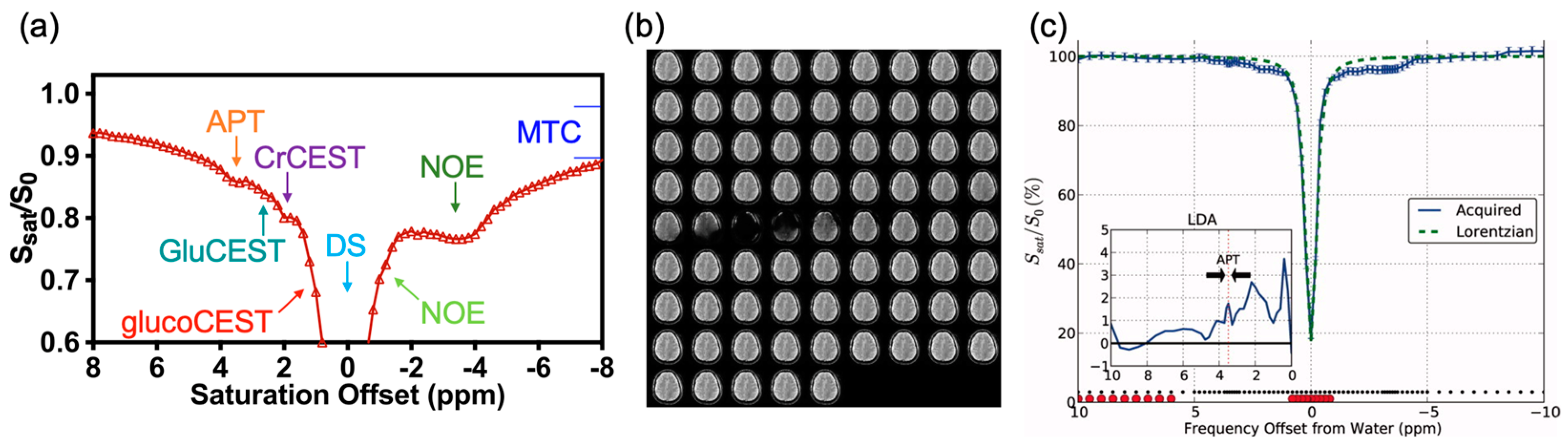
2. CEST Imaging of Brain Tumors
2.1. Endogenous Contrast
2.1.1. APT-Weighted (APTw) Contrast
2.1.2. NOE Contrast
2.2. Glioblastoma and Gliomas (Grade II, III)

| Species | Tumor Type (Grade) | B0 (T) | Analysis Method | CEST Contrast | Molecular/Cellular Changes | Ref. |
|---|---|---|---|---|---|---|
| Rat | Glioma, C6 | 3 | DISC-CEST | APT | Cellular and nuclear atypia | Wu Y. et al., 2019 [92] |
| Rat | Gliosarcoma, 9L | 4.7 | MTRasym | APTw | Cellular proteins and peptides | Zhou Z. et al., 2003 [3] |
| Rat | Gliosarcoma, 9L | 4.7 | MTRasym | APTw | pH | Zhou Z. et al., 2003 [48] |
| Rat | Gliosarcoma, 9L SF188/V + glioma | 4.7 | MTRasym | APTw | Treatment effects (radiation therapy), radiation necrosis, mobile cytosolic proteins, and peptides | Zhou J. et al., 2011 [70] |
| Rat | Gliosarcoma, 9L | 4.7 | MTRasym | APTw NOE (−2.5 to −5 ppm) | Mobile proteins, peptides, lipids, and metabolites | Zhou J. et al., 2013 [43] |
| Rat | U87 | 4.7 | MTRasym | APTw | Treatment effects (radiation therapy), radiation necrosis, cellularity, nuclear atypia, and vacuolation | Hong X. et al., 2014 [69] |
| Rat | GBM | 4.7 | EMR | APT, NOE | Mobile proteins and peptides | Heo HY. et al., 2016 [87] |
| Rat | GBM | 4.7 | MTRREX, AREX, CESTR, CESTRnr | APT, 2 ppm | APT: mobile proteins and peptides, 2 ppm: protein and peptide side-chain amide protons and various amine-related protons | Heo HY. et al., 2017 [89] |
| Rat | U87 | 4.7 | MTRasym | APTw | Amide proton mobile amide proton content or the increased amide proton exchange rate | Lee DH. et al. 2017 [90] |
| EMR | APT, NOE | |||||
| Rat | Glioma | 4.7 | DISC-CEST | APT NOE | APT: intracellular mobile proteins/peptides concentration NOE: aliphatic and olefinic protons | Zhou IY. et al., 2017 [26] |
| Rat | Gliosarcoma, 9L | 4.7 | MTRasym | APTw | NA | Heo H. et al., 2019 [91] |
| EMR | APT, NOE | |||||
| Rat | Gliosarcoma, 9L | 9.4 | AREX | APT, NOE | Protein contents | Xu J. et al., 2014 [93] |
| Rat | Gliosarcoma, 9L | 9.4 | Lorentzian | APT (3.6 ppm) NOE (−3.2 ppm) | Amide proton | Cai K. et al., 2015 [115] |
| 2 ppm | Tumor progression and creatine | |||||
| Rat | Gliosarcoma, 9L; glioma, F98 | 9.4 | Lorentzian | 2 ppm | Creatine and tumor aggressiveness | Cai K. et al., 2017 [116] |
| Rat | Gliosarcoma, 9L | 9.4 | MTRasym, AREX | 3 ppm | Amine and protein | Zhang XY. et al., 2017 [117] |
| Rat | ENU1564 (brain metastasis model) | 9.4 | APTR* | APT | Protein concentration and pH | Ray KJ. et al., 2019 [107] |
| Rat | Gliosarcoma, 9L | 9.4 | Lorentzian | 3 ppm | Glutamate | Debnath A. et al., 2020 [118] |
| Rat | Gliosarcoma, 9L | 9.4 | RPT | NOE (−1.6 ppm) | Phospholipids on cell membranes | Zu Z. et al., 2020 [101] |
| Mouse | GBM, patient cells | 7 | MTRasym | APTw | Proliferation, cellular acidification, and treatment effect (TMZ) | Sagiyama K. et al., 2014 [40] |
| Mouse | Glioma, GL261 | 7 | MTRasym | 3 ppm | Amine, pH, cellularity, and necrosis | Harris RJ. et al., 2015 [38] |
| Mouse | U87MG | 9.4 | AACID | AACID (amide at 3.5 ppm, amine at 2.75 ppm) | Intracellular pH and treatment effect | Albatany M. et al., 2019 [66] |
| Human (n = 10) | GBM (IV), oligodendroglioma (III), LGO (II), LGA (II), Meningioma | 3 | MTRasym | APTw | Cellular protein/peptide and intracellular pH | Jones CK. et al., 2006 [4] |
| Human (n = 9) | GMB (IV), AO (III), AA (III), LGO (II), LGA (II) | 3 | MTRasym | APTw | Glioma grading, cytosolic protein and peptide, and intracellular pH | Zhou J. et al., 2008 [60] |
| Human (n = 12) | GBM (IV), astrocytoma (III), oligodendroglioma (III) | 3 | MTRasym | APTw | Viable tumor core, edema, necrosis, mobile protein, and peptide | Wen Z. et al., 2010 [45] |
| Human (n = 14) | GBM (IV), AA (III), LGO (II), LGA (II), LGOA (II) | 3 | MTRasym | APTw | Protein content | Zhou J. et al., 2013 [42] |
| Human (n = 36) | GBM (IV), AO (III), AA (III), AOA (III), LGA (II), LGO (II), LGOA (II) | 3 | MTRasym | APTw | Glioma grading, necrosis, cell density, and proliferation | Togao O. et al., 2014 [39] |
| Human (n = 25) | Glioma (II–IV) | 3 | MTRasym | 3 ppm | An acidic signature, treatment effect (CRT), and PFS | Harris RJ. et al., 2015 [38] |
| Human (n = 26) | GBM (IV), AA (III), AO (III), LGO (II), LGOA (II) | 3 | MTRasym | APTw | Glioma grading | Sakata A. et al., 2015 [65] |
| Human (n = 13) | GBM (IV), Gliomas (low–grade), meningiomas, lymphoma | 3 | MTRasym | APTw | NA | Togao O. et al., 2015 [36] |
| Human (n = 11) | High–grade glioma | 3 | EMR | APT, NOE | NA | Heo HY. et al., 2016 [119] |
| Human (n = 32) | High–grade glioma Lymphomas | 3 | MTRasym | APTw | Differentiate lymphomas from high-grade glioma and protein | Jiang S. et al., 2016 [64] |
| Human (n = 65) | Glioma (II–IV) | 3 | MTRasym | APTw | Proliferation | Park J. et al., 2016 [32] |
| Human (n = 32) | GBM (IV), AA (III), gliomas (low–grade) | 3 | MTRasym | APTw | Cellularity | Ma B. et al., 2016 [68] |
| Human (n = 65) | Glioma (II–IV) | 3 | MTRasym | APTw | Proliferation | Park J. et al., 2016 [32] |
| Human (n = 32) | GBM (IV), AA (III), gliomas (low–grade) | 3 | MTRasym | APTw | Cellularity | Ma B. et al., 2016 [68] |
| Human (n = 7) | AA (III), LGO (II), LGA (II) | 3 | MTRasym | APTw | NA | Zhang Y. et al., 2016 [88] |
| Human (n = 44) | Glioma (II–IV) | 3 | MTRasym | APTw | Glioma grading and proliferation | Bai Y. et al., 2017 [63] |
| Human (n = 46) | Glioma (II–IV) | 3 | MTRasym | APTw | Glioma grading, protein, and peptide | Choi YS. et al., 2017 [31] |
| Human (n = 24) | Glioma (II–IV), edema | 3 | MTRasym | APTw | Cellularity, proliferation, and glioma grading | Jiang S. et al., 2017 [30] |
| Human (n = 27) | Glioma (II) | 3 | MTRasym | APTw | IDH mutation | Jiang S. et al., 2017 [29] |
| Human (n = 42) | Glioma (II–IV) | 3 | MTRasym | APTw | Glioma grading, proliferation, choline, and N-acetylaspartate | Su C. et al., 2017 [27] |
| Human (n = 18) | GBM (IV) | 3 | MTRasym | APTw | MGMT promoter methylation status | Jiang S. et al., 2018 [24] |
| Human (n = 57) | Meningioma | 3 | MTRasym | APTw | Intracellular proteins and peptides | Joo B. et al., 2018 [23] |
| Human (n = 42) | Glioma (II–IV) | 3 | MTRasym | APTw | MGMT prediction | Su L. et al., 2018 [20] |
| Human (n = 21) | GBM (IV), glioma (II), metastases, meningoma, chronic infarction | 3 | MTRasym | APTw | Proteins and peptides | Sun H. et al., 2018 [120] |
| Human (n = 32) | Glioma (II–IV) | 3 | Z-spectral fitted, | APT | Glioma grading and proliferation | Zhang J. et al., 2018 [19] |
| MTRasym | APTw | |||||
| Human (n = 51) | Glioma (II–IV) | 3 | MTRasym | APTw | Glioma grading and mobile cellular proteins | Zou T. et al., 2018 [62] |
| Human (n = 21) | GBM (IV), gliosarcoma (IV), AA (III), | 3 | MTRasym | APTw | Cellularity, proliferation, tumor recurrence, and a marker for active glioma | Jiang S. et al., 2019 [18] |
| Human (n = 71) | Glioma (III and IV) | 3 | MTRasym | APTw | Overall survival, PFS, and IDH mutation | Joo B. et al., 2019 [17] |
| Human (n = 14) | GBM (IV) | 3 | MTRasym | APTw | IDH and pH | Schure JR. et al., 2019 [108] |
| Lorentzian | APT | |||||
| Human (n = 90) | Glioma (II–IV) | 3 | MTRasym | 3 ppm | Cerebral blood volume and IDH mutation | Wang YL. et al., 2019 [72] |
| Human (n = 26) | Glioma (II, IV) Metastasis | 3 | MTRasym | APTw (3.5±0.4 ppm) | Glioma grading, MGMT, and IDH | Durmo F. et al., 2020 [61] |
| Human (n = 59) | Glioma (II, III) | 3 | MTRasym, machine learning | APTw | IDH1 mutation | Han Y. et al., 2020 [71] |
| Human (n = 54) | GBM (IV) | 3 | MTRasym | APTw | Treatment effect (bevacizumab), 12-month progression, PFS, and CBV | Park J. et al., 2020 [13] |
| Human (n = 30) | Glioma (III, IV) | 3 | MTRasym | APTw | Treatment effect (radiotherapy or CRT), tumor recurrence, and protein | Liu J. et al., 2020 [14] |
| Human (n = 46) | Glioma (II–IV) | 3 | MTRasym | APTw | Cellularity and CBV glioma grading | Schon S. et al., 2020 [59] |
| Human (n = 18) | GBM (IV), AA (III), astrocytoma (III), LGO (II), LGA (II) | 3 | MTRasym | APTw | Cytosolic protein content, mobile proteins, and semisolid macromolecules | Warnert EAH. et al., 2021 [11] |
| Lorentzian | APT | |||||
| Human (n = 51) | Glioma (II–IV) | 3 | MTRasym | APTw | Glioma grading (peptide or protein concentrations), cellularity, proliferation, and IDH mutation | Xu Z. et al., 2021 [9] |
| Human (n = 48) | Glioma (II–IV), Brain metastases | 3 | MTRasym, machine learning | APTw | Protein content | Sartoretti E. et al., 2021 [12] |
| Human (n = 19) | GBM, meningioma, brain metastasis | 3 | QUASS | APT, MT&NOE (−1.5 ppm) | −1.5 ppm: proliferation | Wu Y. et al., 2021 [10] |
| Human (n = 48) | High–grade glioma (III,IV) Low–grade glioma (I,II) | 3 | CESTRnr, EMR | APT | Glioma grading (proteins and peptides) | Zhang H. et al., 2021 [8] |
| Human (n = 81) | H3K27M–mutant associated brainstem glioma | 3 | MTRasym | APTw | H3K27M mutation, proliferation, pH, and protein and peptide metabolism | Zhuo Z. et al., 2021 [6] |
| Human (n = 113) | Glioma (II–IV) | 3 | Lorentzian | APT | Glioma grading (cellularity, mobile protein, and peptides), and IDH mutation | Su C. et al., 2022 [5] |
| 2 ppm | Creatine and 1p/19q co-deletion | |||||
| Human (n = 1) | AA (III) | 7 | MTRasym | −3.5 ppm | Cellular density | Jones CK. et al., 2013 [44] |
| Lorentzian | APT (3.3 to 3.7 ppm) NOE (−2 to −5 ppm) | |||||
| Human (n = 2) | GBM (IV), glioma (II or III) | 7 | MTRasym | −3 ppm | Necrosis and the structural integrity of proteins in cells (protein folding) | Zaiss M. et al., 2013 [121] |
| Human (n = 12) | GBM (IV) | 7 | MTRasym | 3.3 ppm | Protein structures proliferation | Paech D. et al., 2014 [41] |
| Human (n = 15) | GBM (IV) | 7 | MTRasym | 3.3 ppm | Cell density and edema | Paech D. et al., 2015 [37] |
| Human (n = 1) | LGO (II) | 7 | AREX | APT, NOE | NA | Windschuh J. et al., 2015 [35] |
| Human (n = 10) | GBM (IV) | 7 | AREX | 3.5 ppm, NOE | Protein and lipid | Zaiss M. et al., 2015 [34] |
| Human (n = 10) | Gliomas (II–IV) | 7 | MTRasym | APTw | Glioma grading | Heo HY. et al., 2016 [33] |
| EMR | APT (3.3 to 3.7 ppm) NOE (−3.3 to −3.7 ppm) | |||||
| Human (n = 11) | GBM (IV) | 7 | MTRasym, dnsAREX | 3.5 ppm | Amide proton and pH | Zaiss M. et al., 2017 [25] |
| Human (n = 31) | Glioma (II–IV) | 7 | MTRasym, dnsAREX | APT (3.5 ppm) | Glioma grading, IDH mutation, and MGMT promoter methylation status | Paech D. et al., 2018 [22] |
| Human (n = 20) | GBM (IV) | 7 | Lorentzian | NOE | Treatment effect (First-line therapy) | Regnery S. et al., 2018 [21] |
| MTRasym | APTw | |||||
| dnsAREX | APT | |||||
| Human (n = 12) | GBM (IV), LGO (II), LGA (II) | 7 | AREX | NOE | Treatment effect (CRT) | Meissner JE. et al., 2019 [67] |
| dnsAREX | APT | |||||
| Human (n = 26) | GBM (IV), AA (III) | 7 | AREX, dnsAREX | APT | Overall survival and PFS, amino acid, and protein | Paech D. et al., 2019 [16] |
| Human (n = 1) | GBM | 9.4 | Lorentzian | 3.5 ppm, NOE (−1.6, −3.5 ppm), 2 ppm, 2.7 ppm | Proteins and lipids | Zaiss M. et al., 2018 [98] |
2.3. Multiple CEST Contrast in Brain Tumors
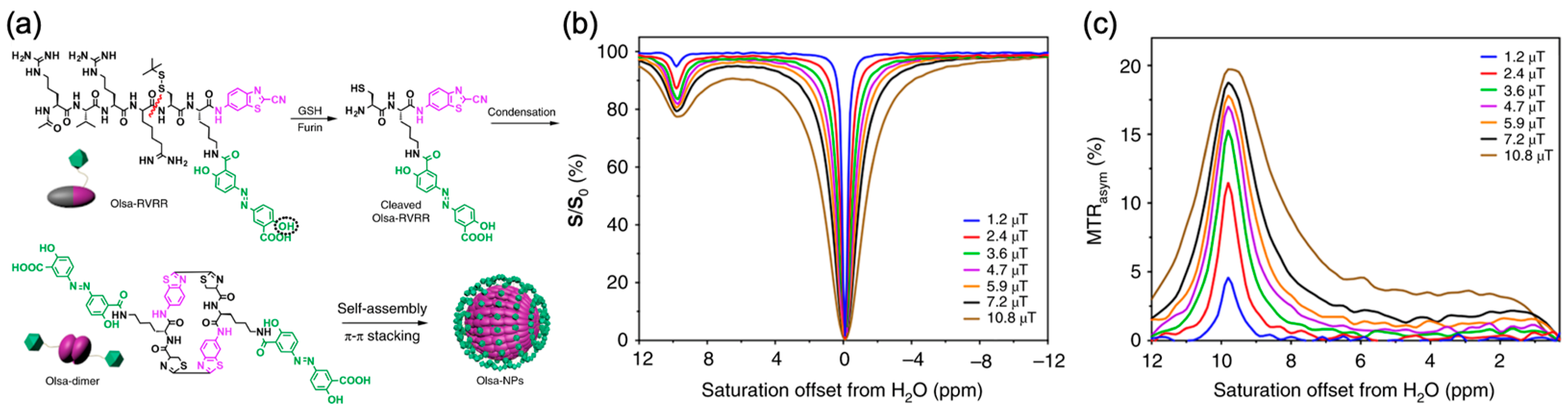
3. Non-Metallic CEST Contrast Agents for Brain Tumor Imaging
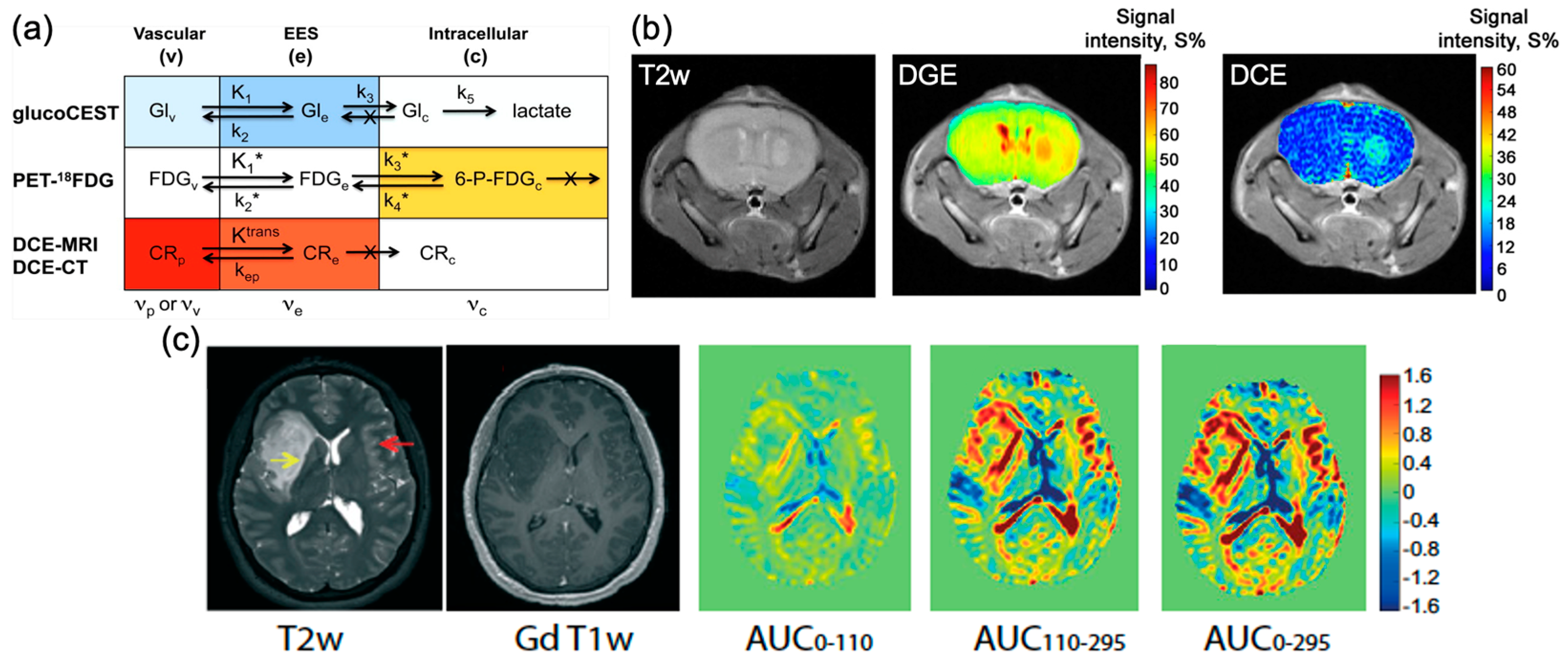
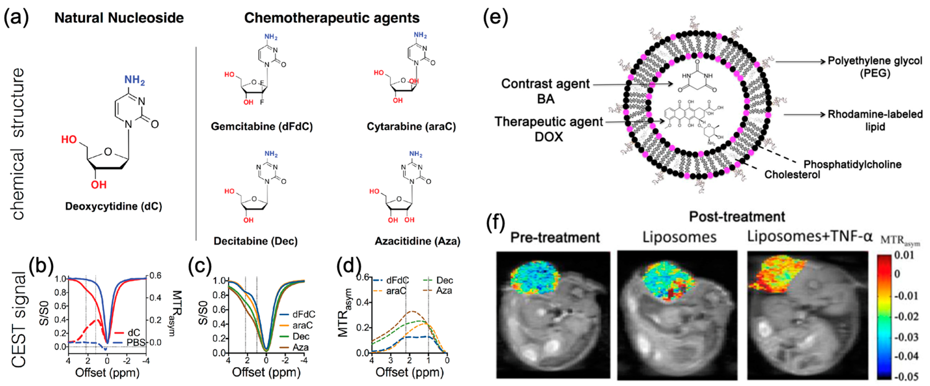
4. Imaging Drugs and Drug Delivery
4.1. Imaging Drugs and Drug Delivery Using CEST MRI

4.2. Theranostic Applications
5. Technical Part
5.1. CEST Acquisition
5.2. CEST Post-Processing
5.2.1. Z-Spectra and B0/B1 Correction
5.2.2. Z-Spectra Analysis
- (1)
- MTRasym analysis
- (2)
- Lorentzian difference analysis (LDA)
- (3)
- Multi-pool Lorentzian fitting
- (4)
- Polynomial and Lorentzian line-shape fitting (PLOF)
- (5)
- Three-offset method
5.2.3. Inverse Z-Spectra Analysis
5.2.4. Deep Learning-Based Analysis Methods
- (1)
- Deep learning-based Z-spectra analysis
- (2)
- Deep learning-based CEST fingerprinting
6. Promises and Challenges
7. Conclusions
Author Contributions
Funding
Institutional Review Board Statement
Informed Consent Statement
Data Availability Statement
Conflicts of Interest
References
- Sanvito, F.; Castellano, A.; Falini, A. Advancements in Neuroimaging to Unravel Biological and Molecular Features of Brain Tumors. Cancers 2021, 13, 424. [Google Scholar] [CrossRef] [PubMed]
- Gritsch, S.; Batchelor, T.T.; Gonzalez Castro, L.N. Diagnostic, therapeutic, and prognostic implications of the 2021 World Health Organization classification of tumors of the central nervous system. Cancer 2022, 128, 47–58. [Google Scholar] [CrossRef] [PubMed]
- Zhou, J.; Lal, B.; Wilson, D.A.; Laterra, J.; van Zijl, P.C. Amide proton transfer (APT) contrast for imaging of brain tumors. Magn. Reson. Med. 2003, 50, 1120–1126. [Google Scholar] [CrossRef] [PubMed]
- Jones, C.K.; Schlosser, M.J.; van Zijl, P.C.; Pomper, M.G.; Golay, X.; Zhou, J. Amide proton transfer imaging of human brain tumors at 3 T. Magn. Reson. Med. 2006, 56, 585–592. [Google Scholar] [CrossRef] [PubMed]
- Su, C.; Xu, S.; Lin, D.; He, H.; Chen, Z.; Damen, F.C.; Ke, C.; Lv, X.; Cai, K. Multi-parametric Z-spectral MRI may have a good performance for glioma stratification in clinical patients. Eur. Radiol. 2022, 32, 101–111. [Google Scholar] [CrossRef] [PubMed]
- Zhuo, Z.; Qu, L.; Zhang, P.; Duan, Y.; Cheng, D.; Xu, X.; Sun, T.; Ding, J.; ** tumour heterogeneity with pulsed 3D CEST MRI in non-enhancing glioma at 3T. MAGMA 2021. [Google Scholar] [CrossRef] [PubMed]
- Sartoretti, E.; Sartoretti, T.; Wyss, M.; Reischauer, C.; van Smoorenburg, L.; Binkert, C.A.; Sartoretti-Schefer, S.; Mannil, M. Amide proton transfer weighted (APTw) imaging based radiomics allows for the differentiation of gliomas from metastases. Sci. Rep. 2021, 11, 5506. [Google Scholar] [CrossRef] [PubMed]
- Park, J.E.; Kim, H.S.; Park, S.Y.; Jung, S.C.; Kim, J.H.; Heo, H.Y. Identification of Early Response to Anti-Angiogenic Therapy in Recurrent Glioblastoma: Amide Proton Transfer-weighted and Perfusion-weighted MRI compared with Diffusion-weighted MRI. Radiology 2020, 295, 397–406. [Google Scholar] [CrossRef] [PubMed]
- Liu, J.; Li, C.; Chen, Y.; Lv, X.; Lv, Y.; Zhou, J.; ** methods for correction of CEST MRI in the presence of temporal B0 field variations. Magn. Reson. Med. 2019, 82, 633–646. [Google Scholar] [CrossRef] [Green Version]
- Stancanello, J.; Terreno, E.; Castelli, D.D.; Cabella, C.; Uggeri, F.; Aime, S. Development and validation of a smoothing-splines-based correction method for improving the analysis of CEST-MR images. Contrast Media Mol. Imaging 2008, 3, 136–149. [Google Scholar] [CrossRef]
- Kim, M.; Gillen, J.; Landman, B.A.; Zhou, J.; Van Zijl, P.C. Water saturation shift referencing (WASSR) for chemical exchange saturation transfer (CEST) experiments. Magn. Reson. Med. Off. J. Int. Soc. Magn. Reson. Med. 2009, 61, 1441–1450. [Google Scholar] [CrossRef] [Green Version]
- Sun, P.Z.; Farrar, C.T.; Sorensen, A.G. Correction for artifacts induced by B0 and B1 field inhomogeneities in pH-sensitive chemical exchange saturation transfer (CEST) imaging. Magn. Reson. Med. Off. J. Int. Soc. Magn. Reson. Med. 2007, 58, 1207–1215. [Google Scholar] [CrossRef]
- Sui, R.; Chen, L.; Li, Y.; Huang, J.; Chan, K.W.; Xu, X.; van Zijl, P.C.; Xu, J. Whole-brain amide CEST imaging at 3T with a steady-state radial MRI acquisition. Magn. Reson. Med. 2021, 86, 893–906. [Google Scholar] [CrossRef]
- Schuenke, P.; Windschuh, J.; Roeloffs, V.; Ladd, M.E.; Bachert, P.; Zaiss, M. Simultaneous map** of water shift and B1 (WASABI)—Application to field-inhomogeneity correction of CEST MRI data. Magn. Reson. Med. 2017, 77, 571–580. [Google Scholar] [CrossRef] [Green Version]
- Khlebnikov, V.; Windschuh, J.; Siero, J.C.; Zaiss, M.; Luijten, P.R.; Klomp, D.W.; Hoogduin, H. On the transmit field inhomogeneity correction of relaxation-compensated amide and NOE CEST effects at 7 T. NMR Biomed. 2017, 30, e3687. [Google Scholar] [CrossRef] [Green Version]
- Sun, P.Z.; Benner, T.; Kumar, A.; Sorensen, A.G. Investigation of optimizing and translating pH-sensitive pulsed-chemical exchange saturation transfer (CEST) imaging to a 3T clinical scanner. Magn. Reson. Med. Off. J. Int. Soc. Magn. Reson. Med. 2008, 60, 834–841. [Google Scholar] [CrossRef] [Green Version]
- Jones, C.K.; Polders, D.; Hua, J.; Zhu, H.; Hoogduin, H.J.; Zhou, J.; Luijten, P.; van Zijl, P.C. In vivo three-dimensional whole-brain pulsed steady-state chemical exchange saturation transfer at 7 T. Magn. Reson. Med. 2012, 67, 1579–1589. [Google Scholar] [CrossRef] [Green Version]
- Zaiss, M.; Schmitt, B.; Bachert, P. Quantitative separation of CEST effect from magnetization transfer and spillover effects by Lorentzian-line-fit analysis of z-spectra. J. Magn. Reson. 2011, 211, 149–155. [Google Scholar] [CrossRef]
- Zhou, I.Y.; Wang, E.; Cheung, J.S.; Zhang, X.; Fulci, G.; Sun, P.Z. Quantitative chemical exchange saturation transfer (CEST) MRI of glioma using Image Downsampling Expedited Adaptive Least-squares (IDEAL) fitting. Sci. Rep. 2017, 7, 1–10. [Google Scholar] [CrossRef]
- Chen, L.; Barker, P.B.; Weiss, R.G.; van Zijl, P.C.; Xu, J. Creatine and phosphocreatine map** of mouse skeletal muscle by a polynomial and Lorentzian line-shape fitting CEST method. Magn. Reson. Med. 2019, 81, 69–78. [Google Scholar] [CrossRef] [Green Version]
- Chen, L.; Wei, Z.; Cai, S.; Li, Y.; Liu, G.; Lu, H.; Weiss, R.G.; van Zijl, P.C.; Xu, J. High-resolution creatine map** of mouse brain at 11.7 T using non-steady-state chemical exchange saturation transfer. NMR Biomed. 2019, 32, e4168. [Google Scholar] [CrossRef]
- Chen, L.; Zeng, H.; Xu, X.; Yadav, N.N.; Cai, S.; Puts, N.A.; Barker, P.B.; Li, T.; Weiss, R.G.; van Zijl, P.C. Investigation of the contribution of total creatine to the CEST Z-spectrum of brain using a knockout mouse model. NMR Biomed. 2017, 30, e3834. [Google Scholar] [CrossRef]
- **, T.; Wang, P.; Zong, X.; Kim, S.G. MR imaging of the amide-proton transfer effect and the pH-insensitive nuclear Overhauser effect at 9.4 T. Magn. Reson. Med. 2013, 69, 760–770. [Google Scholar] [CrossRef] [Green Version]
- Sun, P.Z.; Benner, T.; Copen, W.A.; Sorensen, A.G. Early experience of translating pH-weighted MRI to image human subjects at 3 Tesla. Stroke 2010, 41, S147–S151. [Google Scholar] [CrossRef] [Green Version]
- Zhang, X.Y.; Wang, F.; Li, H.; Xu, J.; Gochberg, D.F.; Gore, J.C.; Zu, Z. Accuracy in the quantification of chemical exchange saturation transfer (CEST) and relayed nuclear Overhauser enhancement (rNOE) saturation transfer effects. NMR Biomed. 2017, 30, e3716. [Google Scholar] [CrossRef]
- Huang, J.; Lai, J.H.; Tse, K.-H.; Cheng, G.W.; Liu, Y.; Chen, Z.; Han, X.; Chen, L.; Xu, J.; Chan, K.W. Deep neural network based CEST and AREX processing: Application in imaging a model of Alzheimer’s disease at 3 T. Magn. Reson. Med. 2022, 87, 1529–1545. [Google Scholar] [CrossRef]
- Zaiss, M.; Xu, J.; Goerke, S.; Khan, I.S.; Singer, R.J.; Gore, J.C.; Gochberg, D.F.; Bachert, P. Inverse Z-spectrum analysis for spillover-, MT-, and T1-corrected steady-state pulsed CEST-MRI–application to pH-weighted MRI of acute stroke. NMR Biomed. 2014, 27, 240–252. [Google Scholar] [CrossRef] [Green Version]
- Cohen, O.; Huang, S.; McMahon, M.T.; Rosen, M.S.; Farrar, C.T. Rapid and quantitative chemical exchange saturation transfer (CEST) imaging with magnetic resonance fingerprinting (MRF). Magn. Reson. Med. 2018, 80, 2449–2463. [Google Scholar] [CrossRef]
- Kim, B.; Schär, M.; Park, H.; Heo, H.-Y. A deep learning approach for magnetization transfer contrast MR fingerprinting and chemical exchange saturation transfer imaging. Neuro Image 2020, 221, 117165. [Google Scholar] [CrossRef]
- LeCun, Y.; Bengio, Y.; Hinton, G. Deep learning. Nature 2015, 521, 436–444. [Google Scholar] [CrossRef]
- Kang, B.; Kim, B.; Schär, M.; Park, H.; Heo, H.Y. Unsupervised learning for magnetization transfer contrast MR fingerprinting: Application to CEST and nuclear Overhauser enhancement imaging. Magn. Reson. Med. 2021, 85, 2040–2054. [Google Scholar] [CrossRef]
- Svolos, P.; Kousi, E.; Kapsalaki, E.; Theodorou, K.; Fezoulidis, I.; Kappas, C.; Tsougos, I. The role of diffusion and perfusion weighted imaging in the differential diagnosis of cerebral tumors: A review and future perspectives. Cancer Imaging 2014, 14, 1–20. [Google Scholar] [CrossRef] [Green Version]
- Field, A.S.; Alexander, A.L.; Wu, Y.C.; Hasan, K.M.; Witwer, B.; Badie, B. Diffusion tensor eigenvector directional color imaging patterns in the evaluation of cerebral white matter tracts altered by tumor. J. Magn. Reson. Imaging Off. J. Int. Soc. Magn. Reson. Med. 2004, 20, 555–562. [Google Scholar] [CrossRef]
- Dangouloff-Ros, V.; Deroulers, C.; Foissac, F.; Badoual, M.; Shotar, E.; Grévent, D.; Calmon, R.; Pagès, M.; Grill, J.; Dufour, C. Arterial spin labeling to predict brain tumor grading in children: Correlations between histopathologic vascular density and perfusion MR imaging. Radiology 2016, 281, 553–566. [Google Scholar] [CrossRef] [Green Version]
- Warmuth, C.; Gunther, M.; Zimmer, C. Quantification of blood flow in brain tumors: Comparison of arterial spin labeling and dynamic susceptibility-weighted contrast-enhanced MR imaging. Radiology 2003, 228, 523–532. [Google Scholar] [CrossRef] [Green Version]
- Heye, A.K.; Culling, R.D.; Hernández, M.d.C.V.; Thrippleton, M.J.; Wardlaw, J.M. Assessment of blood–brain barrier disruption using dynamic contrast-enhanced MRI. A systematic review. Neuro Image Clin. 2014, 6, 262–274. [Google Scholar] [CrossRef] [Green Version]
- Shiroishi, M.S.; Castellazzi, G.; Boxerman, J.L.; D’Amore, F.; Essig, M.; Nguyen, T.B.; Provenzale, J.M.; Enterline, D.S.; Anzalone, N.; Dörfler, A. Principles of T2*-weighted dynamic susceptibility contrast MRI technique in brain tumor imaging. J. Magn. Reson. Imaging 2015, 41, 296–313. [Google Scholar] [CrossRef]
- Law, M.; Yang, S.; Babb, J.S.; Knopp, E.A.; Golfinos, J.G.; Zagzag, D.; Johnson, G. Comparison of cerebral blood volume and vascular permeability from dynamic susceptibility contrast-enhanced perfusion MR imaging with glioma grade. Am. J. Neuroradiol. 2004, 25, 746–755. [Google Scholar]
- Wainwright, D.A.; Nigam, P.; Thaci, B.; Dey, M.; Lesniak, M.S. Recent developments on immunotherapy for brain cancer. Expert Opin. Emerg. Drugs 2012, 17, 181–202. [Google Scholar] [CrossRef] [Green Version]
- Ishimaru, H.; Morikawa, M.; Iwanaga, S.; Kaminogo, M.; Ochi, M.; Hayashi, K. Differentiation between high-grade glioma and metastatic brain tumor using single-voxel proton MR spectroscopy. Eur. Radiol. 2001, 11, 1784–1791. [Google Scholar] [CrossRef]
- Ott, D.; Hennig, J.; Ernst, T. Human brain tumors: Assessment with in vivo proton MR spectroscopy. Radiology 1993, 186, 745–752. [Google Scholar] [CrossRef]
- Dunet, V.; Pomoni, A.; Hottinger, A.; Nicod-Lalonde, M.; Prior, J.O. Performance of 18F-FET versus 18F-FDG-PET for the diagnosis and grading of brain tumors: Systematic review and meta-analysis. Neuro-oncology 2015, 18, 426–434. [Google Scholar] [CrossRef] [Green Version]
- Chao, S.T.; Suh, J.H.; Raja, S.; Lee, S.Y.; Barnett, G. The sensitivity and specificity of FDG PET in distinguishing recurrent brain tumor from radionecrosis in patients treated with stereotactic radiosurgery. Int. J. Cancer 2001, 96, 191–197. [Google Scholar] [CrossRef]
- Herz, K.; Mueller, S.; Perlman, O.; Zaitsev, M.; Knutsson, L.; Sun, P.Z.; Zhou, J.; van Zijl, P.; Heinecke, K.; Schuenke, P. Pulseq-CEST: Towards multi-site multi-vendor compatibility and reproducibility of CEST experiments using an open-source sequence standard. Magn. Reson. Med. 2021, 86, 1845–1858. [Google Scholar] [CrossRef]
- Liu, R.; Zhang, H.; Niu, W.; Lai, C.; Ding, Q.; Chen, W.; Liang, S.; Zhou, J.; Wu, D.; Zhang, Y. Improved chemical exchange saturation transfer imaging with real-time frequency drift correction. Magn. Reson. Med. 2019, 81, 2915–2923. [Google Scholar] [CrossRef]
- Mueller, S.; Stirnberg, R.; Akbey, S.; Ehses, P.; Scheffler, K.; Stöcker, T.; Zaiss, M. Whole brain snapshot CEST at 3T using 3D-EPI: Aiming for speed, volume, and homogeneity. Magn. Reson. Med. 2020, 84, 2469–2483. [Google Scholar] [CrossRef]
- Villano, D.; Romdhane, F.; Irrera, P.; Consolino, L.; Anemone, A.; Zaiss, M.; Dastrù, W.; Longo, D.L. A fast multislice sequence for 3D MRI-CEST pH imaging. Magn. Reson. Med. 2021, 85, 1335–1349. [Google Scholar] [CrossRef]
- Zaiss, M.; Ehses, P.; Scheffler, K. Snapshot-CEST: Optimizing spiral-centric-reordered gradient echo acquisition for fast and robust 3D CEST MRI at 9.4 T. NMR Biomed. 2018, 31, e3879. [Google Scholar] [CrossRef]


Publisher’s Note: MDPI stays neutral with regard to jurisdictional claims in published maps and institutional affiliations. |
© 2022 by the authors. Licensee MDPI, Basel, Switzerland. This article is an open access article distributed under the terms and conditions of the Creative Commons Attribution (CC BY) license (https://creativecommons.org/licenses/by/4.0/).
Share and Cite
Huang, J.; Chen, Z.; Park, S.-W.; Lai, J.H.C.; Chan, K.W.Y. Molecular Imaging of Brain Tumors and Drug Delivery Using CEST MRI: Promises and Challenges. Pharmaceutics 2022, 14, 451. https://doi.org/10.3390/pharmaceutics14020451
Huang J, Chen Z, Park S-W, Lai JHC, Chan KWY. Molecular Imaging of Brain Tumors and Drug Delivery Using CEST MRI: Promises and Challenges. Pharmaceutics. 2022; 14(2):451. https://doi.org/10.3390/pharmaceutics14020451
Chicago/Turabian StyleHuang, Jianpan, Zilin Chen, Se-Weon Park, Joseph H. C. Lai, and Kannie W. Y. Chan. 2022. "Molecular Imaging of Brain Tumors and Drug Delivery Using CEST MRI: Promises and Challenges" Pharmaceutics 14, no. 2: 451. https://doi.org/10.3390/pharmaceutics14020451