
Raman Map**-Based Reverse Engineering Facilitates Development of Sustained-Release Nifedipine Tablet


Abstract
:
1. Introduction
2. Materials and Methods
2.1. Materials
2.2. Raman Map** of Adalat®-L
2.3. Preparation of Nifedipine Sustained-Release Tablets
2.4. Validation of the Prepared Tablets by Raman Map**
2.5. In Vitro Dissolution Studies
2.6. Bioequivalence Studies
3. Results and Discussion
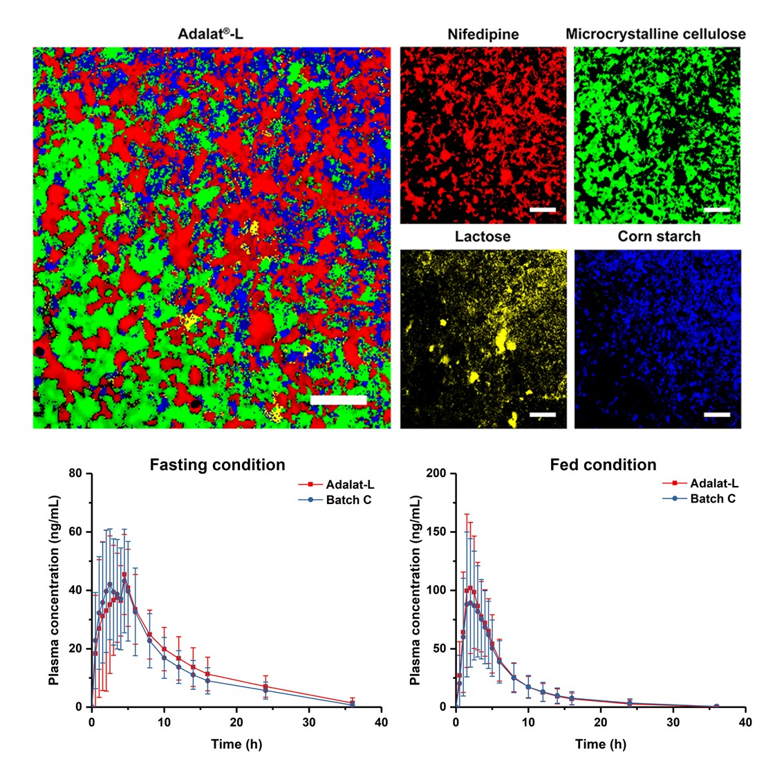
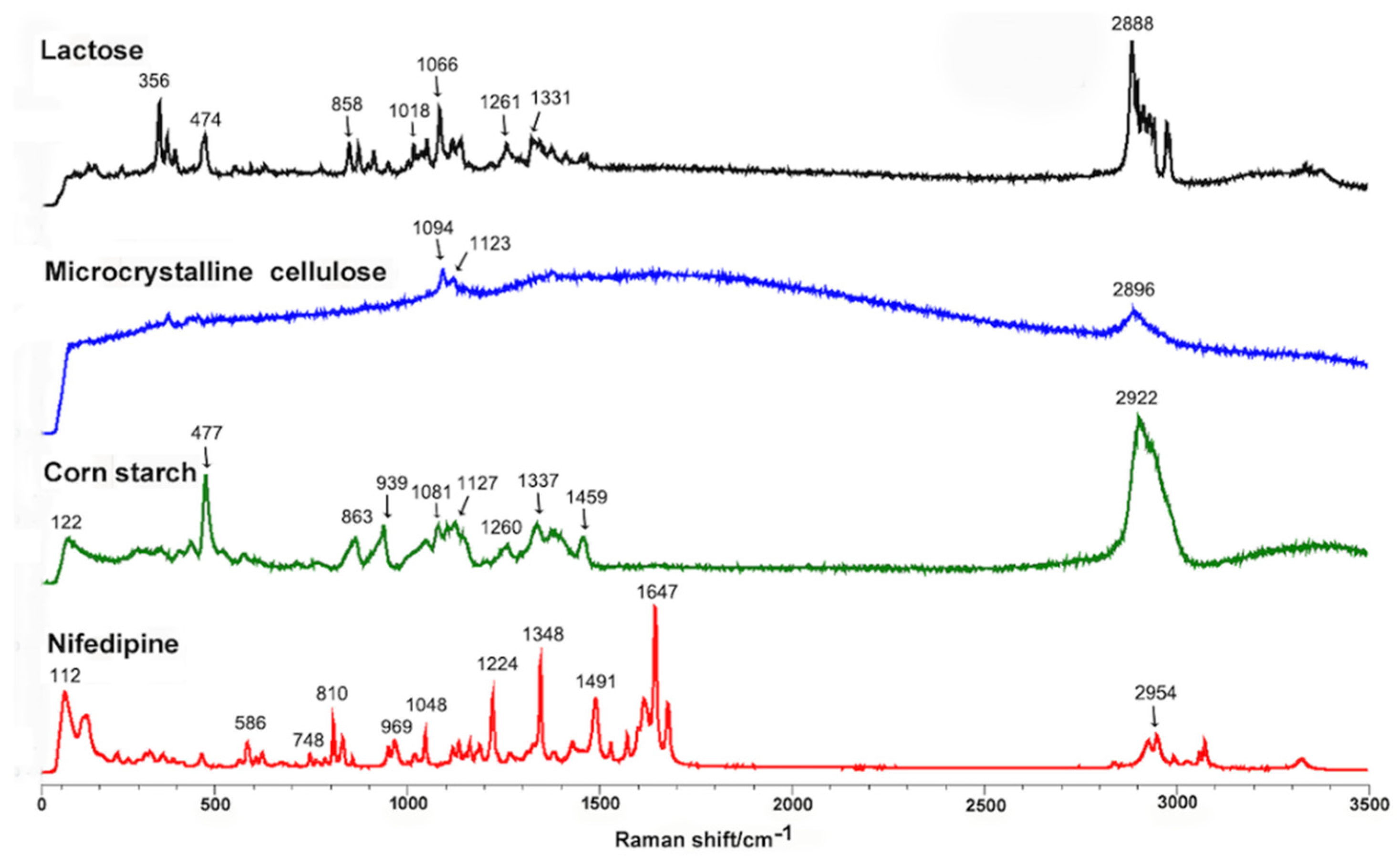
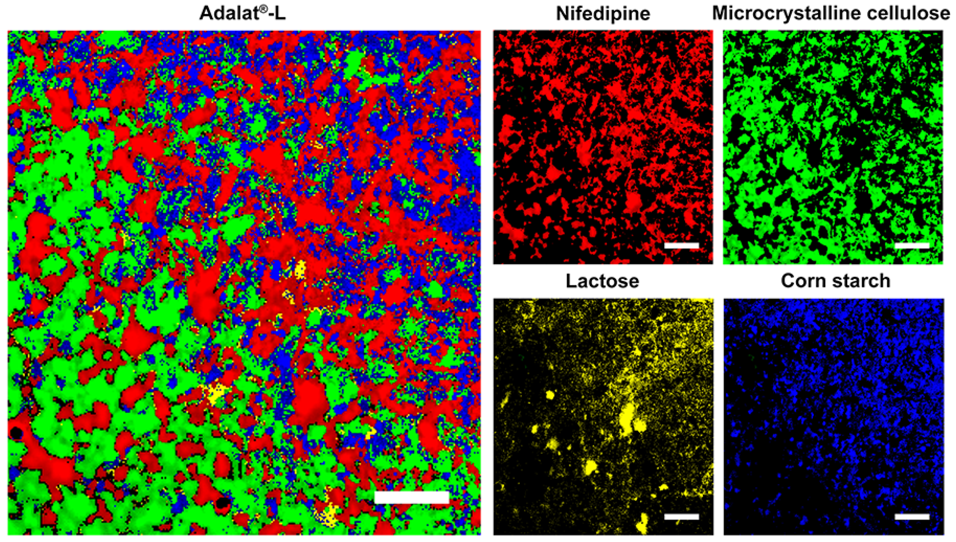
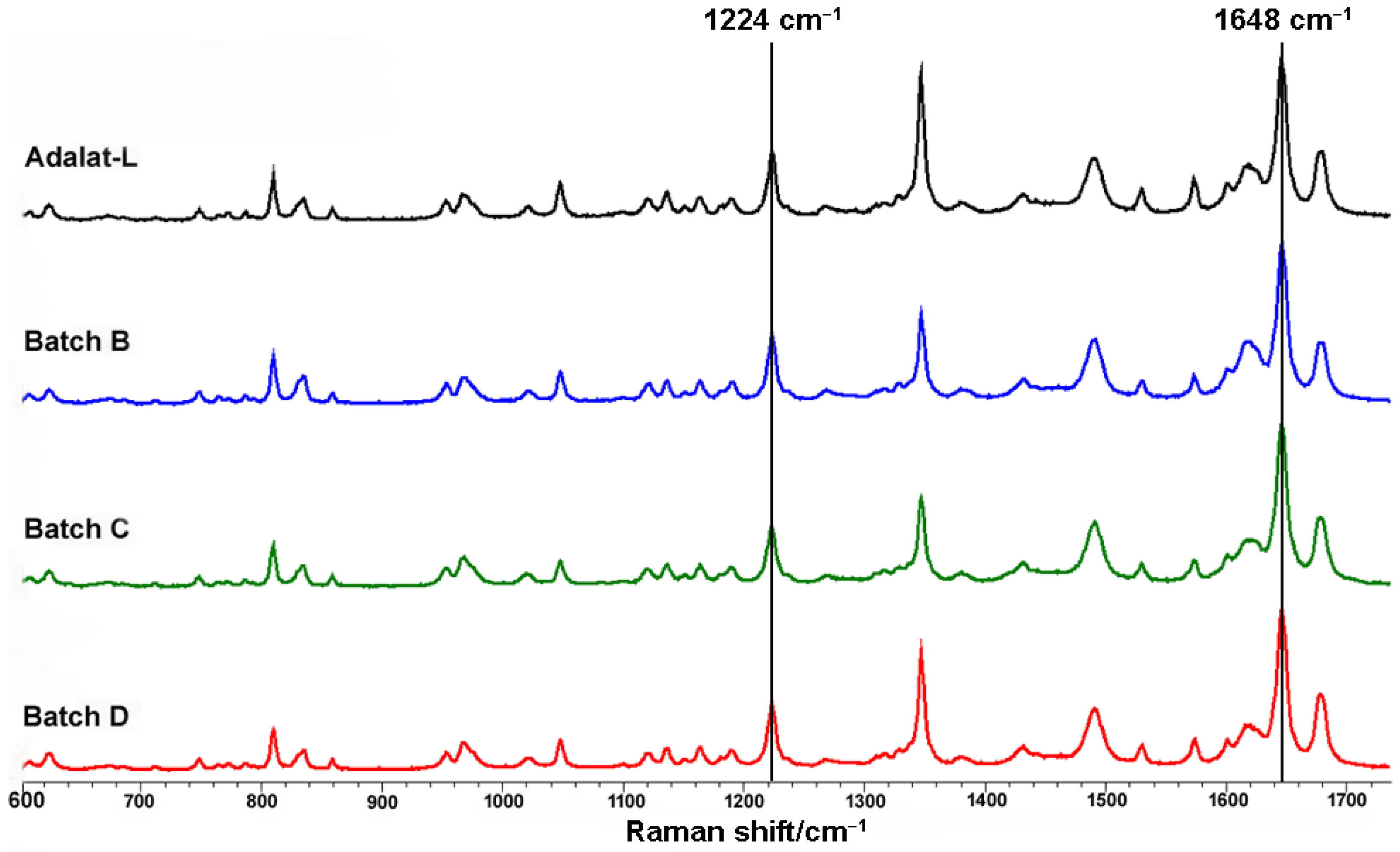
3.1. Analysis of Adalat®-L by Laser Raman Spectroscopy
3.2. Comminution and Particle Size Control of Nifedipine
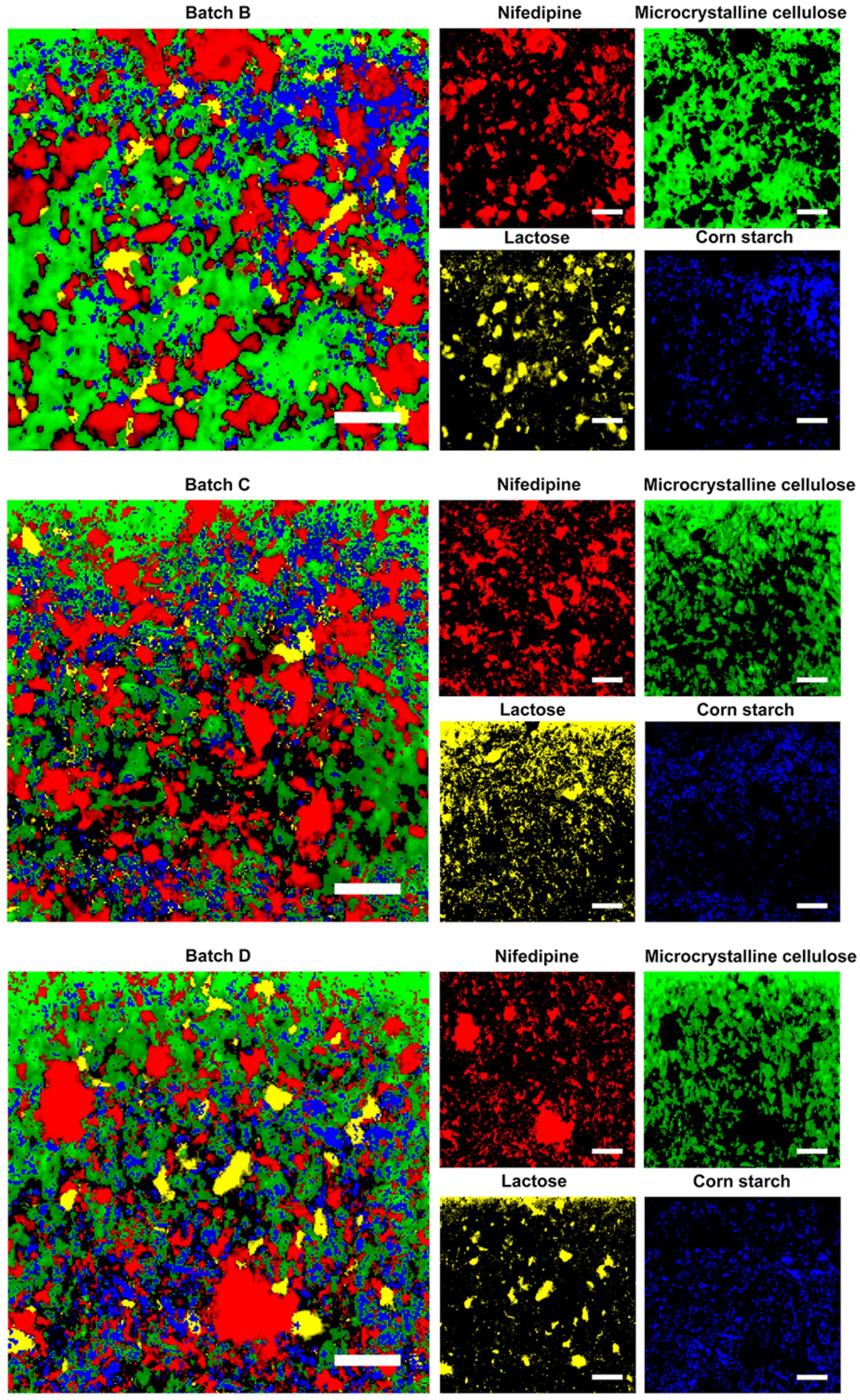
3.3. Validation by Raman Map** of Prepared Tablets
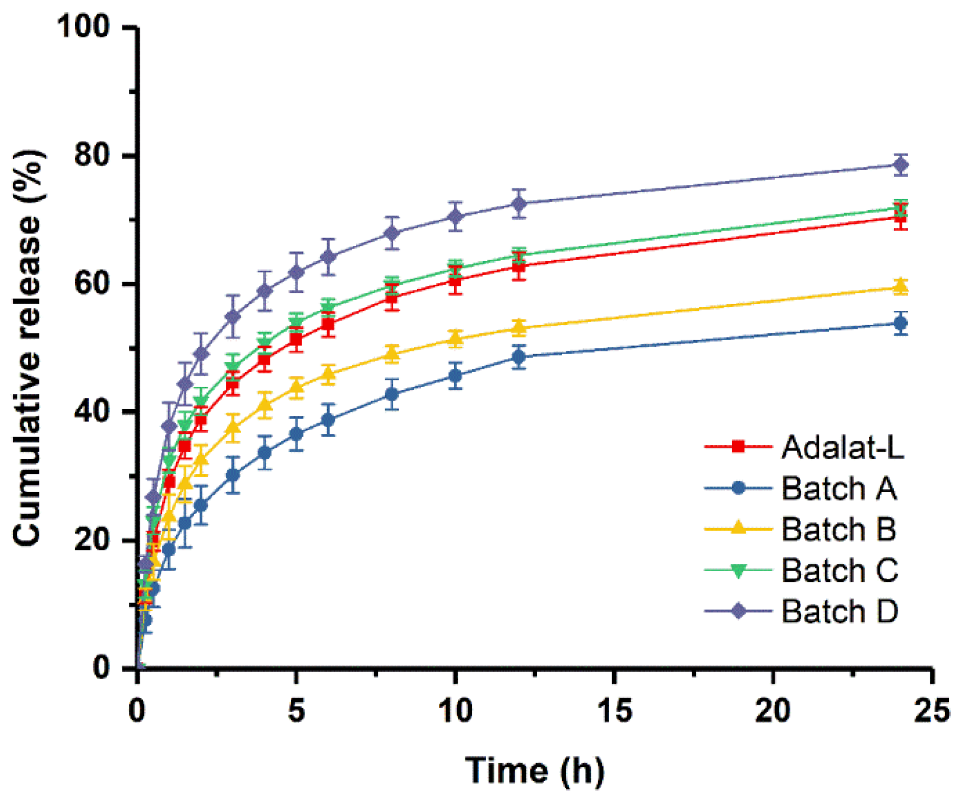
3.4. In Vitro Dissolution Studies
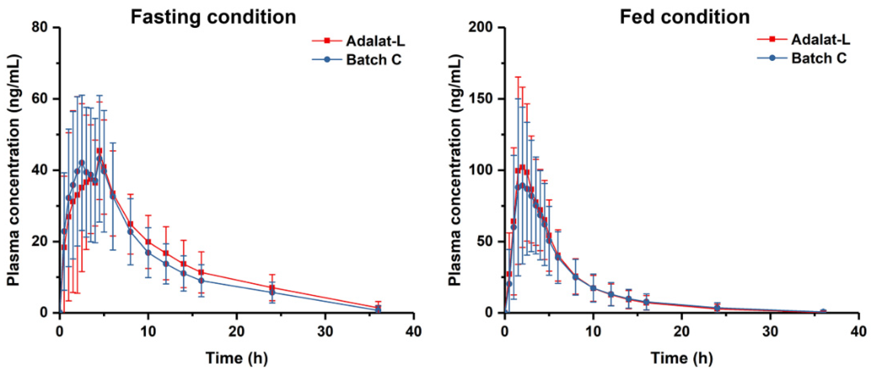
3.5. Bioequivalence Studies
4. Conclusions
Author Contributions
Funding
Institutional Review Board Statement
Informed Consent Statement
Data Availability Statement
Conflicts of Interest
References
- Wouters, O.J.; Kanavos, P.G.; McKee, M. Comparing generic drug markets in Europe and the United States: Prices, volumes, and spending. Milbank Q. 2017, 95, 554–601. [Google Scholar] [CrossRef] [PubMed] [Green Version]
- Holman, A. Is Bioequivalence a Sufficient Measure of Equivalence? J. Leg. Med. 2019, 39, 247–261. [Google Scholar] [CrossRef] [PubMed]
- Mishuk, A.U.; Qian, J.; Howard, J.N.; Harris, I.; Frank, G.; Kiptanui, Z.; Hansen, R. The association between patient sociodemographic characteristics and generic drug use: A systematic review and meta-analysis. J. Manag. Care Spec. Pharm. 2018, 24, 252–264. [Google Scholar] [CrossRef] [PubMed]
- Meredith, P. Bioequivalence and other unresolved issues in generic drug substitution. Clin. Ther. 2003, 25, 2875–2890. [Google Scholar] [CrossRef]
- Yu, L.X. Pharmaceutical quality by design: Product and process development, understanding, and control. Pharm. Res. 2008, 25, 781–791. [Google Scholar] [CrossRef]
- Suarez-Sharp, S.; Li, M.; Duan, J.; Shah, H.; Seo, P. Regulatory experience with in vivo in vitro correlations (IVIVC) in new drug applications. AAPS J. 2016, 18, 1379–1390. [Google Scholar] [CrossRef]
- Lionberger, R.A.; Lee, S.L.; Lee, L.; Raw, A.; Yu, L.X. Quality by design: Concepts for ANDAs. AAPS J. 2008, 10, 268–276. [Google Scholar] [CrossRef] [Green Version]
- Iacocca, R.G.; Burcham, C.L.; Hilden, L.R. Particle engineering: A strategy for establishing drug substance physical property specifications during small molecule development. J. Pharm. Sci. 2010, 99, 51–75. [Google Scholar] [CrossRef]
- Shekunov, B.Y.; Chattopadhyay, P.; Tong, H.H.; Chow, A.H. Particle size analysis in pharmaceutics: Principles, methods and applications. Pharm. Res. 2007, 24, 203–227. [Google Scholar] [CrossRef]
- Overhoff, K.A.; Johnston, K.P.; Williams III, R.O. Improvement of dissolution rate of poorly water soluble drugs using a new particle engineering process: Spray freezing into liquid. ACS Symp. Ser. 2006, 924, 305–319. [Google Scholar] [CrossRef]
- Bukovec, P.; Benkič, P.; Smrkolj, M.; Vrečzer, F. Effect of crystal habit on the dissolution behaviour of simvastatin crystals and its relationship to crystallization solvent properties. Die Pharm. Int. J. Pharm. Sci. 2016, 71, 263–268. [Google Scholar] [CrossRef]
- Hu, J.; Johnston, K.P.; Williams III, R.O. Nanoparticle engineering processes for enhancing the dissolution rates of poorly water soluble drugs. Drug Dev. Ind. Pharm. 2004, 30, 233–245. [Google Scholar] [CrossRef] [PubMed]
- Sadeghi, F.; Garekani, H.A.; Goli, F. Tableting of Eudragit RS and propranolol hydrochloride solid dispersion: Effect of particle size, compaction force, and plasticizer addition on drug release. Drug Dev. Ind. Pharm. 2004, 30, 759–766. [Google Scholar] [CrossRef] [PubMed]
- Rabinow, B.E. Nanosuspensions in drug delivery. Nat. Rev. Drug Discov. 2004, 3, 785–796. [Google Scholar] [CrossRef]
- Eksi-Kocak, H.; Tamer, S.I.; Yilmaz, S.; Eryilmaz, M.; Boyaci, I.H.; Tamer, U. Quantification and spatial distribution of salicylic acid in film tablets using FT-Raman map** with multivariate curve resolution. Asian J. Pharm. Sci. 2018, 13, 155–162. [Google Scholar] [CrossRef]
- Ibrahim, M.; Zhang, J.; Repka, M.; Chen, R. Characterization of the solid physical state of API and its distribution in pharmaceutical hot melt extrudates using terahertz Raman imaging. AAPS PharmSciTech 2019, 20, 62. [Google Scholar] [CrossRef]
- Sacré, P.-Y.; De Bleye, C.; Chavez, P.-F.; Netchacovitch, L.; Hubert, P.; Ziemons, E. Data processing of vibrational chemical imaging for pharmaceutical applications. J. Pharm. Biomed. Anal. 2014, 101, 123–140. [Google Scholar] [CrossRef]
- Ewing, A.V.; Kazarian, S.G. Recent advances in the applications of vibrational spectroscopic imaging and map** to pharmaceutical formulations. Spectrochim. Acta A Mol. Biomol. Spectrosc. 2018, 197, 10–29. [Google Scholar] [CrossRef]
- Cailletaud, J.; De Bleye, C.; Dumont, E.; Sacré, P.-Y.; Netchacovitch, L.; Gut, Y.; Boiret, M.; Ginot, Y.-M.; Hubert, P.; Ziemons, E. Critical review of surface-enhanced Raman spectroscopy applications in the pharmaceutical field. J. Pharm. Biomed. Anal. 2018, 147, 458–472. [Google Scholar] [CrossRef]
- Wang, W.-t.; Zhang, H.; Yuan, Y.; Guo, Y.; He, S.-x. Research progress of Raman spectroscopy in drug analysis. AAPS PharmSciTech 2018, 19, 2921–2928. [Google Scholar] [CrossRef]
- Willett, D.R.; Rodriguez, J.D. Quantitative Raman assays for on-site analysis of stockpiled drugs. Anal. Chim. Acta. 2018, 1044, 131–137. [Google Scholar] [CrossRef]
- Matthews, T.E.; Coffman, C.; Kolwyck, D.; Hill, D.; Dickens, J.E. Enabling robust and rapid raw material identification and release by handheld raman spectroscopy. PDA J. Pharm. Sci. Technol. 2019, 73, 356–372. [Google Scholar] [CrossRef] [PubMed]
- Okotrub, K.A.; Zykova, V.A.; Adichtchev, S.V.; Surovtsev, N.V. Deciphering the orientation of lipid molecules by principal component analysis of Raman map** data. Analyst 2020, 145, 1466–1472. [Google Scholar] [CrossRef] [PubMed]
- Meek, C.; Hoe, J.; Evans, J.; Thurman, R.; Ashworth, L.; Leff, R. Raman Spectroscopy: A Sensitive and Specific Technique for Determining the Accuracy of Compounded Pharmaceutical Formulations. J. Pediatr. Pharmacol. Ther. 2016, 21, 413–418. [Google Scholar] [CrossRef] [Green Version]
- Gallimore, P.J.; Davidson, N.M.; Kalberer, M.; Pope, F.D.; Ward, A.D. 1064 nm Dispersive Raman microspectroscopy and optical trap** of pharmaceutical aerosols. Anal. Chem. 2018, 90, 8838–8844. [Google Scholar] [CrossRef] [PubMed]
- Galata, D.L.; Zsiros, B.; Mészáros, L.A.; Nagy, B.; Szabó, E.; Farkas, A.; Nagy, Z.K. Raman map**-based non-destructive dissolution prediction of sustained-release tablets. J. Pharm. Biomed. Anal. 2022, 212, 114661. [Google Scholar] [CrossRef]
- Navin, C.V.; Tondepu, C.; Toth, R.; Lawson, L.S.; Rodriguez, J.D. Quantitative determinations using portable Raman spectroscopy. J. Pharm. Biomed. Anal. 2017, 136, 156–161. [Google Scholar] [CrossRef] [Green Version]
- Paiva, E.M.; da Silva, V.H.; Poppi, R.J.; Pereira, C.F.; Rohwedder, J.J. Comparison of macro and micro Raman measurement for reliable quantitative analysis of pharmaceutical polymorphs. J. Pharm. Biomed. Anal. 2018, 157, 107–115. [Google Scholar] [CrossRef]
- Ma, X.; Sun, X.; Wang, H.; Wang, Y.; Chen, D.; Li, Q. Raman spectroscopy for Pharmaceutical quantitative analysis by Low-Rank Estimation. Front. Chem. 2018, 6, 400. [Google Scholar] [CrossRef] [Green Version]
- Paiva, E.M.; Ribessi, R.L.; Pereira, C.F.; Rohwedder, J.J.R. Low-frequency Raman spectrophotometer with wide laser illumination on the sample: A tool for pharmaceutical analytical analysis. Spectrochim. Acta A Mol. Biomol. Spectrosc. 2020, 228, 117798. [Google Scholar] [CrossRef]
- Smith, M.; Logan, M.; Bazley, M.; Blanchfield, J.; Stokes, R.; Blanco, A.; McGee, R. A Semi-quantitative method for the detection of fentanyl using surface-enhanced Raman scattering (SERS) with a handheld Raman instrument. J. Forensic Sci. 2021, 66, 505–519. [Google Scholar] [CrossRef] [PubMed]
- Mansouri, M.A.; Sacré, P.-Y.; Coïc, L.; De Bleye, C.; Dumont, E.; Bouklouze, A.; Hubert, P.; Marini, R.; Ziemons, E. Quantitation of active pharmaceutical ingredient through the packaging using Raman handheld spectrophotometers: A comparison study. Talanta 2020, 207, 120306. [Google Scholar] [CrossRef] [PubMed]
- Čapková, T.; Pekárek, T.; Hanulíková, B.; Matějka, P. Application of reverse engineering in the field of pharmaceutical tablets using Raman map** and chemometrics. J. Pharm. Biomed. Anal. 2022, 209, 114496. [Google Scholar] [CrossRef] [PubMed]
- Nakamoto, K.; Urasaki, T.; Hondo, S.; Murahashi, N.; Yonemochi, E.; Terada, K. Evaluation of the crystalline and amorphous states of drug products by nanothermal analysis and Raman imaging. J. Pharm. Biomed. Anal. 2013, 75, 105–111. [Google Scholar] [CrossRef] [PubMed]
- Šašić, S.; Mehrens, S. Raman chemical map** of low-content active pharmaceutical ingredient formulations. III. Statistically optimized sampling and detection of polymorphic forms in tablets on stability. Anal. Chem. 2012, 84, 1019–1025. [Google Scholar] [CrossRef] [PubMed]
- Kang, Y.; Shao, Z.; Wang, Q.; Hu, X.; Yu, D. Quantitation of polymorphic impurity in entecavir polymorphic mixtures using powder X-ray diffractometry and Raman spectroscopy. J. Pharm. Biomed. Anal. 2018, 158, 28–37. [Google Scholar] [CrossRef] [PubMed]
- Dadou, S.M.; El-Barghouthi, M.I.; Antonijevic, M.D.; Chowdhry, B.Z.; Badwan, A.A. Elucidation of the controlled-release behavior of metoprolol succinate from directly compressed xanthan gum/chitosan polymers: Computational and experimental studies. ACS Biomater. Sci. Eng. 2020, 6, 21–37. [Google Scholar] [CrossRef] [PubMed]
- Čapková-Helešicová, T.; Pekárek, T.; Schöngut, M.; Matějka, P. New designed special cells for Raman map** of the disintegration process of pharmaceutical tablets. J. Pharm. Biomed. Anal. 2019, 168, 113–123. [Google Scholar] [CrossRef]
- Esmonde-White, K.A.; Cuellar, M.; Uerpmann, C.; Lenain, B.; Lewis, I.R. Raman spectroscopy as a process analytical technology for pharmaceutical manufacturing and bioprocessing. Anal. Bioanal. Chem. 2017, 409, 637–649. [Google Scholar] [CrossRef] [Green Version]
- Kim, B.; Woo, Y.-A. Coating process optimization through in-line monitoring for coating weight gain using Raman spectroscopy and design of experiments. J. Pharm. Biomed. Anal. 2018, 154, 278–284. [Google Scholar] [CrossRef]
- Lim, Y.-I.; Han, J.; Woo, Y.-A.; Kim, J.; Kang, M.J. Rapid quantitation of atorvastatin in process pharmaceutical powder sample using Raman spectroscopy and evaluation of parameters related to accuracy of analysis. Spectrochim. Acta A Mol. Biomol. Spectrosc. 2018, 200, 26–32. [Google Scholar] [CrossRef] [PubMed]
- Fu, X.; Zhong, L.-m.; Cao, Y.-b.; Chen, H.; Lu, F. Quantitative analysis of excipient dominated drug formulations by Raman spectroscopy combined with deep learning. Anal. Methods 2021, 13, 64–68. [Google Scholar] [CrossRef] [PubMed]
- Hou, H.-Y.; Yang, X.; Mao, Z.-L.; Yao, X.-Y.; Chen, X.-B. Raman study of impurity influence on active center in artemisinin. Spectrochim. Acta A Mol. Biomol. Spectrosc. 2019, 221, 117206. [Google Scholar] [CrossRef] [PubMed]
- Hegasy, A.; Ramsch, K.D. Solid Medicament Formulations Containing Nifedipine, and Processes for Their Preparation. U.S. Patent 5,264,446, 1 June 1992. [Google Scholar]
- Moore, J. Mathematical comparison of dissolution profiles. Pharm. Technol. 1996, 20, 64–75. [Google Scholar]
- Ishida, H.; Wu, T.; Yu, L. Sudden rise of crystal growth rate of nifedipine near Tg without and with polyvinylpyrrolidone. J. Pharm. Sci. 2007, 96, 1131–1138. [Google Scholar] [CrossRef]


| Ingredients | Amount (mg) |
|---|---|
| Nifedipine | 20.0 |
| Polysorbate 80 | 0.4 |
| Microcrystalline cellulose | 26.0 |
| Lactose | 8.0 |
| Corn starch (for blending) | 12.4 |
| Corn starch (for starch slurry) | 12.4 |
| Magnesium stearate | 0.8 |
| SUM | 80.0 |
| D90 (μm) | D50 (μm) | D10 (μm) | |
|---|---|---|---|
| Nifedipine | 150 | 118 | 30.7 |
| Components | Nifedipine | Microcrystalline Cellulose | Lactose | Corn Starch |
|---|---|---|---|---|
| Proportion (%) | 33.94 | 33.02 | 1.40 | 31.65 |
| Batch | Particle Size | ||
|---|---|---|---|
| D (90) (μm) | D (50) (μm) | D (10) (μm) | |
| A | 145.2 ± 0.6 | 69.3 ± 0.6 | 12.1 ± 0.5 |
| B | 96.1 ± 0.7 | 38.2 ± 0.6 | 8.4 ± 0.6 |
| C | 62.0 ± 0.6 | 26.3 ± 0.2 | 5.5 ± 0.3 |
| D | 38.9 ± 0.5 | 18.8 ± 0.4 | 6.1 ± 0.4 |
| Particle Size | |||
|---|---|---|---|
| D (90) (μm) | D (50) (μm) | D (10) (μm) | |
| Adalat®-L | 150 | 118 | 30.7 |
| Batch B | 173 | 118 | 52.2 |
| Batch C | 147 | 121 | 47.5 |
| Batch D | 108 | 103 | 32.7 |
| Condition | Pharmacokinetic Parameters | Mean and Ratio | 90% Confidence Interval | ||
|---|---|---|---|---|---|
| Batch C (T) | Adalat®-L (R) | (T/R)% | |||
| Fasting (N = 28) | Cmax (ng/mL) | 56.4 ± 15.4 | 54.9 ± 16.3 | 102.75 | 92.15~114.56 |
| AUC0–t (ng/mL·h) | 443.4 ± 150.0 | 478.1 ± 156.2 | 92.73 | 86.67~99.21 | |
| AUC0–∞ (ng/mL·h) | 501.6 ± 147.8 | 536.6 ± 162.8 | 93.48 | 87.55~99.80 | |
| Fed (N = 28) | Cmax (ng/mL) | 107.5 ± 46.5 | 119.8 ± 44.1 | 89.67 | 81.95~98.11 |
| AUC0–t (ng/mL·h) | 539.6 ± 303.4 | 569.3 ± 283.0 | 94.78 | 88.39~101.63 | |
| AUC0–∞ (ng/mL·h) | 569.1 ± 314.9 | 597.3 ± 297.3 | 95.28 | 88.97~102.04 | |
Publisher’s Note: MDPI stays neutral with regard to jurisdictional claims in published maps and institutional affiliations. |
© 2022 by the authors. Licensee MDPI, Basel, Switzerland. This article is an open access article distributed under the terms and conditions of the Creative Commons Attribution (CC BY) license (https://creativecommons.org/licenses/by/4.0/).
Share and Cite
Sun, N.; Chang, L.; Lu, Y.; Wu, W. Raman Map**-Based Reverse Engineering Facilitates Development of Sustained-Release Nifedipine Tablet. Pharmaceutics 2022, 14, 1052. https://doi.org/10.3390/pharmaceutics14051052
Sun N, Chang L, Lu Y, Wu W. Raman Map**-Based Reverse Engineering Facilitates Development of Sustained-Release Nifedipine Tablet. Pharmaceutics. 2022; 14(5):1052. https://doi.org/10.3390/pharmaceutics14051052
Chicago/Turabian StyleSun, Ningyun, Liang Chang, Yi Lu, and Wei Wu. 2022. "Raman Map**-Based Reverse Engineering Facilitates Development of Sustained-Release Nifedipine Tablet" Pharmaceutics 14, no. 5: 1052. https://doi.org/10.3390/pharmaceutics14051052