1. Introduction
Titanium dioxide nanoparticles (TiO
2-NPs) are among the most widely used engineered nanostructures of TiO
2. Most of the TiO
2 applications are due to their structural and electronic properties. It consists of mainly three phases which included anatase, rutile, and brookite [
1]. Due to its greater photocatalytic activity [
2], anatase has the highest number of industrial applications compared to rutile and brookite [
3]. At the beginning of the 20th century, mass production of TiO
2-NPs started as a non-toxic substitute for a white dye used in paints. It has various applications in food colorant or white pigment in manufacturing plastics, paper, ink, paints, and welding rod coating material. Ultrafine TiO
2-NPs [
4] are widely used in other industrial products, such as cosmetics, toothpaste, pharmaceuticals, and skincare products [
3,
5,
6,
7]. TiO
2-NPs is an n-type semiconductor with a wide bandgap, having various applications in wastewater treatments dye-sensitized solar cells, photoionized devices, and lithium batteries [
8]. TiO
2-NPs have specific medicinal properties and have been used in many biomedical applications such as artificial hips, dental implants, bone plates, coatings, scaffolds, and also in gene and drug delivery systems.
Interest in the synthesis of TiO
2-NPs and their applications are due to their biocompatibility [
9,
10,
11], antimicrobial properties [
12,
13], high chemical stability, specific surface area, and catalytic activity [
14,
15,
16]. Because of the wide range of applications, TiO
2-NPs have considerable potential for human exposure. People can be exposed to these nanoparticles through oral, respiratory, or dermal routes. TiO
2-NPs have been used in the food industry since 1996 when the US Food and Drug Administration approved it as a food supplement [
17]. TiO
2-NPs are also used for electrostatic properties in terms of electrostatic potentials and ion distributions [
18] for dye-degrading activities, H
2O
2 scavenging, and anticoagulant activities [
19] to enhance the glasses’ radiation protection properties along with bioactive glasses [
20] as nanocarriers for drug delivery [
21] and as a nano-drug for treating diseases [
22]. The idea of agents for two modes of therapy enhancement and image contrast is one field that remains largely unexplored. The principle of two modes of enhancement agents has been validated for radiotherapy enhancement applications by using conventional non-nanoparticle image contrast agents such as iodine [
23,
24,
25].
There have been reports in which TiO
2-NPs can be used in radiation therapy applications for the treatment of cancer due to their absorptive properties and higher refractive index [
23,
24,
25,
26]. The tumor during radiotherapy is exposed to ionizing radiation. As a result, it gives damage to the DNA of the tumor and other biological molecules and ultimately results in the cell’s death.
The inescapable radiation dose administered to the surrounding healthy tissues of the tumor being treated is the intrinsic drawback of radiation therapy. The use of radiotherapy is restricted for some tumors, due to unwanted damage to the surrounding healthy tissue by the dose required to destroy the tumor [
27]. Cheng et al. investigated a hybrid anisotropic nanostructure, composed of gold (Au) doped titanium dioxide (TiO
2), as a radio-sensitizer for radiotherapy of triple-negative breast cancer (TNBC) [
28]. Liang et al. used TiO
2 to enhance the quantum yield in ultra-sound activated sonodynamic therapy (SDT) [
29]. Yang et al. synthesized carbon-doped titanium dioxide to generate ROS to eliminate tumor cells under ultrasound irradiation [
30]. Su et al. construct an active site in a TiO
2 nano catalyst by auger electrons (AEs) for cancer catalytic internal radiotherapy (CIRT) applications [
31]. Contrast-enhanced radiotherapy (CERT) [
32] requires injection of contrast agents to the target volume with high-Z (high atomic number) and kilovoltage X-rays irradiation. Since materials with a high Z content have different absorption properties from those of healthy tissues, therefore the distribution of the absorbed dose can be changed due to their presence. In areas where a contrast agent is present, an increase in localized dose can be obtained without affecting the healthy tissue [
23,
24]. Kilovoltage X-rays energy range is considered the ideal range for CERT [
24] because the change in photo absorbing properties is noticeable at kilovolt energies [
32,
33]. Some improvement has also been observed in contrast by using X-ray energy in the megavolt range provided by medical linear accelerators (Linac) [
34,
35]. TiO
2-NPs with a size in the range of smaller than 100 nm can penetrate the cell membrane and preferably accumulate in cancer cells [
36]. Moreover, it can work as the required contrast agent.
Previously iodine has been used as a contrast agent for computed tomography (CT) imaging [
37]. A conventional CT scanner was used to monitor iodine uptake and tumor progression simultaneously. The pioneer studies for photodynamic therapy and MRI enhancement have missed or ignored the concept of nanoparticles-based enhancement agents [
38,
39]. NPs synthesized for radiotherapy and ionizing radiation would be beneficial to monitor them with CT scanners as it is the standard imaging tool for treatment planning and diagnosis [
40,
41]. These CT scanners are gradually replacing the conventional radiotherapy treatment planning simulator units. A conventional CT scanner may also be updated to deliver therapeutic doses of X-rays by using iodine CERT [
24,
41]. The dose distributions generated by this method is comparable to those obtained with 10 MV therapy and allows simulation, hybrid imaging, and treatment on a similar mechanism [
24]. A similar alternative would be highly desirable for more advanced contrast agents.
This alternative can be sought with TiO2-NPs because it has the potential to be used as a contrast agent, radiotherapy enhancement agent, and have an anti-cancer effect. Although various studies have examined the imaging properties of chemically modified TiO2-NPs, the imaging properties of unmodified TiO2-NPs have not been studied. Thus, the current study is based on the visibility of unmodified TiO2-NPs (synthesized by hydrothermal route) by using CT and kilovoltage X-ray unit to determine their image contrast and radiation enhancement properties.
3. Results
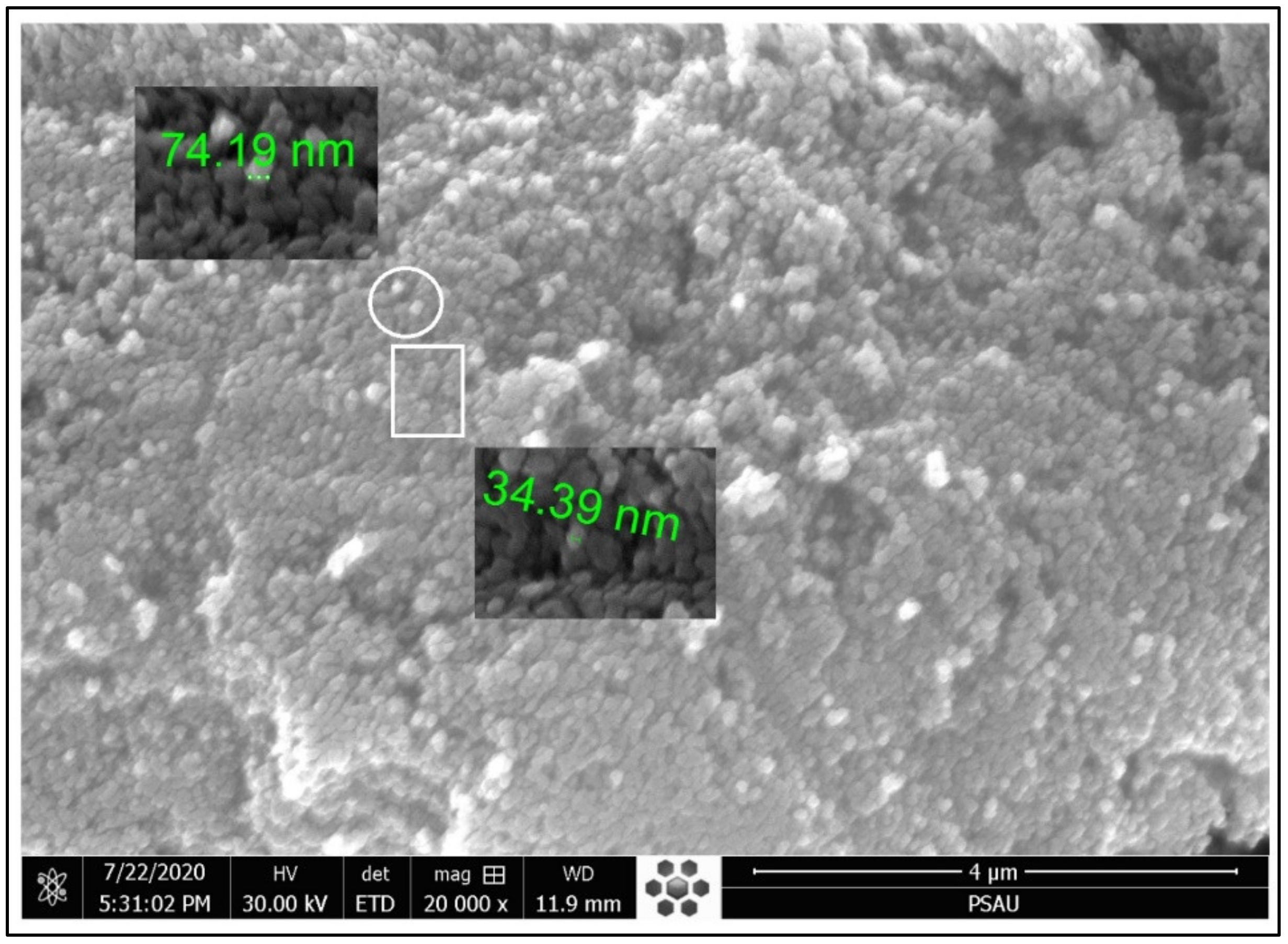
A field emission scanning electron microscope (FE-SEM) is used to observe the apparent morphology of the as-synthesized TiO
2-NPs. The obtained information about the structure or morphology of the as-synthesized TiO
2-NPs is shown in
Figure 1. All the TiO
2-NPs seem like small spheres (nanospheres). The micrograph shows that almost all of the TiO
2-NPs are in the nanometer range. At the bottom, most of the smaller size particles are closely packed alongside others. The smaller the size of the particle, the more at the bottom it lies followed by a bit bigger, etc. In contrast, at the top, the larger size particles are mostly seemed isolated from one another. It is an indication of the top-down approach followed during the synthesis of TiO
2-NPs. Nanoparticles of different sizes (smaller and bigger) can be seen and observed in the current micrograph. Thus, the biggest particles in the sample are placed at the top followed by the smaller and smallest at the bottom. This observation further verifying the top-down approach followed during the synthesis of TiO
2-NPs. The FE-SEM results show that the average particle size is below 100 nm.
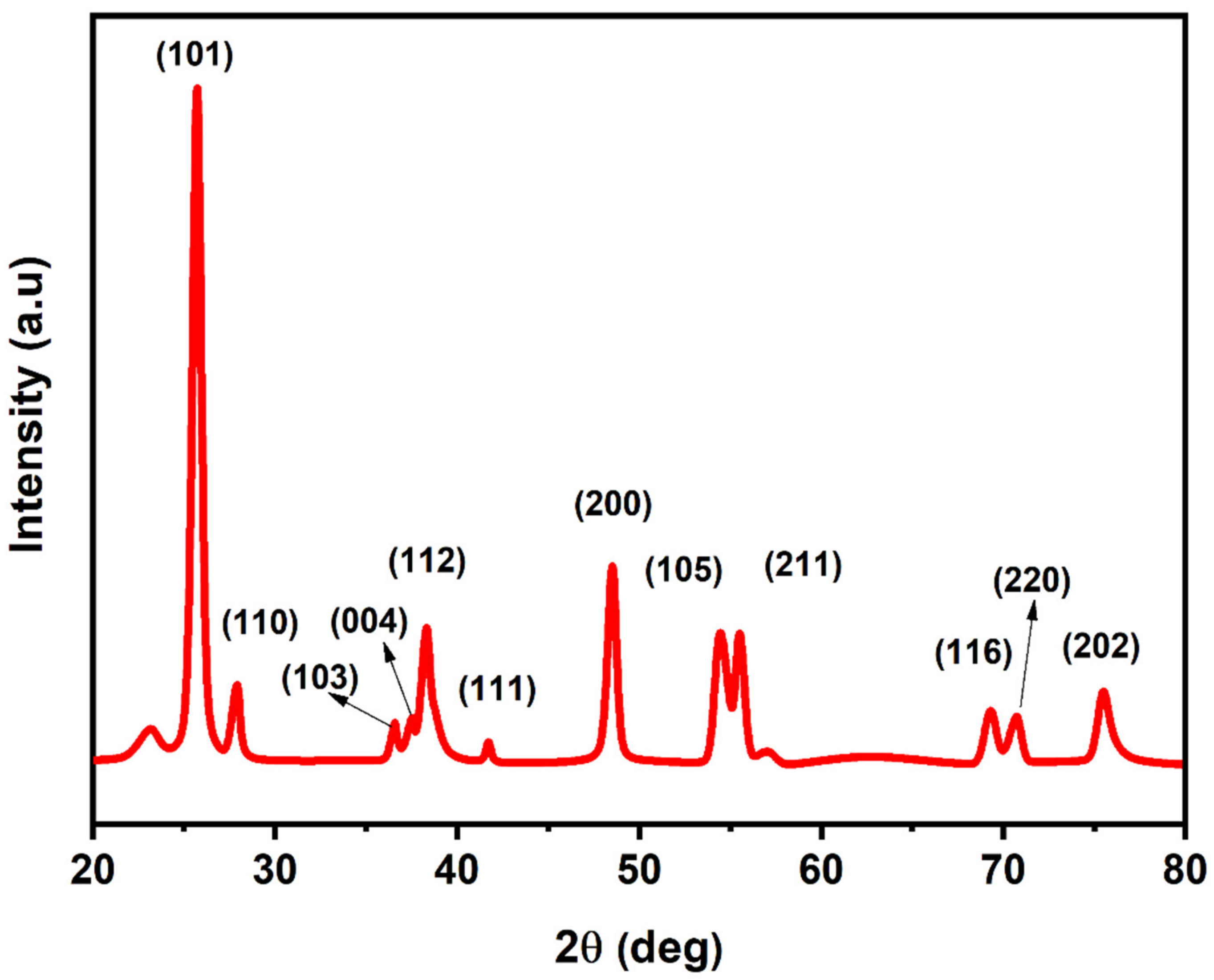
X-ray diffraction pattern of the as-synthesized TiO
2-NPs is shown in
Figure 2. It is an analytical technique used to identify the crystallinity and phase formation of TiO
2-NPs. The XRD study was conducted through the X-ray diffractometer at 298 K by using a nickel-filtered Cu α radiations source (K = 1.5418) at 298 K. The intensity data was attained over a 2θ range of 10–80°. The diffraction peaks were observed at the diffraction angle of 23.17°, 25.71°, 27.92°, 36.57°, 37.53°, 38.32°, 41.71°, 48.52°, 54.46°, 55.51°, 57.09°, 69.30°, 70.71°, and 75.48°, respectively. The crystalline phases of TiO
2-NPs were identified by X’Pert high score by comparing it with the standard data JCPDS card no. 01-075-1537 (Anatase) and 01-082-0514 (Rutile). Accordingly, all the peaks correspond to (101), (110), (103), (004), (112), (111), (200), (105), (211), (220), (116), (220), and (202) planes in the as-synthesized TiO
2. Debye Scherrer equation was used to measure the crystallite size of the synthesized nanoparticles with the help of XRD data of all the prominent peaks according to which the average crystallite size of TiO
2-NPs was 20 nm.
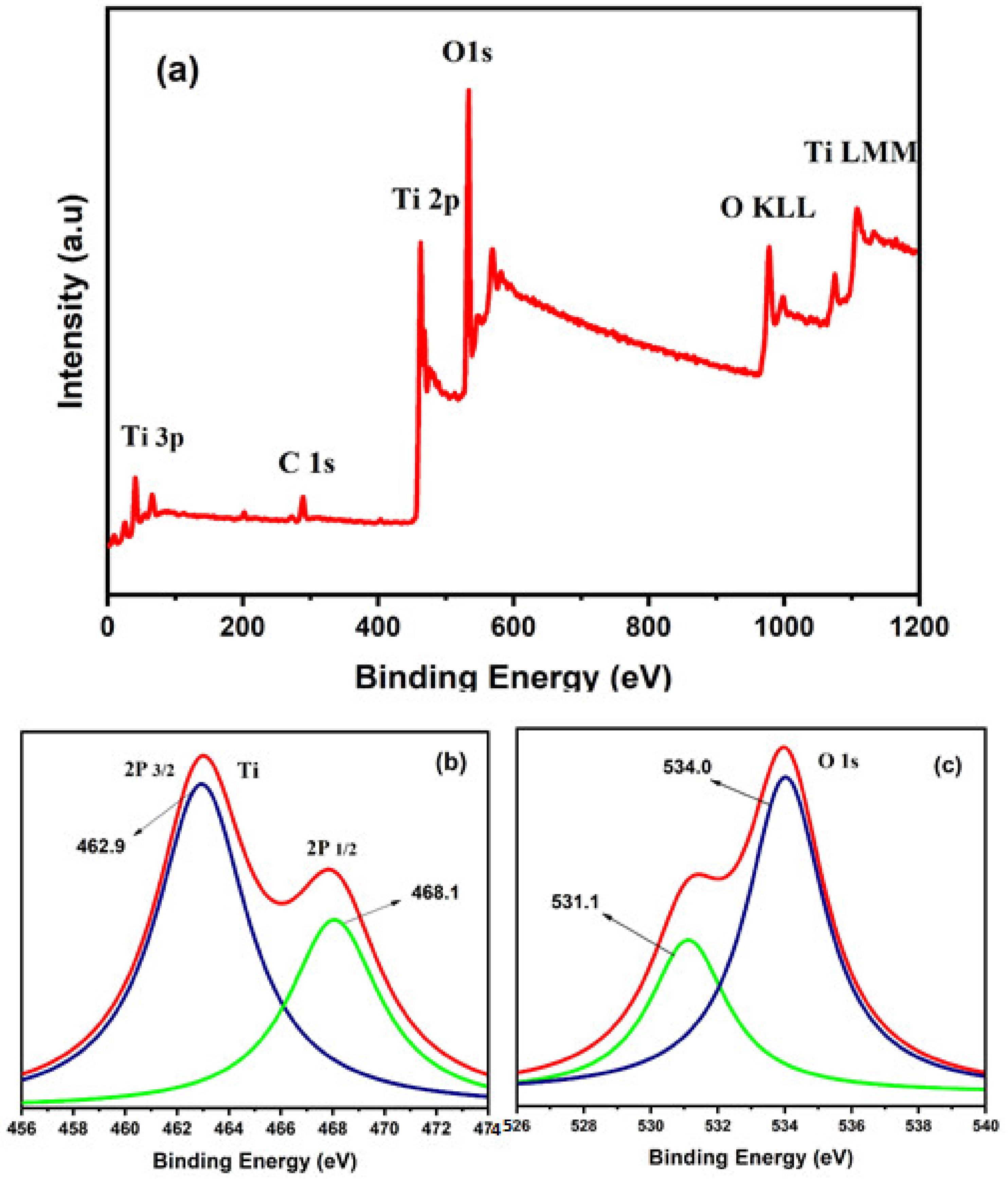
Bonding properties, valence states, and quantitative analysis of the electronic structures of the synthesized TiO
2-NPs were performed by X-ray photoelectron spectroscopy (XPS). In the XPS analysis, Ti 2p and O 1s are observed and no other precursor elements or magnetic contamination are found as shown in
Figure 3. Moreover, the hydrocarbon contamination (C 1s) peak is detected at approximately 285 eV [
43]. The XPS survey with the peaks tagged for the corresponding elements for their binding energies is shown in
Figure 3a. Two sharp peaks are observed in
Figure 3b, which corresponded to the spin-orbit of Ti 2p
3/2 and Ti 2p
1/2 at binding energies of 463 eV and 468 eV for Ti
4+ in TiO
2. No obvious peak shift is observed. This shows that Ti atoms exist in the same oxidation state [
44]. The magnified O 1 s peak is shown in
Figure 3c. The peak observed at 533 eV is interpreted to be due to bulk oxygen in the TiO
2 [
45]. The full width at half maximum (FWHM) is approximately similar in all spectra (1.35 eV). In
Figure 3a, escorting to the higher binding energy side of the peak involves many components resulting from TiO
2 surface hydroxylation (when it is mounted the electrode is exposed to air). These are attributable to both acidic and fundamental hydroxyl groups and absorbed water at the outermost surface. We observe the O 1s spectrum similar to those recorded on untreated (as grown) single crystal of anatase [
46] and rutile exposed to vapor and liquid water [
47,
48].
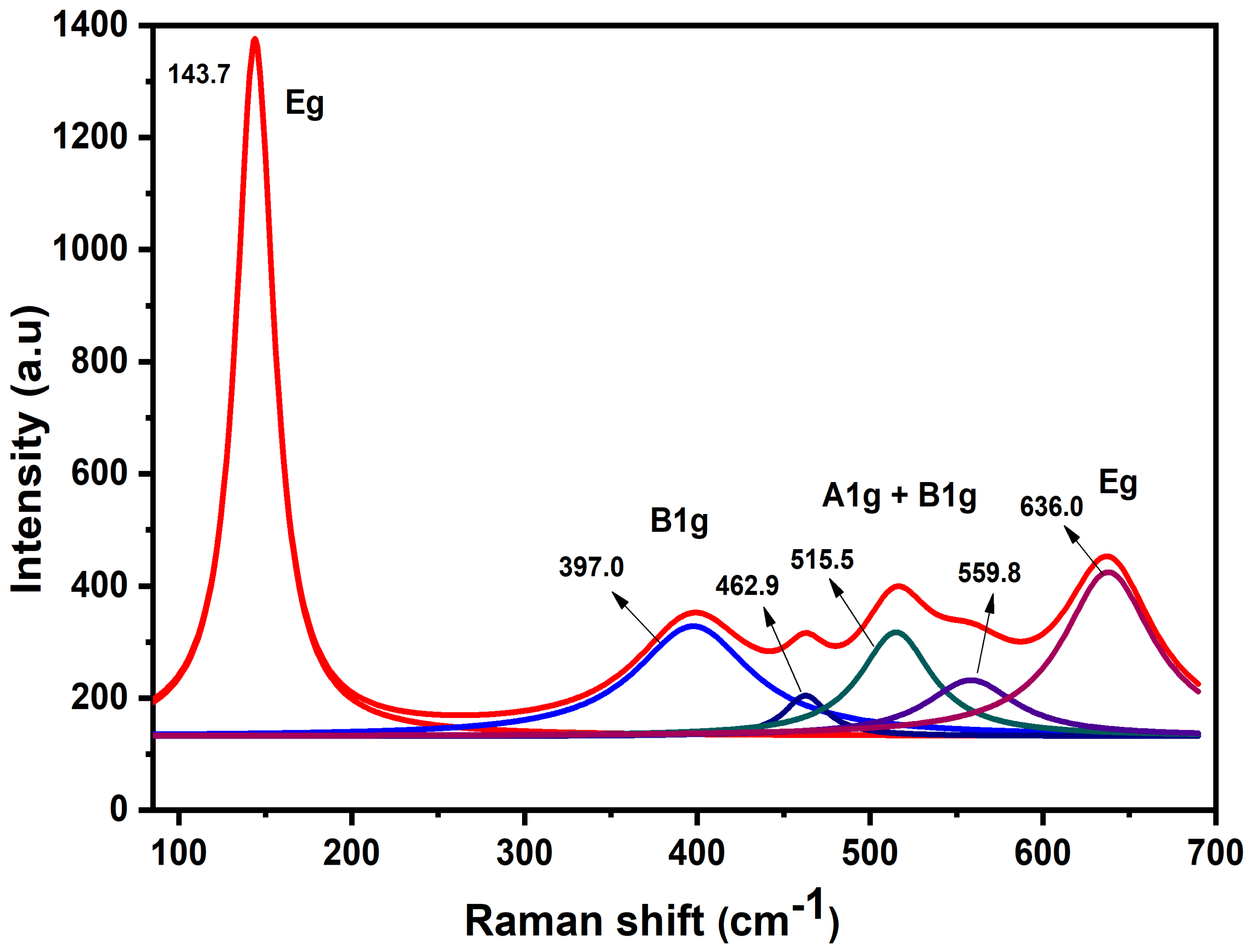
Figure 4 shows the Raman spectrum of the as-synthesized TiO
2-NPs. The intense E
g mode appears at 143.7 cm
−1 [
49]. B
1g, (A
1g + B
1g) and E
g peaks appear at 399 cm
−1, 516 cm
−1, and 637 cm
–1 respectively. In TiO
2 vibrational modes are due to the symmetric, asymmetric, and bending vibration of the Ti–O–Ti bond [
50,
51]. The peak tagged by Eg stands for Ti–O stretching mode, whereas the peaks located at 516 cm
–1 and 399 cm
–1 modes refer to Ti–O stretching mode (the only O is moving) and O–Ti–O bending mode (Ti is moving) [
51,
52]. Phonons near the center of the Brillouin zone (BZ) contribute to the inelastic dispersion of incident radiations only in a perfect infinite crystal. A greater part of the BZ will efficiently participate in scattering processes as the size of the crystal vary in the nanoscale due to the weakening of the selection rule at q
0 ≈ 0. Thus, a change in the Raman frequency peaks and the shape of the Raman band can be observed [
53].
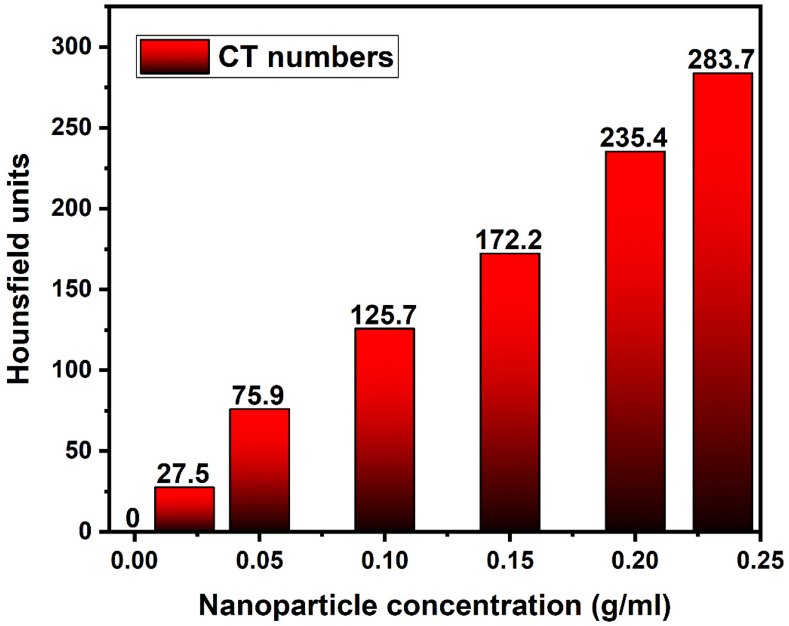
Figure 5 indicates the CT numbers of the different concentrations of TiO
2-NPs that were scanned. With the addition of NPs concentration, the CT numbers of the solutions improve roughly linearly. The uncertainty of the measurements in the Hounsfield unit (HU) is given by the standard deviation. The CT numbers were ranging from 0 ± 3 HU (without NPs) to 283.7 ± 3 HU (0.235 g/mL). A value of (27.5 ± 3, 75.9 ± 3, 125.7 ± 3, 172.2 ± 3, 235.4 ± 3, and 283.7 ± 3) HU is given by a (0.02, 0.05, 0.1, 0.15, 0.2, 0.235) g/mL concentration of NPs. This concentration is higher than the noise value typically reported by most CT scanners of about 5 HU [
37]. These findings show that the existence of TiO
2-NPs even at low concentrations induces observable variations in the scanned media that can be observed on a traditional CT scanner.
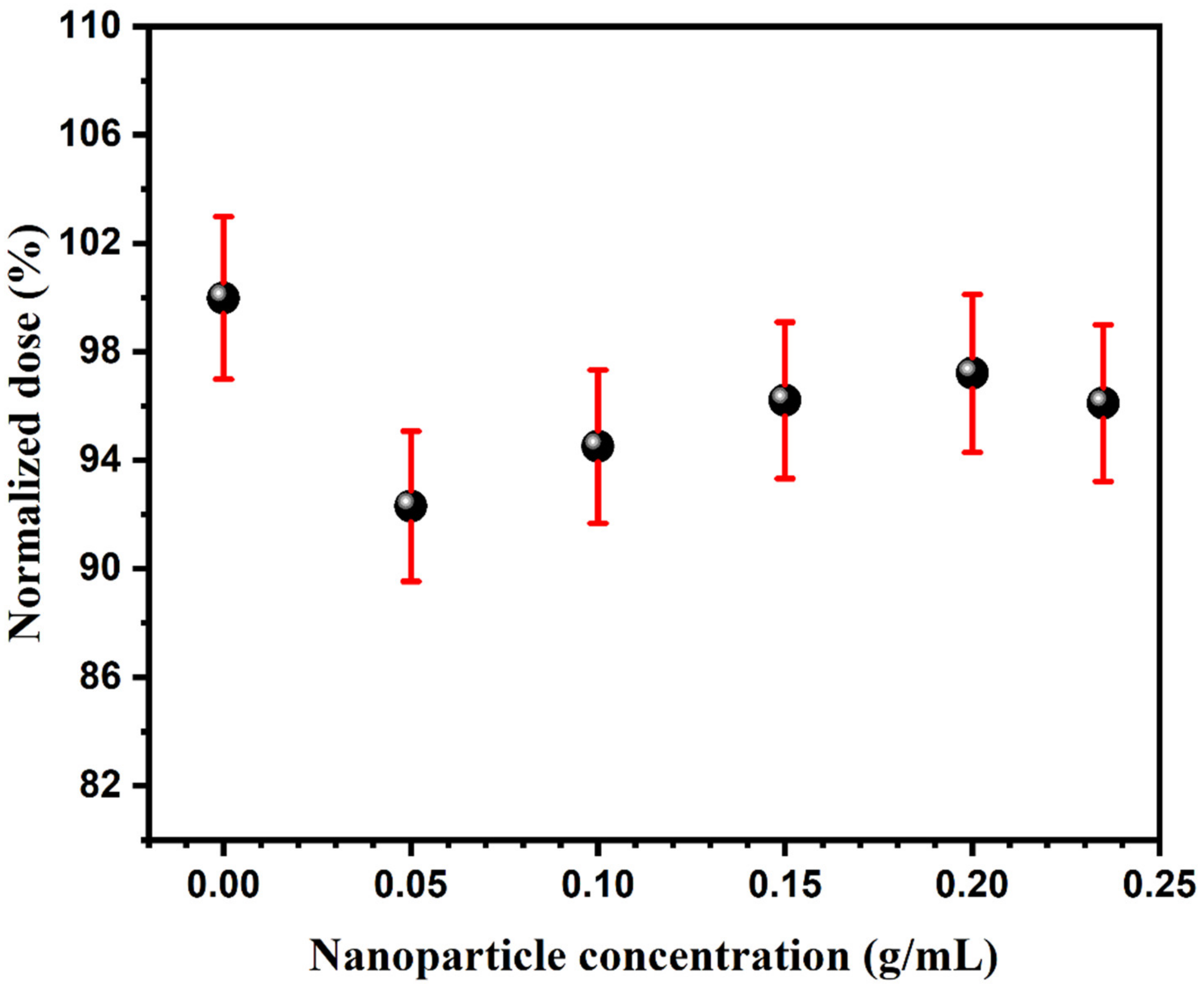
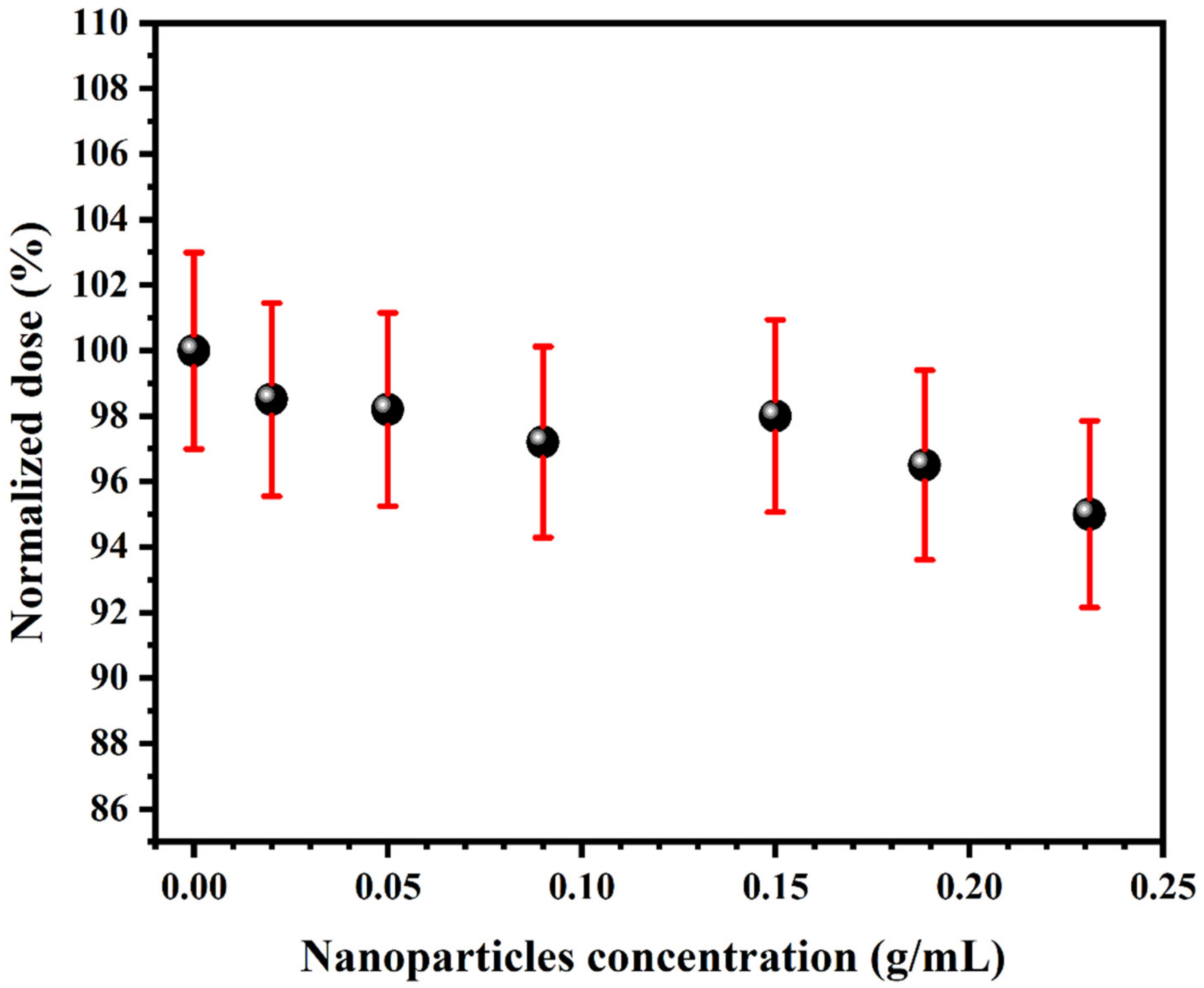
In
Figure 6 and
Figure 7, the surface doses are reported as a function of NPs concentration by the EBT2 film pieces. The enhancement is not observed at surface dose for both energies. The measured dose is maximum in both cases only for the solution having no NPs. The measured dose at 50 kVp falls approximately linearly with the concentration of NPs. Changes found during measurements of dose for all concentrations of NPs within 4% of the measurement error for a beam energy of 125 kVp. For the vial that does not contain nanoparticles, none of the obtained values reflects more than 97% of the dose measured. The decrease in surface dose at higher concentrations of NPs can be associated with the absorption of backscattered radiation by the solution of NPs near the surface without a sequential increase in the dose to compensate for this.


 ,
, 


