1. Introduction
Severe acute respiratory syndrome coronavirus 2 (SARS-CoV-2), the causal agent of the coronavirus disease 19 (COVID-19), has infected more than 95 million people [
1], at the time of this writing. Never before in contemporary history has humankind faced an infectious disease at this scale. In response to this unprecedented challenge, a vast number of methods to screen for SARS-CoV-2 infection have been proposed [
2]. These diagnostic methods are based on the identification of SARS-CoV-2 antigens, the amplification of SARS-CoV-2 nucleic acids [
3,
4], or the detection of anti-SARS-CoV-2 antibodies [
5]. Antigen-based methods are particularly useful for massive screening efforts: their cost is relatively low, they are easily implementable and portable, and their specificity is high (~100%); however, their sensitivity is often lower than that of amplification-based methods (~30–81%) [
6,
7,
8]. Amplification-based methods are highly accurate (sensitivities above 95% and specificities above 85%); indeed, quantitative versions of the reverse-transcription and polymerase chain reaction (RT-qPCR) amplification methods are the gold standard for diagnostics for SARS-CoV-2 infection [
2]. However, the feasibility of massive testing using RT-qPCR has been seriously challenged by COVID-19. Alternative amplification methods (strategies based mainly on Loop-mediated amplification methods (LAMP)) may enable accurate and massive SARS-CoV-2 detection (even from saliva) during the first 11 days of infection [
9,
10]. The methods based on the detection of anti-SARS-CoV-2 antibodies in blood (or serological methods) are poorly suited for diagnosis of infection during the first two weeks [
11]. However, they are precisely suitable for evaluating the immune reaction to SARS-CoV-2 infection in a subject (whether symptomatic or asymptomatic) after 14 days of infection.
Massive worldwide serological testing is needed to determine the relevant epidemiological indicators related to COVID-19 infection, including the extent of the exposure, the ratio of symptomatic to asymptomatic infected persons, and the duration and extent of immunity after infection [
12,
13,
14,
15]. Moreover, as vaccines are developed, tested in animal models and humans, and applied to open populations, we will depend on assays for reliable and quantitative characterization of the immune responses associated with the administration of a vaccine to determine the level of immunization conferred [
12,
16].
Fortunately, the time kinetics of the various immunoglobulins (IgAs, IgMs, and IgGs) produced against SARS-CoV-2 in COVID-19 patients has been well described in recent reports [
17]. For instance, we know that the determination of IgGs 15 days after viral exposure is a good indicator of a previous infection. Several semi-automated serological assays are commercially available to determine the likelihood of infection [
11,
18,
19]. Most established commercial platforms perform well, in terms of accurate prediction of infection in convalescent patients, when the analysis is performed 15 days (or more) after a possible contact [
19]. However, despite this reliability, automated serological platforms are expensive when compared to other techniques, such as regular enzyme-linked immunoassays (ELISAs).
ELISAs continue to be the most reliable and widely used method for characterization of the amount of antibodies developed against a specific antigen [
19,
20]. Laboratories around the world, and particularly in develo** countries, depend on traditional ELISAs to conduct widespread serological testing. Therefore, reliable and cost-effective antigens for ELISA testing are greatly needed.
In the context of COVID-19 research, a limited number of reports have been published on the development and characterization of SARS-CoV-2 antigens for ELISAs [
21,
22,
23,
24]. The spike protein (S) [
25,
26] and the nucleocapsid protein (N) [
27] of SARS-CoV-2 have been used for COVID-19 serological diagnostics [
22]. However, only a few detailed reports have been made available on the characterization of ELISAs for the identification of anti-SARS-CoV-2 antibodies [
5,
15,
21,
22]. Most of these reports describe transient mammalian cell expression [
21,
23,
28] of the entire spike protein of SARS-CoV-2, or a fraction of the spike protein containing the RBD or receptor-binding domain [
28].
Here, we report the production of an antigen inspired by the structure of the receptor-binding domain (RBD) [
28] of the spike protein of SARS-CoV-2. This antigen is produced by a bacterial culture of
Escherichia coli, which enables massive production at low cost [
29]. In addition, we characterize and contrast the performance of two ELISA versions, involving (a) direct attachment of the antigen to the surface of plates or (b) the use of a bed of anti-histidine antibodies (anti-his-mediated ELISA) to engineer the reactive surface.
2. Materials and Methods
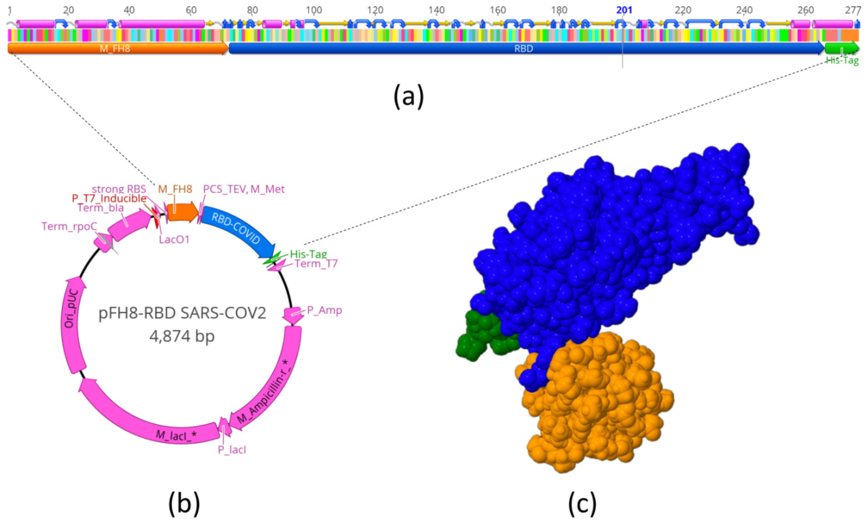
2.1. Design of S-RBDN318-V510 and Prediction of Its 3D Structure
We used the Geneious 11.1.5 software (Biomatters, Ltd., Auckland, New Zealand) to design the vector pFH8-RBD SARS-CoV-2 that contained the RBD (region of 193 aa from N318 to V510) of the SARS-CoV-2 spike protein. The construct also contained a region for the expression of M FH8 [
30], an enterokinase restriction site, as well as a histidine tag (his-tag). Histidine and FH8 tags were added for use in the purification process. The histidine tag (his-tag) provides an additional handle for separation using his-tag affinity columns (loaded with divalent ions such as Ni
+2). In addition, antigenic proteins containing histidine tags can be fixed to surfaces through anti-histidine antibodies to enable ELISA serological assays using his-tagged antigens [
29,
31].
The 3D structure of the RBD protein with tags was predicted using the software I-TASSER server (University of Michigan,Ann Arbor, MI, USA).
Figure 1a schematically shows the sequence that we used to encode and produce the MFH8-RBD
Spike-HisTag protein (S-RBD
N318-V510 for short). This expression cassette was inserted in a plasmid for expression in
E. coli.
Figure 1c shows the molecular 3D structure of this product, as predicted by molecular structure simulations.
2.2. Cloning and Transformation
The full spike coding sequence was synthesized by GenScript (Piscataway, NJ, USA) and was used to obtain the sequences comprising the RBD. This sequence was cloned in an expression vector (ATUM, Newark, CA, USA) regulated by a T7 promoter (IPTG-inducible) using a SapI restriction site and T4 DNA ligase (New England Biolabs, Ipswich, MA, USA). The expression vector was transformed into chemical-competent E. coli BL21 strain C41 cells (Lucigen Corporation, Middleton, WI, USA) to obtain producer clones. High-producer clones were further cultured using Luria–Bertani (LB) medium in Erlenmeyer flasks, and recombinant expression was induced using isopropyl β-d-1-thiogalactopyranoside (IPTG).
2.3. RBD Production in Erlenmeyer Flasks
The highest-producer clone was selected and cultured in Luria–Bertani broth containing 50 µg/mL ampicillin (LB-Amp) in 2 L Erlenmeyer flasks. For the initial growth, 200 mL of LB-Amp broth was maintained overnight at 37 °C with 250 rpm agitation in an orbital shaker (VWR International, Radnor, PA, USA). After 12 h of culture, cells were harvested using a Z36 HK centrifuge (Hermle Labortechnik, Wehingen, Germany) at 5000× g for 10 min. The cell pellet was then resuspended in fresh LB-Amp broth containing 1 mM isopropyl β-d-1-thiogalactopyranoside (IPTG) to induce RBD production. The induction was conducted at 30 °C with agitation at 250 rpm for 8–12 h. After induction, cells were recovered by centrifugation at 5000× g for 10 min at 4 °C. Cell pellets were kept at −20 °C until further processing.
2.4. S-RBDN318-V510 Recovery and Purification
Pellets from IPTG-induced cells were re-suspended in phosphate-buffered saline (PBS) (pH = 7.4) containing 100 mM NaCl in a proportion of 7.5 mL per gram of cells (wet weight). The cells were then disrupted in an EmulsiFlex-C3 high-pressure homogenizer (Avestin, Ottawa, ON, Canada). The process comprised three cycles, with the first cycle set to reach 5000 psi and the following two cycles performed at 20,000 psi. Cell lysates were centrifuged at 15,000× g for 30 min at 4 °C in a Z36 HK centrifuge. The pellet containing the inclusion body (IB) was re-suspended in IB wash buffer (PBS, pH = 7.4, 1mM EDTA, 500 mM NaCl, 2 M urea, and 2% Triton X-100) at a ratio of 25 mL per g of IB pellet (wet weight), centrifuging to recover the pellet, washing with PBS, and re-suspending in IB solubilization buffer (PBS, pH = 8.0, 500 mM NaCl, 8 M Urea, 2.5 mM 2-mercaptoethanol, and 10 mM imidazole).
The S-RBDN318-V510 protein was purified by immobilized metal-affinity chromatography (IMAC), using a HiTrap™ column (GE Healthcare, UK) packed with 1 mL Ni2+ charged agarose Ni-NTA Superflow (Quiagen, Germany) in an Akta Pure system (GE Healthcare, Chicago, IL, USA) chromatography system. The degree of purity of S-RBDN318-V510 was estimated from SDS-PAGE protein profiles using Image J, an open source software for scanning densitometry analysis.
A dual-phase separation strategy was implemented. Phase A consisted of 20 mM PBS, 300 mM NaCl, and 20 mM imidazole, pH = 7.4, and phase B was 20 mM PBS, pH = 7.4, 300 mM NaCl, and 300 mM imidazole at pH = 7.4. The purification protocol was set with an initial equilibrium of 10 column volumes (CV) of phase A and a flow rate of 1 mL/min. A 5 mL sample of protein was injected at a flow rate of 0.5 mL/min. After sample injection, a washing step of 8 CV was set at a flow rate of 1 mL/min, followed by elution with 3 CV of a linear gradient from 0% to 80% of phase B, then 10 CV of 20%/80% phase A/B, and finally 5 CV of 100% phase B, all at a flow rate of 1 mL/min. Finally, to prepare for further purifications, the column was re-equilibrated with 5 CV of phase A at 1 mL/min. The fraction containing the protein of interest was recovered based on the chromatogram and stored at 4 °C.
2.5. ELISA Assays
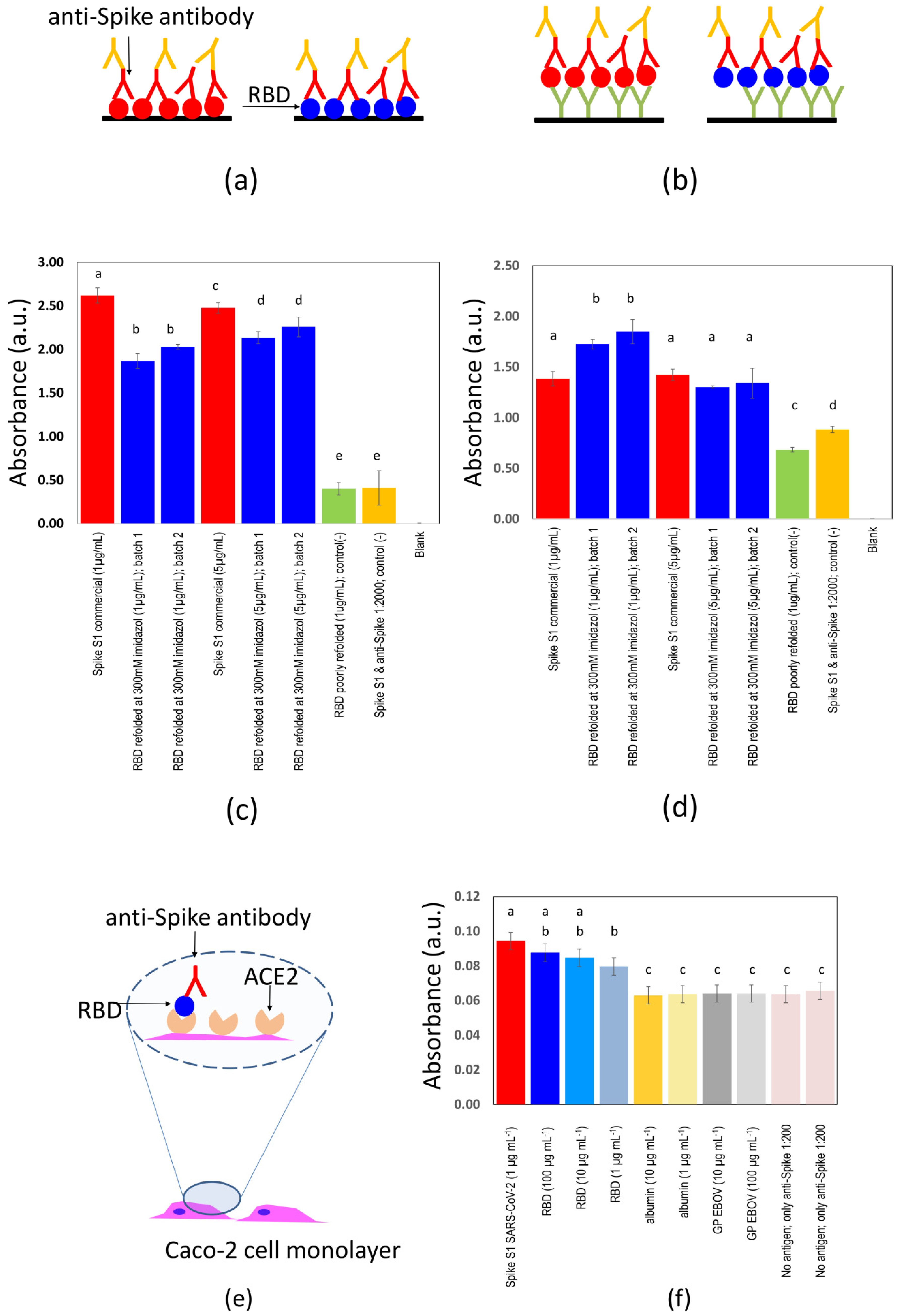
We developed and characterized two ELISA strategies/formats for the evaluation of presence of specific anti-SARS-CoV-2 antibodies (
Figure 2a,b). Standard commercial 96-well micro-assay plates (CorningH, Maxisorp™; Waltham, MA, USA) were used.
In the first format, 100 µL of 1 µg/mL RBD in PBS was dispensed into each well and incubated for 8 h at 4 °C, followed by addition of 100 µL 5% skim milk in PBS and further incubation for 1 h at room temperature, and then three washes with PBS containing 0.05% Tween-20TM. Rabbit anti-spike-SARS-CoV-2 pAb (100 µL; 1:2000 dilution; Sino Biological Inc., Wayne, PA, USA) was then added incubated for 1 h at room temperature, followed by three washes with PBS containing 0.05% Tween-20TM. The presence of rabbit antibodies was revealed by adding donkey anti-rabbit-HRP (100 µL, 1:5000 dilution; Pierce, Rockford, IL, USA), followed by three washes with PBS containing 0.05% Tween-20TM.
The horseradish peroxidase (HRP) was then detected by adding 100 µL 1-StepTM Ultra TMB-ELISA (Pierce, Rockford IL, USA) until a blue color was observed. The reaction was stopped by adding 100 µL 1M H2SO4, and the absorbance was measured at 450 nm in a BioTek microplate reader (Winooski, VT, US).
The second ELISA format consisted of first sensitizing the plate wells with mouse anti-histidine pAb (100 µL, 1:1000 dilution; Bio-Rad Laboratories, Inc., Hercules, CA, USA) and incubating for 8 h at 4 °C, then blocking with skim milk for 1 h at room temperature, followed by three washes with PBS containing 0.05% Tween-20TM. The plates were then incubated with RBD for 1 h at room temperature. All subsequent steps were as described for the first ELISA format.
2.6. ELISA Testing of Serum Samples
We performed ELISA experiments using samples of sera from non-exposed individuals and convalescent, positive volunteers. Five samples of sera from COVID-19 non-exposed individuals were collected from volunteers at Hospital San José (Nuevo León, México), from June 2009 to October 2009, during pandemic Influenza A/H1N1/2009. Fifty-five samples of sera from convalescent patients previously confirmed as COVID-19 (+) by RT-qPCR were collected at Alfa Medical S.A. de C.V. (Monterrey, N.L., México). Samples were collected from patients after obtaining informed and signed written consent and in complete observance of good clinical practices, the principles stated in the Helsinki Declaration, and applicable lab operating procedures at Hospital Alfa. Every precaution was taken to protect the privacy of sample donors and the confidentiality of their personal information. The experimental protocol was approved on 20 May 2020 by a named institutional committee (Alfa Medical Center, Research Committee; resolution AMCCI-TECCOVID-001).
Two different ELISA strategies were contrasted. In the first format (direct ELISA), 100 µL of 1 µg/mL RBD in PBS was dispensed into each well of 96-well plates and incubated for 8 h at 4 °C, followed by blocking with 100 µL 5% skim milk in PBS and incubation for 1 h at room temperature, and three washes with PBS containing 0.05% Tween-20TM. Different dilutions (1:5, 1:50, 1:100, and 1:200; 100 µL) of serum from volunteers were added per well, incubated for 1 h at room temperature and then washed three times with PBS containing 0.05% Tween-20TM. The best results were observed when 1:100 dilutions were used. The presence of human IgG was detected by adding goat anti-human IgG HRP (100 µL; 1:10,000 dilution; Pierce Biotechnology Inc., IL, USA) and incubating for 1 h at room temperature, followed by three washes with PBS containing 0.05% Tween-20TM and detection with 1-StepTM Ultra TMB-ELISA.
In the second format (sandwich ELISA), RBD (100 µL; 1 µg/mL in PBS) was dispensed in each well and incubated for 1 h at room temperature followed by three washes with PBS containing 0.05% Tween-20TM. Then 100 µL of different dilutions (1:5, 1:50, 1:100, and 1:200) of serum from volunteers were added per well, incubated for 1 h at room temperature and subsequently washed three times with PBS containing 0.05% Tween-20TM. The presence of human IgG was again detected with goat anti-human IgG HRP, and the remaining steps were conducted as described for the first ELISA format.
2.7. Binding between S-RBDN318-V510 Protein and the ACE2 Receptor in Caco-2 Cells
We conducted experiments in Caco-2 cells (ATCC® HTB-37) to assess the specific binding between S-RBDN318-V510 and the angiotensin-converting enzyme 2 (ACE2). Toward that aim, we established monolayer cultures of Caco-2 cells in 96-well plates. At confluence, culture medium was removed, and cells were covered with paraformaldehyde, incubated for 10 minutes at room temperature for fixation, added with 100 µL 5% skim milk in PBS, further incubated for 1 h at room temperature, and washed three times with PBS containing 0.05% Tween-20TM. Subsequently, 100 µL of a solution of RBD in PBS were added to the fixed cell monolayer. Three different RBD concentrations were assayed (i.e., 1, 10, and 100 µg/mL). Albumin and glycoprotein (GP) from the Ebola virus were used as negative controls. All proteins were incubated on the Caco-2 monolayer for 1 h at room temperature and then washed three times with PBS containing 0.05% Tween-20TM. A rabbit anti-spike-SARS-CoV-2 pAb (100 µL; 1:1000 dilution; Sino Biological Inc., PA, USA) was then added and incubated for 1 h at room temperature, followed by three washes with PBS containing 0.05% Tween-20TM. The presence of rabbit antibodies was revealed by adding donkey anti-rabbit HRP (100 µL, 1:10,000 dilution; Pierce, Rockford, IL, USA), followed by three washes with PBS containing 0.05% Tween-20TM. HRP was detected by adding 100 µL 1-StepTM Ultra TMB-ELISA (Pierce, Rockford, IL, USA) until blue was observed. The reaction was stopped by adding 100 µL 1M H2SO4, and the absorbance was measured at 450 nm in a BioTek microplate reader (VT, US).
2.8. Antigenicity Assessment of Peptide S-RBDN318-V510 in Mice
We explored the use of peptide S-RBDN318-V510 as an antigen in a reduced set of in vivo experiments in mice. We subcutaneously injected six mice with 20 µg of the purified peptide S-RBDN318-V510 dissolved in HEPES. Three mice was used as a negative control (i.e., injected with only HEPES). We collected blood from the tails of our animals 14 days after injection and then conducted ELISA experiments using the scheme of ELISAs mediated by the use of a layer of anti-histidine antibodies.
This experiment complied with the 3Rs (replacement, reduction, and refinement) in animal model research. The experimental scheme was approved by the Research and Ethics Committee of Alpha Medical Center, at Nuevo-León, México (Approval AMCCI-TECOVID-001; 12 May 2020). To honor the lives of these animals, their sera were used not only to establish the efficacy of the peptide S-RBDN318-V510 as an antigen for serological testing but also its potential as an ingredient for vaccines. These results will be presented in a follow-up report.
4. Discussion
Arguably, antigens are one of the most important reagents that clinicians will require while facing COVID-19 pandemics in the months to come. Complete COVID-19 ELISA kits are commercially available, but their cost (approximately 8 USD per reaction, per well) still limits massive implementation, particularly in develo** economies. The current value of commercially available S1-derived SARS-CoV-2 antigens is approximately 7 USD µg−1, which is also prohibitive for most laboratories for large-scale screening of COVID-19-seropositive subjects.
Here, we report methods for the production of a portion of the S1 fraction of the SARS-CoV-2 spike protein that contains the receptor-binding domain for the angiotensin II human receptor. We engineered an expression construct for the recombinant production of the RBD of the S protein of SARS-CoV-2. Specifically, we selected the region of the S-RBD between the residues N318 and V510 of the consensus sequence of the S protein of SARS-CoV-2. In a recent report, a similar fraction of the SARS-CoV-2 spike protein (from residues 331 to 510) containing the RBD has been transiently expressed in HEK293 cells [
28]. That peptide successfully recognized the ACE receptor, the native target of the RBD of the spike protein [
28].
We chose Escherichia coli as an expression host, and we describe a straightforward process, amenable to widespread implementation, for the production and purification of the S-RBDN318-V510 protein. The fact that our antigen is expressed in bacterial systems greatly facilitates its production and paves the way to scaling up. We show that antigen production of 1.5 mg per L is feasible, even when using non-agitated Erlenmeyer flasks and non-instrumented bioreactors. This production level is already attractive, since production can be completed in 24 h.
However, the use of a bacterial system to express an effective antigen is not always feasible. Bacterial systems lack the capacity to conduct post-translational modifications (e.g., glycosylation), and glycosylation is often required for functionality. Therefore, recombinant proteins produced in bacterial systems may exhibit lower functionality than analogous proteins produced in eukaryotic systems (e.g., CHO or HEK cells).
Here, we investigate whether the S-RBDN318-V510 protein, when produced in a bacterial system, still retains its ability to bind anti-RBD antibodies. Our results suggest that this is the case; S-RBDN318-V510 performs satisfactorily as an antigen in ELISA assays when compared with commercial versions of the spike protein.
From these results, however, it should not be directly inferred that protein S-RBD
N318-V510 retains its capability for effective binding to the ACE2 receptor. We conducted experiments in which we exposed Caco-2 cells (a cell line we use routinely in our lab that natively possesses ACE2) to S-RBD
N318-V510. In parallel experiments, we exposed Caco-2 cells to other proteins (i.e., albumin, a recombinant version of the GP protein from the Ebola virus) to investigate for unspecific attachment. The results of these experiments suggest the specific attachment of S-RBD
N318-V510 to the ACE2 receptor in Caco-2 cells (
Figure 2e,f).
The binding of the S-RBDN318-V510 protein to Caco-2 surfaces was significantly higher than that of the anti-spike pAbs to Caco-2 surfaces. Moreover, the binding of S-RBDN318-V510 to Caco-2 cells was not dose dependent in the window of concentrations of our experiments (i.e., 1 to 100 µg mL−1). However, the binding of the full-length spike protein to Caco-2 cells is higher than that of S-RBDN318-V510 at the lowest concentration tested (i.e., 1 µg mL−1). At higher concentrations of S-RBDN318-V510, that is, 10- or 100-fold higher, the binding to Caco-2 cells equals that exhibited by the full-length spike. Altogether, this suggests that the S-RBDN318-V510 produced in bacteria retains its ability to bind ACE2 but with a lower affinity than the full-length spike expressed in eukaryotic cells.
We describe two different ELISA formats in our experiments (
Figure 2a,b). In a direct format, the S-RBD
N318-V510 protein is directly attached to the surface of the ELISA plate. In a second format, a layer of anti-histidine antibodies mediates the binding between the plate surface and the antigen. In a series of ELISA experiments, we explored the type of ELISA format and the use of different concentrations of the protein S-RBD
N318-V510. In these ELISA experiments, we include two controls. The first negative control (green bars) consists of an antigen deficiently folded (in glycerol and PBS). The second negative control consists of the properly folded S-RBD
N318-V510 antigen but added to a highly diluted solution of commercial anti-spike 1 antibody (1:20,000) to simulate a negative sample. A blank control, where only PBS is added, was also included (absorbance reading ~0.0). The readings obtained from sandwich ELISAs are closer to those obtained when a commercial spike S1 protein is used as antigen. While we believe that the sandwich ELISA will produce similar results to those obtained when commercial antigens are used, the direct ELISA still allows discrimination between samples that contain or do not contain anti-spike antibodies and at a lower cost.
In experiments using human sera from patients possibly exposed to SARS-CoV-2, the results of both ELISA formats were highly consistent (
Figure 3a–e). As shown earlier, anti-his-mediated ELISAs yielded similar results, regardless of the use of the full-length spike protein or S-RBD
N318-V510. Therefore, we assumed that the anti-his-mediated results correlated well with ELISAs conducted with the full spike protein and can be taken as a reference for determining the sensibility and specificity of direct ELISAs performed using S-RBD
N318-V510. When this is done, the selectivity and specificity of the direct S-RBD
N318-V510 format are 97.2% and 52.0%, respectively, when a threshold value of normalized absorbance of 1.10 is used. If a threshold value of normalized absorbance of 1.25 is used instead, the values of selectivity and specificity are 97.2% and 68.0%, respectively. The overall accuracy of the direct S-RBD
N318-V510 ELISA test (i.e., the overall consistency of the results with respect to the anti-his S-RBD
N318-V510 ELISA) was 81.8% and 85.45% when the thresholds were set at 1.10 and 1.25, respectively. Overall, these results suggest that the anti-his S-RBD
N318-V510 ELISA is more consistent with full-length spike ELISAs. However, direct S-RBD
N318-V510 ELISA can be used in serological testing (further reducing the cost) with only a minimum sacrifice of selectivity but with an increased probability of false positives.
5. Conclusions
Here we report protocols for the recombinant production of the protein S-RBDN318-V510, a short peptide of ~33 kDa inspired by the receptor binding domain of the spike protein of SARS-CoV-2, in bacterial systems. We cloned the construct for the production of S-RBDN318-V510 in E. coli BL21 strain C41. Moreover, we demonstrate that a simple purification method, based on the use of his-tag-mediated affinity chromatography and refolding using imidazole solutions, leads to the production of a functional antigen that can be used to discriminate between samples that contain and do not contain anti-spike SARS-CoV-2 antibodies. In ELISA experiments using commercial anti-spike antibodies or actual sera from patients, this protein performs similarly to commercially available antigens based on the expression of larger segments of the spike protein.
Our aim was to enable the widespread use of this simple process to produce a cost-effective SARS-CoV-2 antigen. We believe that a lab-scale manufacturing operation based on the process described here may allow the production of gram amounts per month of an antigen of satisfactory quality to enable wide-scale screening projects in open populations.
This will be particularly useful in the present and near future. Massive serological testing is needed to determine the extent of the exposure to SARS-CoV-2 among regions, the overall ratio of symptomatic to asymptomatic infected persons (which has not been clearly established yet), and the duration and extent of immunity after infection. As the vaccination is deployed, we will also depend on anti-SARS-CoV-2 antibody assays for the reliable, scalable, and cost-effective quantification of the extent of immunity conferred to populations.

 ,
, 


