
Small Bowel Malignancies in Patients Undergoing Capsule Endoscopy for Iron Deficiency Anemia


Abstract
:1. Introduction
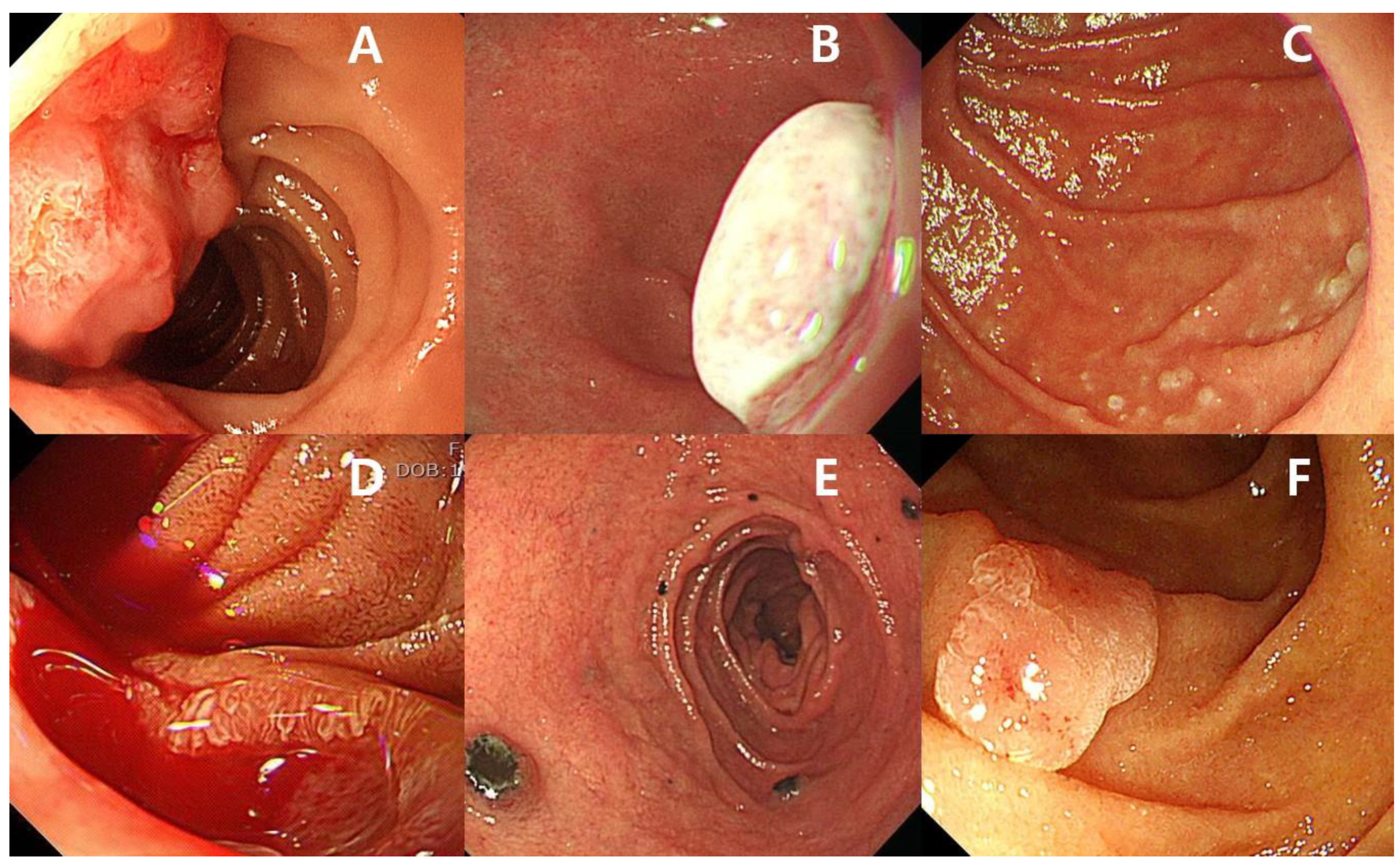
2. Small Bowel Malignancies
3. Gastrointestinal Tract Evaluation of Patients with Iron Deficiency Anemia
4. Role of Capsule Endoscopy in Patients with Iron Deficiency Anemia
5. Diagnostic Yield of Capsule Endoscopy in Patients with Iron Deficiency Anemia
6. Complications of Capsule Endoscopy
7. Conclusions
Author Contributions
Funding
Institutional Review Board Statement
Informed Consent Statement
Data Availability Statement
Conflicts of Interest
References
- Ohmiya, N. Management of Obscure Gastrointestinal Bleeding: Comparison of Guidelines between Japan and Other Countries. Dig. Endosc. 2020, 32, 204–218. [Google Scholar] [CrossRef] [Green Version]
- Teshima, C.W.; Kuipers, E.J.; van Zanten, S.V.; Mensink, P.B. Double Balloon Enteroscopy and Capsule Endoscopy for Obscure Gastrointestinal Bleeding: An Updated Meta-Analysis. J. Gastroenterol. Hepatol. 2011, 26, 796–801. [Google Scholar] [CrossRef]
- Hosoe, N.; Takabayashi, K.; Ogata, H.; Kanai, T. Capsule Endoscopy for Small-Intestinal Disorders: Current Status. Dig. Endosc. 2019, 31, 498–507. [Google Scholar] [CrossRef] [PubMed]
- Raju, G.S.; Gerson, L.; Das, A.; Lewis, B. American Gastroenterological Association (Aga) Institute Medical Position Statement on Obscure Gastrointestinal Bleeding. Gastroenterology 2007, 133, 1694–1696. [Google Scholar] [CrossRef] [PubMed]
- Raju, G.S.; Gerson, L.; Das, A.; Lewis, B. American Gastroenterological Association (Aga) Institute Technical Review on Obscure Gastrointestinal Bleeding. Gastroenterology 2007, 133, 1697–1717. [Google Scholar] [CrossRef] [PubMed] [Green Version]
- Pennazio, M.; Spada, C.; Eliakim, R.; Keuchel, M.; May, A.; Mulder, C.J.; Rondonotti, E.; Adler, S.N.; Albert, J.; Baltes, P.; et al. Small-Bowel Capsule Endoscopy and Device-Assisted Enteroscopy for Diagnosis and Treatment of Small-Bowel Disorders: European Society of Gastrointestinal Endoscopy (Esge) Clinical Guideline. Endoscopy 2015, 47, 352–376. [Google Scholar] [CrossRef] [Green Version]
- Rockey, D.C.; Altayar, O.; Falck-Ytter, Y.; Kalmaz, D. Aga Technical Review on Gastrointestinal Evaluation of Iron Deficiency Anemia. Gastroenterology 2020, 159, 1097–1119. [Google Scholar] [CrossRef] [PubMed]
- Lazaridis, L.D.; Tziatzios, G.; Toth, E.; Beaumont, H.; Dray, X.; Eliakim, R.; Ellul, P.; Fernandez-Urien, I.; Keuchel, M.; Panter, S.; et al. Implementation of European Society of Gastrointestinal Endoscopy (Esge) Recommendations for Small-Bowel Capsule Endoscopy into Clinical Practice: Results of an Official Esge Survey. Endoscopy 2021, 53, 970–980. [Google Scholar] [CrossRef]
- Islam, R.S.; Leighton, J.A.; Pasha, S.F. Evaluation and Management of Small-Bowel Tumors in the Era of Deep Enteroscopy. Gastrointest. Endosc. 2014, 79, 732–740. [Google Scholar] [CrossRef]
- Kim, E.R. Roles of Capsule Endoscopy and Device-Assisted Enteroscopy in the Diagnosis and Treatment of Small-Bowel Tumors. Clin. Endosc. 2020, 53, 410–416. [Google Scholar] [CrossRef]
- Ihara, Y.; Torisu, T.; Moriyama, T.; Umeno, J.; Hirano, A.; Okamoto, Y.; Hori, Y.; Yamamoto, H.; Kitazono, T.; Esaki, M. Endoscopic Features of Gastrointestinal Stromal Tumor in the Small Intestine. Intest. Res. 2019, 17, 398–403. [Google Scholar] [CrossRef]
- Yoo, A.Y.; Lee, B.J.; Kim, W.S.; Kim, S.M.; Kim, S.H.; Joo, M.K.; Kim, H.J.; Park, J.J. Clinicopathological Features of Small Bowel Tumors Diagnosed by Video Capsule Endoscopy and Balloon-Assisted Enteroscopy: A Single Center Experience. Clin. Endosc. 2021, 54, 85–91. [Google Scholar] [CrossRef]
- Pourmand, K.; Itzkowitz, S.H. Small Bowel Neoplasms and Polyps. Curr. Gastroenterol. Rep. 2016, 18, 23. [Google Scholar] [CrossRef]
- Paski, S.C.; Semrad, C.E. Small Bowel Tumors. Gastrointest. Endosc. Clin. N. Am. 2009, 19, 461–479. [Google Scholar] [CrossRef] [PubMed]
- Van Leerdam, M.E.; Roos, V.H.; van Hooft, J.E.; Dekker, E.; Jover, R.; Kaminski, M.F.; Latchford, A.; Neumann, H.; Pellisé, M.; Saurin, J.C.; et al. Endoscopic Management of Polyposis Syndromes: European Society of Gastrointestinal Endoscopy (Esge) Guideline. Endoscopy 2019, 51, 877–895. [Google Scholar] [CrossRef] [PubMed] [Green Version]
- Locher, C.; Batumona, B.; Afchain, P.; Carrère, N.; Samalin, E.; Cellier, C.; Aparicio, T.; Becouarn, Y.; Bedenne, L.; Michel, P.; et al. Small Bowel Adenocarcinoma: French Intergroup Clinical Practice Guidelines for Diagnosis, Treatments and Follow-up (Snfge, Ffcd, Gercor, Unicancer, Sfcd, Sfed, Sfro). Dig. Liver Dis. 2018, 50, 15–19. [Google Scholar] [CrossRef] [PubMed] [Green Version]
- Aparicio, T.; Zaanan, A.; Mary, F.; Afchain, P.; Manfredi, S.; Evans, T.R. Small Bowel Adenocarcinoma. Gastroenterol. Clin. N. Am. 2016, 45, 447–457. [Google Scholar] [CrossRef]
- Kastrinos, F.; Syngal, S. Inherited Colorectal Cancer Syndromes. Cancer J. 2011, 17, 405–415. [Google Scholar] [CrossRef] [Green Version]
- Genta, R.M.; Feagins, L.A. Advanced Precancerous Lesions in the Small Bowel Mucosa. Best Pract. Res. Clin. Gastroenterol. 2013, 27, 225–233. [Google Scholar] [CrossRef]
- Maguire, A.; Sheahan, K. Primary Small Bowel Adenomas and Adenocarcinomas-Recent Advances. Virchows Arch. 2018, 473, 265–273. [Google Scholar] [CrossRef]
- Sharaiha, R.Z.; Cohen, M.S.; Reimers, L.; Khashab, M.A.; Giardiello, F.M.; Neugut, A.I. Sporadic Duodenal Adenoma and Association with Colorectal Neoplasia: A Case-Control Study. Dig. Dis. Sci. 2014, 59, 2523–2528. [Google Scholar] [CrossRef] [PubMed]
- Chung, W.C.; Lee, B.I.; Roh, S.Y.; Kwak, J.W.; Hwang, S.M.; Ko, Y.H.; Oh, J.H.; Cho, H.; Chae, H.S.; Cho, Y.S. Increased Prevalence of Colorectal Neoplasia in Korean Patients with Sporadic Duodenal Adenomas: A Case-Control Study. Gut Liver 2011, 5, 432–436. [Google Scholar] [CrossRef] [PubMed] [Green Version]
- Dariusz, A.; Jochen, R. Increased Prevalance of Colorectal Adenoma in Patients with Sporadic Duodenal Adenoma. Eur. J. Gastroenterol. Hepatol. 2009, 21, 816–818. [Google Scholar] [CrossRef] [PubMed]
- Goddard, A.F.; James, M.W.; McIntyre, A.S.; Scott, B.B. Guidelines for the Management of Iron Deficiency Anaemia. Gut 2011, 60, 1309–1316. [Google Scholar] [CrossRef] [Green Version]
- Almilaji, O.; Smith, C.; Surgenor, S.; Clegg, A.; Williams, E.; Thomas, P.; Snook, J. Refinement and Validation of the Idiom Score for Predicting the Risk of Gastrointestinal Cancer in Iron Deficiency Anaemia. BMJ Open Gastroenterol. 2020, 7, e000403. [Google Scholar] [CrossRef]
- James, M.W.; Chen, C.M.; Goddard, W.P.; Scott, B.B.; Goddard, A.F. Risk Factors for Gastrointestinal Malignancy in Patients with Iron-Deficiency Anaemia. Eur. J. Gastroenterol. Hepatol. 2005, 17, 1197–1203. [Google Scholar] [CrossRef]
- Looker, A.C.; Dallman, P.R.; Carroll, M.D.; Gunter, E.W.; Johnson, C.L. Prevalence of Iron Deficiency in the United States. JAMA 1997, 277, 973–976. [Google Scholar] [CrossRef]
- Allen, L.H. Pregnancy and Iron Deficiency: Unresolved Issues. Nutr. Rev. 1997, 55, 91–101. [Google Scholar] [CrossRef]
- Snook, J.; Bhala, N.; Beales, I.L.P.; Cannings, D.; Kightley, C.; Logan, R.P.; Pritchard, D.M.; Sidhu, R.; Surgenor, S.; Thomas, W.; et al. British Society of Gastroenterology Guidelines for the Management of Iron Deficiency Anaemia in Adults. Gut 2021, 70, 2030–2051. [Google Scholar] [CrossRef]
- Fireman, Z.; Zachlka, R.; Mouch, S.A.; Kopelman, Y. The Role of Endoscopy in the Evaluation of Iron Deficiency Anemia in Premenopausal Women. Isr. Med. Assoc. J. 2006, 8, 88–90. [Google Scholar]
- Annibale, B.; Lahner, E.; Chistolini, A.; Gallucci, C.; di Giulio, E.; Capurso, G.; Luana, O.; Monarca, B.; Fave, G.D. Endoscopic Evaluation of the Upper Gastrointestinal Tract Is Worthwhile in Premenopausal Women with Iron-Deficiency Anaemia Irrespective of Menstrual Flow. Scand. J. Gastroenterol. 2003, 38, 239–245. [Google Scholar] [CrossRef]
- Carter, D.; Levi, G.; Tzur, D.; Novis, B.; Avidan, B. Prevalence and Predictive Factors for Gastrointestinal Pathology in Young Men Evaluated for Iron Deficiency Anemia. Dig. Dis. Sci. 2013, 58, 1299–1305. [Google Scholar] [CrossRef]
- Niv, E.; Elis, A.; Zissin, R.; Naftali, T.; Novis, B.; Lishner, M. Iron Deficiency Anemia in Patients without Gastrointestinal Symptoms—A Prospective Study. Fam. Pract. 2005, 22, 58–61. [Google Scholar] [CrossRef] [PubMed] [Green Version]
- Yun, G.W.; Yang, Y.J.; Song, I.C.; Park, K.U.; Baek, S.W.; Yun, H.J.; Kim, S.; Jo, D.Y.; Lee, H.J. A Prospective Evaluation of Adult Men with Iron-Deficiency Anemia in Korea. Intern. Med. 2011, 50, 1371–1375. [Google Scholar] [CrossRef] [Green Version]
- Fairweather-Tait, S.J.; Wawer, A.A.; Gillings, R.; Jennings, A.; Myint, P.K. Iron Status in the Elderly. Mech. Ageing Dev. 2014, 136–137, 22–28. [Google Scholar] [CrossRef] [PubMed] [Green Version]
- Wawer, A.A.; Jennings, A.; Fairweather-Tait, S.J. Iron Status in the Elderly: A Review of Recent Evidence. Mech. Ageing Dev. 2018, 175, 55–73. [Google Scholar] [CrossRef] [PubMed] [Green Version]
- Joosten, E. Iron Deficiency Anemia in Older Adults: A Review. Geriatr. Gerontol. Int. 2018, 18, 373–379. [Google Scholar] [CrossRef] [Green Version]
- Fisher, L.; Krinsky, M.L.; Anderson, M.A.; Appalaneni, V.; Banerjee, S.; Ben-Menachem, T.; Cash, B.D.; Decker, G.A.; Fanelli, R.D.; Friis, C.; et al. The Role of Endoscopy in the Management of Obscure Gi Bleeding. Gastrointest. Endosc. 2010, 72, 471–479. [Google Scholar] [CrossRef]
- Cheung, D.Y.; Choi, M.G. Current Advance in Small Bowel Tumors. Clin. Endosc. 2011, 44, 13–21. [Google Scholar] [CrossRef]
- Marmo, R.; Rotondano, G.; Piscopo, R.; Bianco, M.A.; Cipolletta, L. Meta-Analysis: Capsule Enteroscopy vs. Conventional Modalities in Diagnosis of Small Bowel Diseases. Aliment. Pharmacol. Ther. 2005, 22, 595–604. [Google Scholar] [CrossRef]
- Triester, S.L.; Leighton, J.A.; Leontiadis, G.I.; Fleischer, D.E.; Hara, A.K.; Heigh, R.I.; Shiff, A.D.; Sharma, V.K. A Meta-Analysis of the Yield of Capsule Endoscopy Compared to Other Diagnostic Modalities in Patients with Obscure Gastrointestinal Bleeding. Am. J. Gastroenterol. 2005, 100, 2407–2418. [Google Scholar] [CrossRef]
- Kwack, W.G.; Lim, Y.J. Current Status and Research into Overcoming Limitations of Capsule Endoscopy. Clin. Endosc. 2016, 49, 8–15. [Google Scholar] [CrossRef]
- Soon, A.; Cohen, B.L.; Groessl, E.J.; Ho, S.B. Long-Term Outcomes and Prognostic Factors for Patients with Endoscopy-Negative Iron Deficiency. Dig. Dis. Sci. 2013, 58, 488–495. [Google Scholar] [CrossRef]
- Melmed, G.Y.; Lo, S.K. Capsule Endoscopy: Practical Applications. Clin. Gastroenterol. Hepatol. 2005, 3, 411–422. [Google Scholar] [CrossRef]
- Koulaouzidis, A.; Rondonotti, E.; Giannakou, A.; Plevris, J.N. Diagnostic Yield of Small-Bowel Capsule Endoscopy in Patients with Iron-Deficiency Anemia: A Systematic Review. Gastrointest. Endosc. 2012, 76, 983–992. [Google Scholar] [CrossRef] [PubMed]
- Olano, C.; Pazos, X.; Avendaño, K.; Calleri, A.; Ketzoian, C. Diagnostic Yield and Predictive Factors of Findings in Small-Bowel Capsule Endoscopy in the Setting of Iron-Deficiency Anemia. Endosc. Int. Open 2018, 6, E688–E693. [Google Scholar] [CrossRef] [Green Version]
- Enns, R.A.; Hookey, L.; Armstrong, D.; Bernstein, C.N.; Heitman, S.J.; Teshima, C.; Leontiadis, G.I.; Tse, F.; Sadowski, D. Clinical Practice Guidelines for the Use of Video Capsule Endoscopy. Gastroenterology 2017, 152, 497–514. [Google Scholar] [CrossRef] [PubMed] [Green Version]
- Syngal, S.; Brand, R.E.; Church, J.M.; Giardiello, F.M.; Hampel, H.L.; Burt, R.W. Acg Clinical Guideline: Genetic Testing and Management of Hereditary Gastrointestinal Cancer Syndromes. Am. J. Gastroenterol. 2015, 110, 223–262. [Google Scholar] [CrossRef] [PubMed] [Green Version]
- Spigelman, A.D.; Williams, C.B.; Talbot, I.C.; Domizio, P.; Phillips, R.K. Upper Gastrointestinal Cancer in Patients with Familial Adenomatous Polyposis. Lancet 1989, 2, 783–785. [Google Scholar] [CrossRef]
- Bülow, S.; Christensen, I.J.; Højen, H.; Björk, J.; Elmberg, M.; Järvinen, H.; Lepistö, A.; Nieuwenhuis, M.; Vasen, H. Duodenal Surveillance Improves the Prognosis after Duodenal Cancer in Familial Adenomatous Polyposis. Colorectal Dis. 2012, 14, 947–952. [Google Scholar] [CrossRef] [PubMed]
- Giardiello, F.M.; Brensinger, J.D.; Tersmette, A.C.; Goodman, S.N.; Petersen, G.M.; Booker, S.V.; Cruz-Correa, M.; Offerhaus, J.A. Very High Risk of Cancer in Familial Peutz-Jeghers Syndrome. Gastroenterology 2000, 119, 1447–1453. [Google Scholar] [CrossRef] [Green Version]
- Sidhu, R.; Sanders, D.S.; Kapur, K.; Leeds, J.S.; McAlindon, M.E. Factors Predicting the Diagnostic Yield and Intervention in Obscure Gastrointestinal Bleeding Investigated Using Capsule Endoscopy. J. Gastrointest. Liver Dis. 2009, 18, 273–278. [Google Scholar]
- Sidhu, P.S.; McAlindon, M.E.; Drew, K.; Sidhu, R. The Utility of Capsule Endoscopy in Patients under 50 Years of Age with Recurrent Iron Deficiency Anaemia: Is the Juice Worth the Squeeze? Gastroenterol. Res. Pract. 2015, 2015, 948574. [Google Scholar] [CrossRef]
- Koulaouzidis, A.; Yung, D.E.; Lam, J.H.; Smirnidis, A.; Douglas, S.; Plevris, J.N. The Use of Small-Bowel Capsule Endoscopy in Iron-Deficiency Anemia Alone; Be Aware of the Young Anemic Patient. Scand. J. Gastroenterol. 2012, 47, 1094–1100. [Google Scholar] [CrossRef]
- Yung, D.E.; Rondonotti, E.; Giannakou, A.; Avni, T.; Rosa, B.; Toth, E.; Lucendo, A.J.; Sidhu, R.; Beaumont, H.; Ellul, P.; et al. Capsule Endoscopy in Young Patients with Iron Deficiency Anaemia and Negative Bidirectional Gastrointestinal Endoscopy. United Eur. Gastroenterol. J. 2017, 5, 974–981. [Google Scholar] [CrossRef]
- Johnston, C.A.; Yung, D.E.; Joshi, A.; Plevris, J.N.; Koulaouzidis, A. Small Bowel Malignancy in Patients Undergoing Capsule Endoscopy at a Tertiary Care Academic Center: Case Series and Review of the Literature. Endosc. Int. Open 2017, 5, E463–E470. [Google Scholar] [CrossRef] [PubMed] [Green Version]
- Hamaker, M.E.; Acampo, T.; Remijn, J.A.; van Tuyl, S.A.; Pronk, A.; van der Zaag, E.S.; Paling, H.A.; Smorenburg, C.H.; de Rooij, S.E.; van Munster, B.C. Diagnostic Choices and Clinical Outcomes in Octogenarians and Nonagenarians with Iron-Deficiency Anemia in the Netherlands. J. Am. Geriatr. Soc. 2013, 61, 495–501. [Google Scholar] [CrossRef] [PubMed]
- Nahon, S.; Lahmek, P.; Barclay, F.; Macaigne, G.; Poupardin, C.; Jounnaud, V.; Delas, N.; Lesgourgues, B. Long-Term Follow-up and Predictive Factors of Recurrence of Anemia in a Cohort of 102 Very Elderly Patients Explored for Iron-Deficiency Anemia. J. Clin. Gastroenterol. 2008, 42, 984–990. [Google Scholar] [CrossRef]
- Girelli, C.M.; Maiero, S.; Porta, P.; Cannizzaro, R. Small Bowel Capsule Endoscopy Performance in Octogenarians: A Case-Control Study. J. Gerontol. A Biol. Sci. Med. Sci. 2011, 66, 68–73. [Google Scholar] [CrossRef] [PubMed]
- Hakim, F.A.; Alexander, J.A.; Huprich, J.E.; Grover, M.; Enders, F.T. Ct-Enterography May Identify Small Bowel Tumors Not Detected by Capsule Endoscopy: Eight Years Experience at Mayo Clinic Rochester. Dig. Dis. Sci. 2011, 56, 2914–2919. [Google Scholar] [CrossRef] [PubMed]
- Baichi, M.M.; Arifuddin, R.M.; Mantry, P.S. Small-Bowel Masses Found and Missed on Capsule Endoscopy for Obscure Bleeding. Scand. J. Gastroenterol. 2017, 42, 1127–1132. [Google Scholar] [CrossRef] [PubMed]
- Postgate, A.; Despott, E.; Burling, D.; Gupta, A.; Phillips, R.; O’Beirne, J.; Patch, D.; Fraser, C. Significant Small-Bowel Lesions Detected by Alternative Diagnostic Modalities after Negative Capsule Endoscopy. Gastrointest. Endosc. 2008, 68, 1209–1214. [Google Scholar] [CrossRef] [PubMed]
- Milano, A.; Balatsinou, C.; Filippone, A.; Caldarella, M.P.; Laterza, F.; Lapenna, D.; Pierdomenico, S.D.; Pace, F.; Cuccurullo, F.; Neri, M. A Prospective Evaluation of Iron Deficiency Anemia in the Gi Endoscopy Setting: Role of Standard Endoscopy, Videocapsule Endoscopy, and Ct-Enteroclysis. Gastrointest. Endosc. 2011, 73, 1002–1008. [Google Scholar] [CrossRef] [PubMed]
- Riccioni, M.E.; Urgesi, R.; Spada, C.; Cianci, R.; Pelecca, G.; Bizzotto, A.; Costamagna, G. Unexplained Iron Deficiency Anaemia: Is It Worthwhile to Perform Capsule Endoscopy? Dig. Liver Dis. 2010, 42, 560–566. [Google Scholar] [CrossRef]
- Muhammad, A.; Pitchumoni, C.S. Evaluation of Iron Deficiency Anemia in Older Adults: The Role of Wireless Capsule Endoscopy. J. Clin. Gastroenterol. 2009, 43, 627–631. [Google Scholar] [CrossRef]
- Romeo, S.; Neri, B.; Mossa, M.; Riccioni, M.E.; Scucchi, L.; Sena, G.; Potenza, S.; Petruzziello, C.; Biancone, L. Diagnostic Yield of Small Bowel Capsule Endoscopy in Obscure Gastrointestinal Bleeding: A Real-World Prospective Study. Intern. Emerg. Med. 2021. Online ahead of print. [Google Scholar] [CrossRef]
- Singeap, A.M.; Cojocariu, C.; Girleanu, I.; Huiban, L.; Sfarti, C.; Cuciureanu, T.; Chiriac, S.; Stanciu, C.; Trifan, A. Clinical Impact of Small Bowel Capsule Endoscopy in Obscure Gastrointestinal Bleeding. Medicina 2020, 56, 548. [Google Scholar] [CrossRef]
- Stone, J.; Grover, K.; Bernstein, C.N. The Use of Capsule Endoscopy for Diagnosis of Iron Deficiency Anemia: A Retrospective Analysis. J. Clin. Gastroenterol. 2020, 54, 452–458. [Google Scholar] [CrossRef]
- Kunihara, S.; Oka, S.; Tanaka, S.; Tsuboi, A.; Otani, I.; Chayama, K. Management of Occult Obscure Gastrointestinal Bleeding Patients Based on Long-Term Outcomes. Ther. Adv. Gastroenterol. 2018, 11, 1–9. [Google Scholar] [CrossRef] [Green Version]
- Sealock, R.J.; Thrift, A.P.; El-Serag, H.B.; Sellin, J. Long-Term Follow up of Patients with Obscure Gastrointestinal Bleeding Examined with Video Capsule Endoscopy. Medicine 2018, 97, e11429. [Google Scholar] [CrossRef]
- Holleran, G.E.; Barry, S.A.; Thornton, O.J.; Dobson, M.J.; McNamara, D.A. The Use of Small Bowel Capsule Endoscopy in Iron Deficiency Anaemia: Low Impact on Outcome in the Medium Term Despite High Diagnostic Yield. Eur. J. Gastroenterol. Hepatol. 2013, 25, 327–332. [Google Scholar] [CrossRef] [PubMed]
- Tong, J.; Svarta, S.; Ou, G.; Kwok, R.; Law, J.; Enns, R. Diagnostic Yield of Capsule Endoscopy in the Setting of Iron Deficiency Anemia without Evidence of Gastrointestinal Bleeding. Can. J. Gastroenterol. 2012, 26, 687–690. [Google Scholar] [CrossRef] [PubMed] [Green Version]
- Laine, L.; Sahota, A.; Shah, A. Does Capsule Endoscopy Improve Outcomes in Obscure Gastrointestinal Bleeding? Randomized Trial Versus Dedicated Small Bowel Radiography. Gastroenterology 2010, 138, 1673–1680. [Google Scholar] [CrossRef] [PubMed]
- Kim, S.; Kedia, P.S.; Jaffe, D.L.; Ahmad, N.A. Impact of Capsule Endoscopy Findings on Patient Outcomes. Dig. Dis. Sci. 2009, 54, 2441–2448. [Google Scholar] [CrossRef] [PubMed]
- Apostolopoulos, P.; Liatsos, C.; Gralnek, I.M.; Giannakoulopoulou, E.; Alexandrakis, G.; Kalantzis, C.; Gabriel, P.; Kalantzis, N. The Role of Wireless Capsule Endoscopy in Investigating Unexplained Iron Deficiency Anemia after Negative Endoscopic Evaluation of the Upper and Lower Gastrointestinal Tract. Endoscopy 2006, 38, 1127–1132. [Google Scholar] [CrossRef] [PubMed]
- Van Tuyl, S.A.; Van Noorden, J.T.; Kuipers, E.J.; Stolk, M.F. Results of Videocapsule Endoscopy in 250 Patients with Suspected Small Bowel Pathology. Dig. Dis. Sci. 2006, 51, 900–905. [Google Scholar] [CrossRef] [PubMed]
- Wang, Y.C.; Pan, J.; Liu, Y.W.; Sun, F.Y.; Qian, Y.Y.; Jiang, X.; Zou, W.B.; **a, J.; Jiang, B.; Ru, N.; et al. Adverse Events of Video Capsule Endoscopy over the Past Two Decades: A Systematic Review and Proportion Meta-Analysis. BMC Gastroenterol. 2020, 20, 364. [Google Scholar] [CrossRef]
- Rezapour, M.; Amadi, C.; Gerson, L.B. Retention Associated with Video Capsule Endoscopy: Systematic Review and Meta-Analysis. Gastrointest. Endosc. 2017, 85, 1157–1168. [Google Scholar] [CrossRef]
- Bandorski, D.; Kurniawan, N.; Baltes, P.; Hoeltgen, R.; Hecker, M.; Stunder, D.; Keuchel, M. Contraindications for Video Capsule Endoscopy. World J. Gastroenterol. 2016, 22, 9898–9908. [Google Scholar] [CrossRef]
- Fernández-Urién, I.; Carretero, C.; González, B.; Pons, V.; Caunedo, Á.; Valle, J.; Redondo-Cerezo, E.; López-Higueras, A.; Valdés, M.; Menchen, P.; et al. Incidence, Clinical Outcomes, and Therapeutic Approaches of Capsule Endoscopy-Related Adverse Events in a Large Study Population. Rev. Esp. Enferm. Dig. 2015, 107, 745–752. [Google Scholar] [CrossRef] [Green Version]
- Rondonotti, E.; Spada, C.; Adler, S.; May, A.; Despott, E.J.; Koulaouzidis, A.; Panter, S.; Domagk, D.; Fernandez-Urien, I.; Rahmi, G.; et al. Small-Bowel Capsule Endoscopy and Device-Assisted Enteroscopy for Diagnosis and Treatment of Small-Bowel Disorders: European Society of Gastrointestinal Endoscopy (Esge) Technical Review. Endoscopy 2018, 50, 423–446. [Google Scholar] [CrossRef] [Green Version]
- Harrington, C.; Rodgers, C. The Longest Duration of Retention of a Video Capsule. BMJ Case Rep. 2014. [Google Scholar] [CrossRef]
- Bhattarai, M.; Bansal, P.; Khan, Y. Longest Duration of Retention of Video Capsule: A Case Report and Literature Review. World J. Gastrointest. Endosc. 2013, 5, 352–355. [Google Scholar] [CrossRef]
- Cheon, J.H.; Kim, Y.S.; Lee, I.S.; Chang, D.K.; Ryu, J.K.; Lee, K.J.; Moon, J.S.; Park, C.H.; Kim, J.O.; Shim, K.N.; et al. Can We Predict Spontaneous Capsule Passage after Retention? A Nationwide Study to Evaluate the Incidence and Clinical Outcomes of Capsule Retention. Endoscopy 2007, 39, 1046–1052. [Google Scholar] [CrossRef]
- Vanfleteren, L.; van der Schaar, P.; Goedhard, J. Ileus Related to Wireless Capsule Retention in Suspected Crohn’s Disease: Emergency Surgery Obviated by Early Pharmacological Treatment. Endoscopy 2009, 41 (Suppl. S2), E134–E135. [Google Scholar] [CrossRef] [Green Version]
- Mitsui, K.; Fujimori, S.; Tanaka, S.; Ehara, A.; Omori, J.; Akimoto, N.; Maki, K.; Suzuki, M.; Kosugi, Y.; Ensaka, Y.; et al. Retrieval of Retained Capsule Endoscopy at Small Bowel Stricture by Double-Balloon Endoscopy Significantly Decreases Surgical Treatment. J. Clin. Gastroenterol. 2016, 50, 141–146. [Google Scholar] [CrossRef] [PubMed]
- Van Weyenberg, S.J.; Van Turenhout, S.T.; Bouma, G.; Van Waesberghe, J.H.; Van der Peet, D.L.; Mulder, C.J.; Jacobs, M.A. Double-Balloon Endoscopy as the Primary Method for Small-Bowel Video Capsule Endoscope Retrieval. Gastrointest. Endosc. 2010, 71, 535–541. [Google Scholar] [CrossRef] [PubMed]
- Harris, L.A.; Hansel, S.L.; Rajan, E.; Srivathsan, K.; Rea, R.; Crowell, M.D.; Fleischer, D.E.; Pasha, S.F.; Gurudu, S.R.; Heigh, R.I.; et al. Capsule Endoscopy in Patients with Implantable Electromedical Devices Is Safe. Gastroenterol. Res. Pract. 2013, 2013, 959234. [Google Scholar] [CrossRef] [PubMed] [Green Version]
- Cuschieri, J.R.; Osman, M.N.; Wong, R.C.; Chak, A.; Isenberg, G.A. Small Bowel Capsule Endoscopy in Patients with Cardiac Pacemakers and Implantable Cardioverter Defibrillators: Outcome Analysis Using Telemetry Review. World J. Gastrointest. Endosc. 2010, 4, 87–93. [Google Scholar] [CrossRef] [PubMed]
- Despott, E.J.; O’Rourke, A.; Anikin, V.; Davison, C.; Panter, S.; Bromley, J.; Plaice, J.; Corbett, M.; Fraser, C. Tracheal Aspiration of Capsule Endoscopes: Detection, Management, and Susceptibility. Dig. Dis. Sci. 2012, 57, 1973–1974. [Google Scholar] [CrossRef]
- Lucendo, A.J.; González-Castillo, S.; Fernández-Fuente, M.; de Rezende, L.C. Tracheal Aspiration of a Capsule Endoscope: A New Case Report and Literature Compilation of an Increasingly Reported Complication. Dig. Dis. Sci. 2011, 56, 2758–2762. [Google Scholar] [CrossRef]
- Parker, C.; Davison, C.; Panter, S. Tracheal Aspiration of a Capsule Endoscope: Not Always a Benign Event. Dig. Dis. Sci. 2012, 57, 1727–1728. [Google Scholar] [CrossRef]
- Yung, D.E.; Plevris, J.N.; Koulaouzidis, A. Short Article: Aspiration of Capsule Endoscopes: A Comprehensive Review of the Existing Literature. Eur. J. Gastroenterol. Hepatol. 2017, 29, 428–434. [Google Scholar] [CrossRef]
- Juanmartiñena Fernández, J.F.; Fernández-Urien, I.; Vila Costas, J.J. Asymptomatic Bronchial Aspiration of Capsule Endoscope: A Significant Complication. Rev. Esp. Enferm. Dig. 2016, 108, 605. [Google Scholar] [CrossRef] [Green Version]
- Amarna, M.; Vanlandingham, A.; Brahmbhatt, P.; Roy, T.M.; Byrd, R.P., Jr. Late Presentation of Capsule Endoscope Aspiration with Successful Extraction by Flexible Bronchoscopy Utilizing a Snare Wire Loop. Endoscopy 2015, 47 (Suppl. S1), E6–E7. [Google Scholar] [CrossRef] [Green Version]
- Arroyo-Mercado, F.M.; Martinez, M. Asymptomatic Tracheal Aspiration of Capsule Endoscopy in a Patient without Dysphagia. ACG Case Rep. J. 2019, 6, e00237. [Google Scholar] [CrossRef]
- Singh, R.A.; Pai, K.R.; Satur, C.M. Delayed Presentation of an Aspirated Capsule Endoscope. J. Surg. Case Rep. 2013, 9. [Google Scholar] [CrossRef] [PubMed] [Green Version]
- Holden, J.P.; Dureja, P.; Pfau, P.R.; Schwartz, D.C.; Reichelderfer, M.; Judd, R.H.; Danko, I.; Iyer, L.V.; Gopal, D.V. Endoscopic Placement of the Small-Bowel Video Capsule by Using a Capsule Endoscope Delivery Device. Gastrointest. Endosc. 2007, 65, 842–847. [Google Scholar] [CrossRef] [PubMed]
- Carey, E.J.; Heigh, R.I.; Fleischer, D.E. Endoscopic Capsule Endoscope Delivery for Patients with Dysphagia, Anatomical Abnormalities, or Gastroparesis. Gastrointest. Endosc. 2004, 59, 423–426. [Google Scholar] [CrossRef]
- Ito, S.; Hotta, K.; Imai, K.; Ono, H. Overtube-Assisted Placement of a Capsule Endoscope in a Patient with a Swallowing Disorder. Endoscopy 2016, 48 (Suppl. S1), E47–E48. [Google Scholar] [CrossRef] [Green Version]


| Reference | Year | Total Patients with IDA | Small Bowel Malignancy (N) | Small Bowel Malignancy (%) |
|---|---|---|---|---|
| Romeo et al. [66] | 2021 | 25 | 0 | 0 |
| Singeap et al. [67] | 2020 | 76 | 2 | 2.63 |
| Stone et al. [68] | 2020 | 620 | 2 | 0.32 |
| Kunihara et al. [69] | 2018 | 357 | 8 | 2.24 |
| Olano et al. [46] | 2018 | 118 | 4 | 3.39 |
| Sealock et al. [70] | 2018 | 75 | 0 | 0 |
| Johnston et al. [56] | 2017 | 805 | 5 | 0.62 |
| Yung et al. [55] | 2017 | 220 | 8 | 3.64 |
| Sidhu et al. [53] | 2015 | 971 | 16 | 1.65 |
| Holleran et al. [71] | 2013 | 64 | 0 | 0 |
| Koulaouzidis et al. [54] | 2012 | 221 | 2 | 0.90 |
| Tong et al. [72] | 2012 | 97 | 0 | 0 |
| Milano et al. [63] | 2011 | 45 | 4 | 8.89 |
| Laine et al. [73] | 2010 | 40 | 0 | 0 |
| Riccioni et al. [64] | 2010 | 138 | 8 | 5.80 |
| Kim et al. [74] | 2009 | 25 | 0 | 0 |
| Muhammad et al. [65] | 2009 | 231 | 0 | 0 |
| Apostolopoulos et al. [75] | 2006 | 51 | 2 | 3.92 |
| Van Tuyl et al. [76] | 2006 | 150 | 4 | 2.67 |
Publisher’s Note: MDPI stays neutral with regard to jurisdictional claims in published maps and institutional affiliations. |
© 2021 by the authors. Licensee MDPI, Basel, Switzerland. This article is an open access article distributed under the terms and conditions of the Creative Commons Attribution (CC BY) license (https://creativecommons.org/licenses/by/4.0/).
Share and Cite
Kim, S.H.; Kim, J.W. Small Bowel Malignancies in Patients Undergoing Capsule Endoscopy for Iron Deficiency Anemia. Diagnostics 2022, 12, 91. https://doi.org/10.3390/diagnostics12010091
Kim SH, Kim JW. Small Bowel Malignancies in Patients Undergoing Capsule Endoscopy for Iron Deficiency Anemia. Diagnostics. 2022; 12(1):91. https://doi.org/10.3390/diagnostics12010091
Chicago/Turabian StyleKim, Su Hwan, and Ji Won Kim. 2022. "Small Bowel Malignancies in Patients Undergoing Capsule Endoscopy for Iron Deficiency Anemia" Diagnostics 12, no. 1: 91. https://doi.org/10.3390/diagnostics12010091