
Recent Advances in Imaging Polypoidal Choroidal Vasculopathy with Swept-Source Optical Coherence Tomography Angiography

Abstract
:1. Introduction
2. Presentation of PCV on SS-OCTA
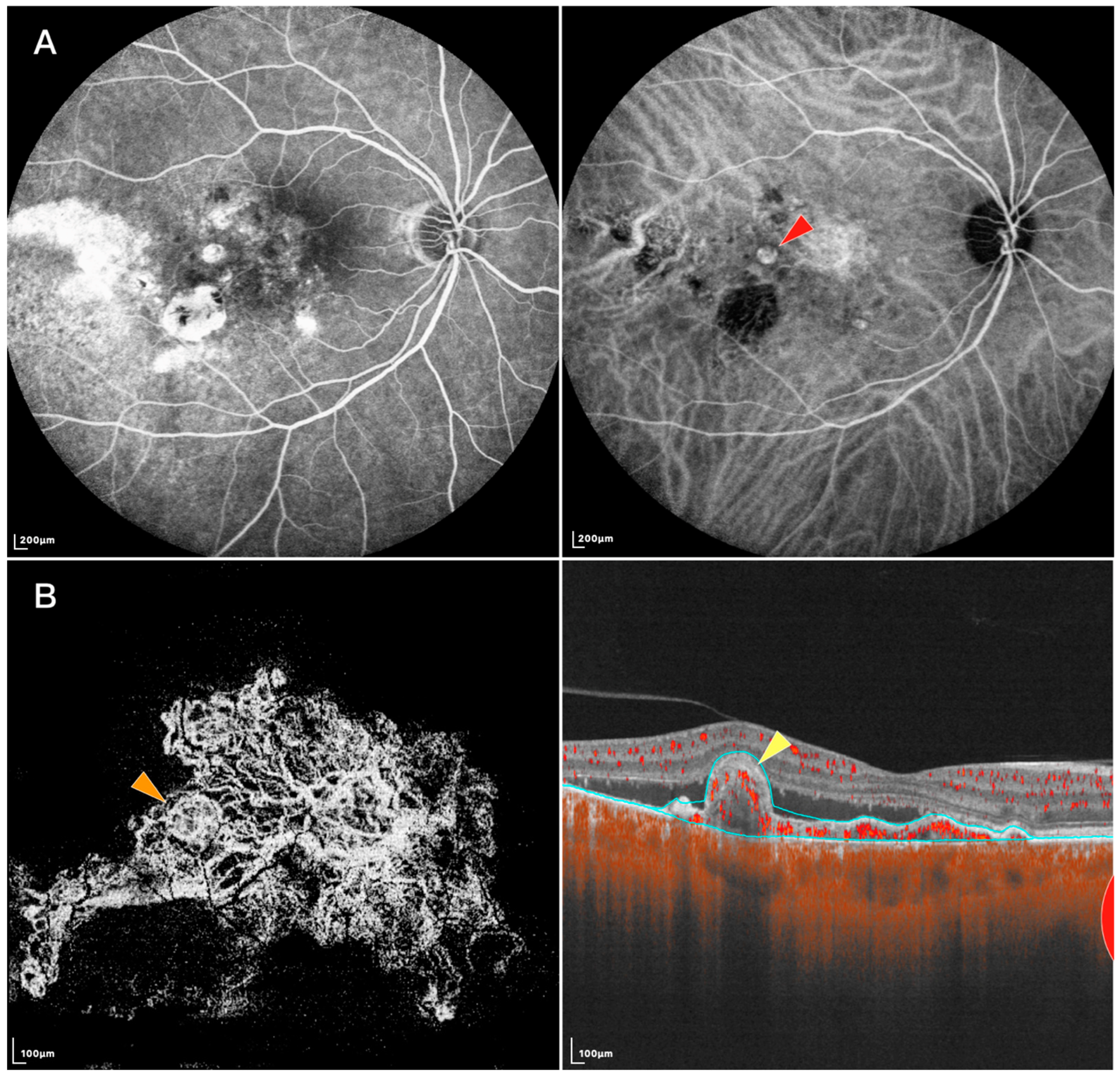
2.1. Presentation of Polypoidal Lesions on SS-OCTA
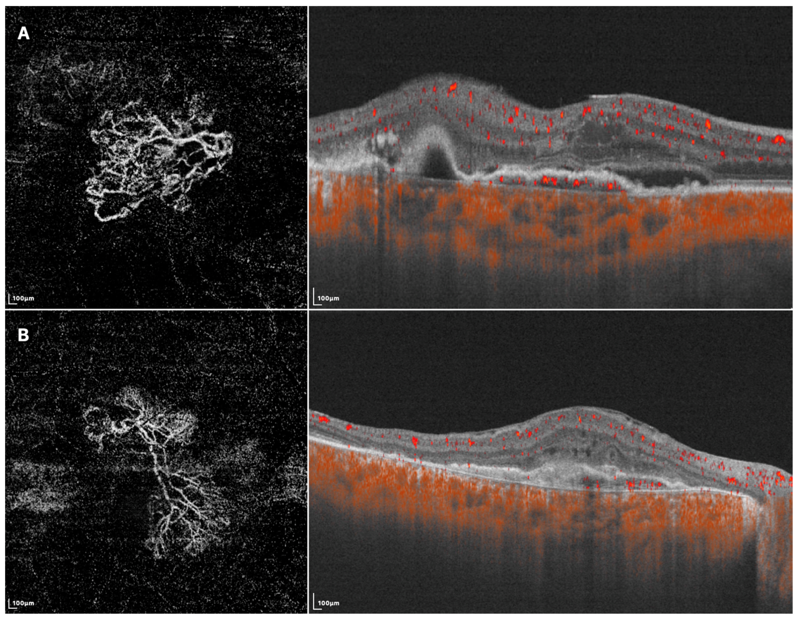
2.2. Presentation of Branching Vascular Networks on SS-OCTA
2.3. Choroidal Changes of PCV on SS-OCTA
3. Clinical Management of PCV with SS-OCTA
3.1. Diagnosis of PCV with SS-OCTA
3.2. Follow up of PCV with SS-OCTA
4. Current Problems and Conclusions
Author Contributions
Funding
Institutional Review Board Statement
Informed Consent Statement
Data Availability Statement
Conflicts of Interest
References
- Yannuzzi, L.A.; Sorenson, J.; Spaide, R.F.; Lipson, B. Idiopathic polypoidal choroidal vasculopathy (IPCV). Retina 1990, 10, 1–8. [Google Scholar] [CrossRef]
- Ryu, G.; Moon, C.; van Hemert, J.; Sagong, M. Quantitative analysis of choroidal vasculature in polypoidal choroidal vasculopathy using ultra-widefield indocyanine green angiography. Sci. Rep. 2020, 10, 18272. [Google Scholar] [CrossRef]
- Zhao, J.; Chandrasekaran, P.R.; Cheong, K.X.; Wong, M.; Teo, K. New Concepts for the Diagnosis of Polypoidal Choroidal Vasculopathy. Diagnostics 2023, 13, 1680. [Google Scholar] [CrossRef]
- Inoue, M.; Balaratnasingam, C.; Freund, K.B. Optical Coherence Tomography Angiography of Polypoidal Choroidal Vasculopathy and Polypoidal Choroidal Neovascularization. Retina 2015, 35, 2265–2274. [Google Scholar] [CrossRef]
- Jia, Y.; Tan, O.; Tokayer, J.; Potsaid, B.; Wang, Y.; Liu, J.J.; Kraus, M.F.; Subhash, H.; Fujimoto, J.G.; Hornegger, J.; et al. Split-spectrum amplitude-decorrelation angiography with optical coherence tomography. Opt. Express 2012, 20, 4710–4725. [Google Scholar] [CrossRef] [PubMed] [Green Version]
- Tan, C.S. The Role of Optical Coherence Tomography Angiography in Polypoidal Choroidal Vasculopathy. JAMA Ophthalmol. 2017, 135, 1316–1317. [Google Scholar] [CrossRef] [PubMed]
- Kim, J.Y.; Kwon, O.W.; Oh, H.S.; Kim, S.H.; You, Y.S. Optical coherence tomography angiography in patients with polypoidal choroidal vasculopathy. Graefe’s Arch. Clin. Exp. Ophthalmol. 2016, 254, 1505–1510. [Google Scholar] [CrossRef]
- Tomiyasu, T.; Nozaki, M.; Yoshida, M.; Ogura, Y. Characteristics of Polypoidal Choroidal Vasculopathy Evaluated by Optical Coherence Tomography Angiography. Investig. Ophthalmol. Vis. Sci. 2016, 57, 324–330. [Google Scholar] [CrossRef] [PubMed]
- Miller, A.R.; Roisman, L.; Zhang, Q.; Zheng, F.; Dias, J.R.D.O.; Yehoshua, Z.; Schaal, K.B.; Feuer, W.; Gregori, G.; Chu, Z.; et al. Comparison Between Spectral-Domain and Swept-Source Optical Coherence Tomography Angiographic Imaging of Choroidal Neovascularization. Investig. Ophthalmol. Vis. Sci. 2017, 58, 1499–1505. [Google Scholar] [CrossRef] [PubMed]
- Rebhun, C.B.; Moult, E.M.; Novais, E.A.; Moreira-Neto, C.; Ploner, S.B.; Louzada, R.N.; Lee, B.; Baumal, C.R.; Fujimoto, J.G.; Duker, J.S.; et al. Polypoidal Choroidal Vasculopathy on Swept-Source Optical Coherence Tomography Angiography with Variable Interscan Time Analysis. Transl. Vis. Sci. Technol. 2017, 6, 4. [Google Scholar] [CrossRef] [Green Version]
- Yasuno, Y.; Miura, M.; Kawana, K.; Makita, S.; Sato, M.; Okamoto, F.; Yamanari, M.; Iwasaki, T.; Yatagai, T.; Oshika, T. Visualization of sub-retinal pigment epithelium morphologies of exudative macular diseases by high-penetration optical coherence tomography. Investig. Ophthalmol. Vis. Sci. 2009, 50, 405–413. [Google Scholar] [CrossRef] [PubMed]
- Lim, F.P.; Loh, B.K.; Cheung, C.M.; Lim, L.S.; Chan, C.M.; Wong, D.W.K. Evaluation of focal choroidal excavation in the macula using swept-source optical coherence tomography. Eye 2014, 28, 1088–1094. [Google Scholar] [CrossRef] [PubMed] [Green Version]
- Ting, D.S.; Cheung, G.C.; Lim, L.S.; Yeo, I.Y.S. Comparison of swept source optical coherence tomography and spectral domain optical coherence tomography in polypoidal choroidal vasculopathy. Clin. Exp. Ophthalmol. 2015, 43, 815–819. [Google Scholar] [CrossRef]
- Dansingani, K.K.; Balaratnasingam, C.; Naysan, J.; Freund, K.B. En face imaging of pachychoroid spectrum disorders with swept-source optical coherence tomography. Retina 2016, 36, 499–516. [Google Scholar] [CrossRef]
- Bakthavatsalam, M.; Ng, D.S.; Lai, F.H.; Tang, F.Y.; Brelén, M.E.; Tsang, C.W.; Lai, T.; Cheung, C. Choroidal structures in polypoidal choroidal vasculopathy, neovascular age-related maculopathy, and healthy eyes determined by binarization of swept source optical coherence tomographic images. Graefe’s Arch. Clin. Exp. Ophthalmol. 2017, 255, 935–943. [Google Scholar] [CrossRef]
- Dong, J.; Chen, R.; Zhao, H.; Zhu, Y. COVID-19 and ocular complications: A review of ocular manifestations, diagnostic tools, and prevention strategies. Adv. Ophthalmol. Pract. Res. 2023, 3, 33–38. [Google Scholar] [CrossRef]
- Wang, M.; Zhou, Y.; Gao, S.S.; Liu, W.; Huang, Y.; Huang, D.; Jia, Y. Evaluating Polypoidal Choroidal Vasculopathy with Optical Coherence Tomography Angiography. Investig. Ophthalmol. Vis. Sci. 2016, 57, 526–532. [Google Scholar] [CrossRef]
- Chan, S.Y.; Wang, Q.; Wang, Y.X.; Shi, X.H.; Jonas, J.B.; Bin Wei, W. Polypoidal choroidal vasculopathy upon optical coherence tomographic angiography. Retina 2018, 38, 1187–1194. [Google Scholar] [CrossRef]
- Chi, Y.T.; Yang, C.H.; Cheng, C.K. Optical Coherence Tomography Angiography for Assessment of the 3-Dimensional Structures of Polypoidal Choroidal Vasculopathy. JAMA Ophthalmol. 2017, 135, 1310–1316. [Google Scholar] [CrossRef]
- Yu, W.; Yang, J.; Sui, W.; Qu, B.; Huang, P.; Chen, Y. Association of genetic variants in the receptor for advanced glycation end products gene with diabetic retinopathy: A meta-analysis. Medicine 2016, 95, e4463. [Google Scholar] [CrossRef]
- Alshahrani, S.T.; Al Shamsi, H.N.; Kahtani, E.S.; Ghazi, N.G. Spectral-domain optical coherence tomography findings in polypoidal choroidal vasculopathy suggest a type 1 neovascular growth pattern. Clin. Ophthalmol. 2014, 8, 1689–1695. [Google Scholar] [CrossRef] [Green Version]
- Otsuji, T.; Tsumura, A.; Takahashi, K.; Sho, K.; Nagai, Y.; Fukuchi, T.; Arisawa, A.; Matsumura, M. Evaluation of cases of polypoidal choroidal vasculopathy showing classic choroidal neovascularization in their natural course. Nippon Ganka Gakkai Zasshi 2006, 110, 454–461. [Google Scholar]
- Alasil, T.; Ferrara, D.; Adhi, M.; Brewer, E.; Kraus, M.F.; Baumal, C.R.; Hornegger, J.; Fujimoto, J.G.; Witkin, A.J.; Reichel, E.; et al. En Face Imaging of the Choroid in Polypoidal Choroidal Vasculopathy Using Swept-Source Optical Coherence Tomography. Am. J. Ophthalmol. 2015, 159, 634–643.e632. [Google Scholar] [CrossRef]
- Srour, M.; Querques, G.; Souied, E.H. Optical Coherence Tomography Angiography of Idiopathic Polypoidal Choroidal Vasculopathy. Dev. Ophthalmol. 2016, 56, 71–76. [Google Scholar] [CrossRef]
- Zhan, Z.; Sun, L.; **, C.; Yang, Y.; Hu, A.; Tang, M.; Wang, Z.; Ding, X. Comparison between non-visualized polyps and visualized polyps on optical coherence tomography angiography in polypoidal choroidal vasculopathy. Graefe’s Arch. Clin. Exp. Ophthalmol. 2019, 257, 2349–2356. [Google Scholar] [CrossRef]
- Liang, S.; Shi, X.; Rosenfeld, P.J.; Li, X. Type 2 choroidal neovascularisation in polypoidal choroidal vasculopathy: A retrospective case series. Br. J. Ophthalmol. 2018, 102, 1570–1574. [Google Scholar] [CrossRef]
- Cheung, C.M.G.; Yanagi, Y.; Mohla, A.; Lee, S.Y.; Mathur, R.; Chan, C.M.; Yeo, I.; Wong, T.Y. Characterization and differentiation of polypoidal choroidal vasculopathy using swept source optical coherence tomography angiography. Retina 2017, 37, 1464–1474. [Google Scholar] [CrossRef] [PubMed]
- Singh, S.R.; Goyal, P.; Parameswarappa, D.C.; Goud, A.; Chhablani, J. Angiographic features of polypoidal choroidal vasculopathy using indocyanine green angiography and optical coherence tomography angiography: A comparative study. Eur. J. Ophthalmol. 2020, 30, 1076–1081. [Google Scholar] [CrossRef] [PubMed]
- Azar, G.; Vasseur, V.; Lahoud, C.; Favard, C.; De Bats, F.; Cochereau, I.; Yachvitz, A.; Mauget-Faÿsse, M. Polypoidal Choroidal Vasculopathy Diagnosis and Neovascular Activity Evaluation Using Optical Coherence Tomography Angiography. Biomed. Res. Int. 2021, 2021, 1637377. [Google Scholar] [CrossRef]
- Bo, Q.; Yan, Q.; Shen, M.; Song, M.; Sun, M.; Yu, Y.; Rosenfeld, P.J.; Wang, F.; Sun, X. Appearance of Polypoidal Lesions in Patients with Polypoidal Choroidal Vasculopathy Using Swept-Source Optical Coherence Tomographic Angiography. JAMA Ophthalmol. 2019, 137, 642–650. [Google Scholar] [CrossRef] [PubMed]
- Fujita, A.; Kataoka, K.; Takeuchi, J.; Nakano, Y.; Horiguchi, E.; Kaneko, H.M.; Ito, Y.M.; Terasaki, H.M. Diagnostic characteristics of polypoidal choroidal vasculopathy based on b-scan swept-source optical coherence tomography angiography and its interrater agreement compared with indocyanine green angiography. Retina 2020, 40, 2296–2303. [Google Scholar] [CrossRef]
- Kim, K.; Yang, J.; Feuer, W.; Gregori, G.; Kim, E.S.; Rosenfeld, P.J.; Yu, S.-Y. A Comparison Study of Polypoidal Choroidal Vasculopathy Imaged with Indocyanine Green Angiography and Swept-Source Optical Coherence Tomography Angiography. Am. J. Ophthalmol. 2020, 217, 240–251. [Google Scholar] [CrossRef]
- Shen, M.; Bo, Q.; Song, M.; Jiang, X.; Yehoshua, Z.; Gregori, G.; Sun, X.; Wang, F.; Rosenfeld, P.J. Replacement of polyps with type 1 macular neovascularization in polypoidal choroidal vasculopathy imaged with swept source OCT angiography. Am. J. Ophthalmol. Case Rep. 2021, 22, 101057. [Google Scholar] [CrossRef] [PubMed]
- Yuzawa, M.; Mori, R.; Kawamura, A. The origins of polypoidal choroidal vasculopathy. Br. J. Ophthalmol. 2005, 89, 602–607. [Google Scholar] [CrossRef] [PubMed] [Green Version]
- Akaza, E.; Matsumoto, Y.; Yuzawa, M. Pulsation in polypoidal choroidal vasculopathy. Nippon Ganka Gakkai Zasshi 2006, 110, 288–292. [Google Scholar] [PubMed]
- Chen, Y.; Yang, Z.; ** technology for retinal diseases. Expert Rev. Med. Devices 2018, 15, 875–882. [Google Scholar] [CrossRef] [PubMed]
- Zhou, H.; Bacci, T.; Freund, K.B.; Wang, R.K. Three-dimensional segmentation and depth-encoded visualization of choroidal vasculature using swept-source optical coherence tomography. Exp. Biol. Med. 2021, 246, 2238–2245. [Google Scholar] [CrossRef] [PubMed]
- Sho, K.; Takahashi, K.; Yamada, H.; Wada, M.; Nagai, Y.; Otsuji, T.; Nishikawa, M.; Mitsuma, Y.; Yamazaki, Y.; Matsumura, M.; et al. Polypoidal choroidal vasculopathy: Incidence, demographic features, and clinical characteristics. Arch. Ophthalmol. 2003, 121, 1392–1396. [Google Scholar] [CrossRef] [Green Version]
- De Carlo, T.E.; Kokame, G.T.; Shantha, J.G.; Lai, J.C.; Wee, R. Spectral-Domain Optical Coherence Tomography Angiography for the Diagnosis and Evaluation of Polypoidal Choroidal Vasculopathy. Ophthalmologica 2018, 239, 103–109. [Google Scholar] [CrossRef]


| Author | Year | Included Eyes | PL Morphology and Locality | BVN Morphology and Locality | PL Detection Rate | BVN Detection Rate |
|---|---|---|---|---|---|---|
| Kishida et al. [69] | 2014 | 17 (-) | Similar to the early phase of ICGA images. (Between the Bruch’s membrane and RPE) | - (-) | 100% | - |
| Cheung et al. [27] | 2017 | 54 (68.5% treated) | Variable in size and may appear as bright round lesions or round lesions with a bright outline but dark lumen. (-) | Variable in size but correlated closely with the location and shape of BVN on ICGA. (-) | 77.80% | 87% |
| Rebhun et al. [10] | 2017 | 7 (100% treated) | Non-uniform blood flow inside a single polyp, with some, appearing slower at the center of the polyp and faster close to the lesion walls. (-) | Blood flow is slower than that of retinal vessels. BVN with larger trunks had faster blood flow inside. (-) | 85.70% | 85.70% |
| Bo et al. [30] | 2019 | 23 (65.2% treated) | Tangled vessels. (Associated with type I or type II NV) | - (Associated with type I or type II NV) | 100% (With manual segmentation) | 100% (With manual segmentation) |
| Fujita et al. [31] | 2020 | 54 (All untreated) | Mostly tangled structures, including coil-like structures. (All inside PED and mostly Located at the margin of the BVNs) | Tangled vascular networks. (-) | B-scan: 94.7% | En face: 72.2% B-scan: 87.0% |
| Kim et al. [32] | 2020 | 31 (All untreated) | Mostly tangled vessels with a variety of configurations. (-) | No significant difference in lesion area measurements between ICGA and SS-OCTA. (-) | 100% (With manual segmentation) | 100% (With manual segmentation) |
| Singh et al. [28] | 2020 | 46 (All untreated) | Hyper-reflective or hypo-reflective with a hyper-reflective border. (-) | OCTA could better delineate the margins of small BVNs. (-) | 69.57% (With manual segmentation) | 100% (With manual segmentation) |
| Azar et al. [29] | 2021 | 14 (-) | Some polyps present with a halo or a hyper-reflective round structure surrounded by a hypo-intense halo. (Above Bruch’s membrane) | Loose or dense pattern. (Between the RPE and Bruch’s membrane) | - | 100% |
| Shen et al. [33] | 2021 | 5 (-) | Tangled vascular structure. (Beneath the PED) | - (-) | - | - |
| Wang et al. [43] | 2021 | 30 (-) | Cluster-like structure at the edge of a BVN. (-) | - (-) | 96.7% (With manual segmentation) | - |
| Arias et al. [41] | 2021 | 22 (72.7% treated) | Active disease: poorly defined shape surrounded by a hypo-reflective halo. Inactive cases: well-defined circular shape. (-) | - (-) | B-scan: 100% | - |
Disclaimer/Publisher’s Note: The statements, opinions and data contained in all publications are solely those of the individual author(s) and contributor(s) and not of MDPI and/or the editor(s). MDPI and/or the editor(s) disclaim responsibility for any injury to people or property resulting from any ideas, methods, instructions or products referred to in the content. |
© 2023 by the authors. Licensee MDPI, Basel, Switzerland. This article is an open access article distributed under the terms and conditions of the Creative Commons Attribution (CC BY) license (https://creativecommons.org/licenses/by/4.0/).
Share and Cite
Gu, X.; Zhao, X.; Zhao, Q.; Wang, Y.; Chen, Y. Recent Advances in Imaging Polypoidal Choroidal Vasculopathy with Swept-Source Optical Coherence Tomography Angiography. Diagnostics 2023, 13, 2458. https://doi.org/10.3390/diagnostics13142458
Gu X, Zhao X, Zhao Q, Wang Y, Chen Y. Recent Advances in Imaging Polypoidal Choroidal Vasculopathy with Swept-Source Optical Coherence Tomography Angiography. Diagnostics. 2023; 13(14):2458. https://doi.org/10.3390/diagnostics13142458
Chicago/Turabian StyleGu, **ngwang, **nyu Zhao, Qing Zhao, Yuelin Wang, and Youxin Chen. 2023. "Recent Advances in Imaging Polypoidal Choroidal Vasculopathy with Swept-Source Optical Coherence Tomography Angiography" Diagnostics 13, no. 14: 2458. https://doi.org/10.3390/diagnostics13142458