
U-Net-Based Models towards Optimal MR Brain Image Segmentation

 , , , , ,
Abstract
:1. Introduction
- Addressing the recent techniques focused on brain tumor segmentation based on U-Net architecture as the backbone, along with its variants.
- Highlighting the major trends and patterns in the research that may help to guide future work in the field by summarizing the cutting-edge techniques in one place.
- Providing a comparative analysis of the most recent relevant literature results and other experimental results to observe the improvements achieved by the incremental research.
1.1. Brain MRI Segmentation
1.2. Before U-Net
- Architecture: traditional deep learning models, such as fully convolutional networks (FCNs) or convolutional neural networks (CNNs), typically have a simpler architecture compared to U-Net-based models.
- Training Data: U-Net-based models are specifically designed to work well with medical imaging data, which often have higher resolutions and more complex structures than natural images. Meanwhile, traditional deep learning models may struggle to handle complex data and may need to be fine-tuned to work well with medical imaging data.
- Performance: U-Net-based models have been shown to perform better than traditional deep learning models on brain tumor segmentation tasks, particularly on datasets with limited training data.
- Small objects segmentation: U-Net-based models have the capacity to handle small structural objects in the image, which is an important aspect in brain tumor segmentation where small tumors need to be segmented.
2. U-Net and U-Net Expansions towards Optimized DL Models for Segmentations
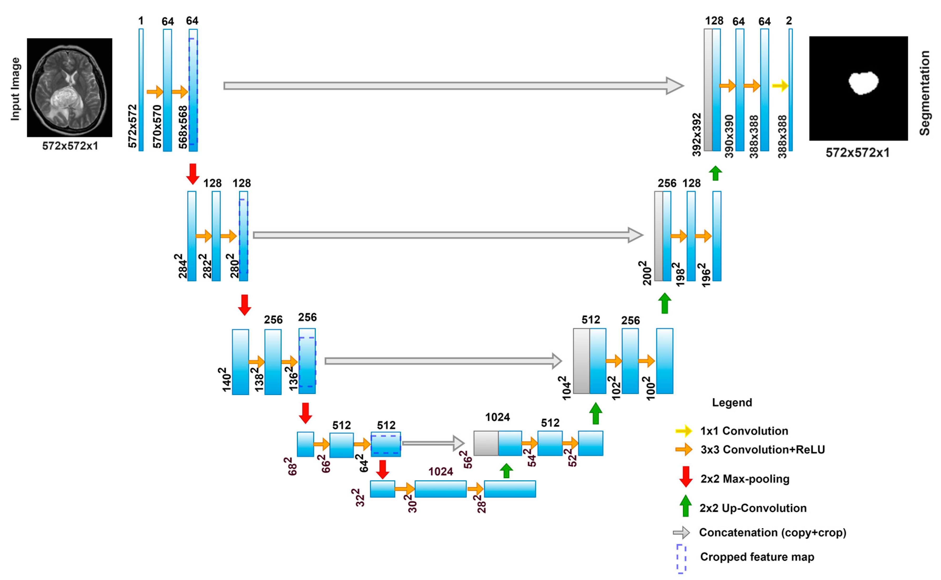
2.1. U-Net
U-Net Workflow
- The Contracting Path
- The Expansion Path
- Training
2.2. 3D U-Net
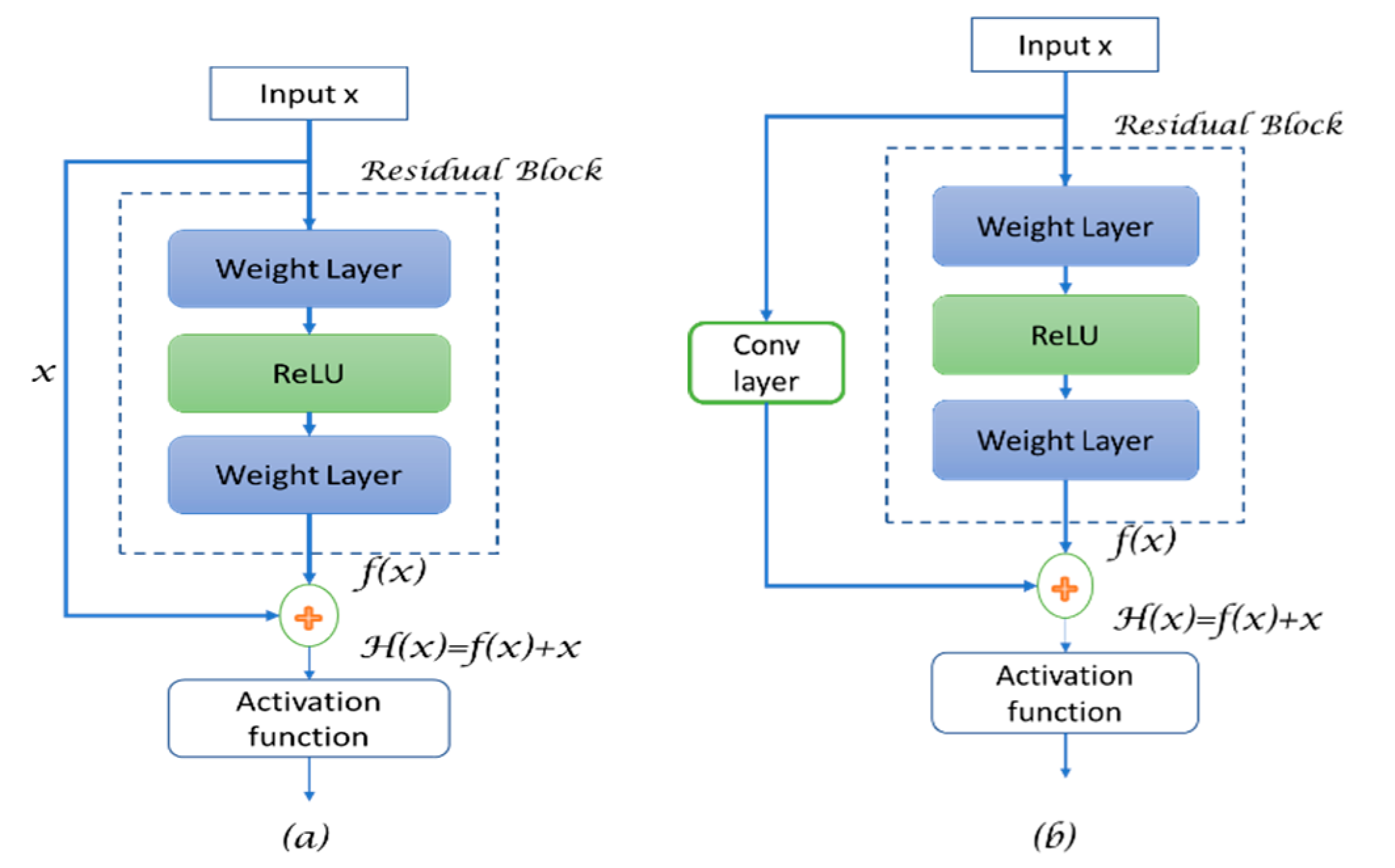
2.3. Residual U-Net
- R(X) refers to the residual map**,
- h(x) is referred to as the identity map function after applying the convolution operation,
- x+1 is the input for the next layer, and
- f(.) is the activation function.
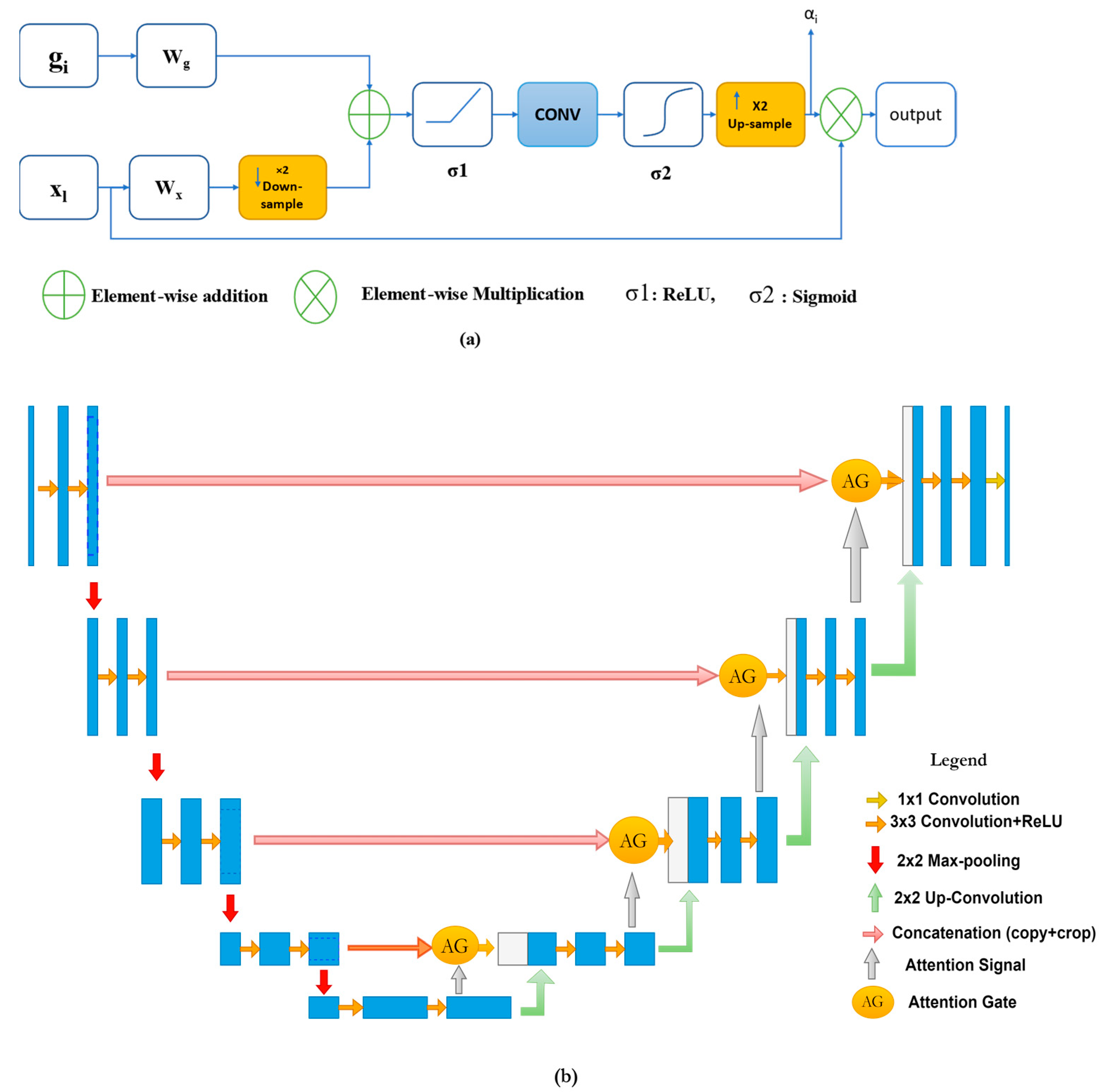
2.4. Attention U-Net
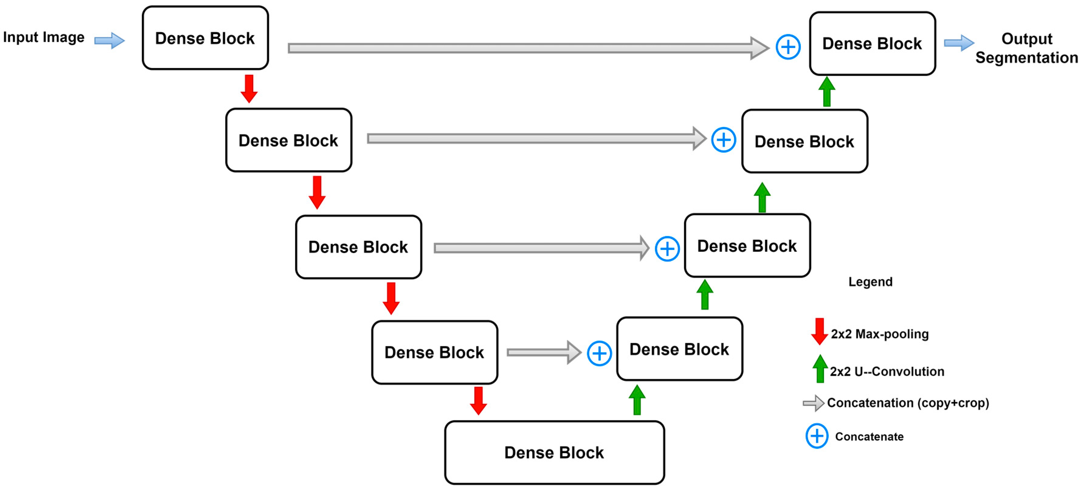
2.5. Dense U-Net
2.6. U-Net++
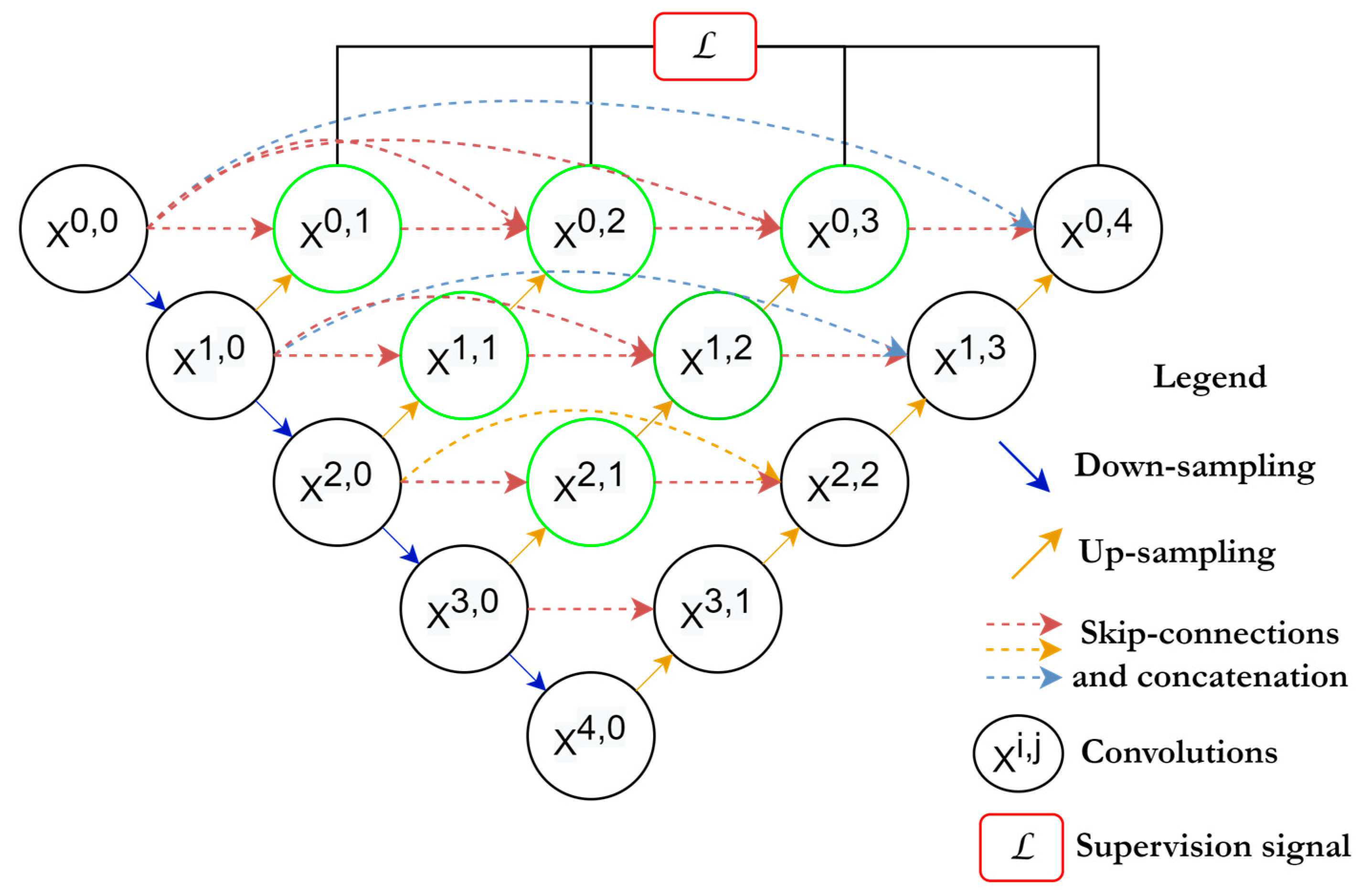
2.7. U-Net 3+
2.8. Adversarial U-Net
2.9. Other Well-Known Architectures Based on U-Net
3. Materials and Methods
3.1. Loss Functions
3.1.1. Cross-Entropy Loss
3.1.2. Dice Loss Function
3.1.3. IoU Loss
3.1.4. Tversky Loss
3.1.5. Hausdorff Distance Loss
3.2. Evaluation Metrics
3.2.1. Dice Coefficient
3.2.2. Jaccard Index/Intersection over Union (IoU)
3.2.3. Hausdorff Distance (HD)
3.2.4. Sensitivity and Specificity
3.3. Comparison and Analysis
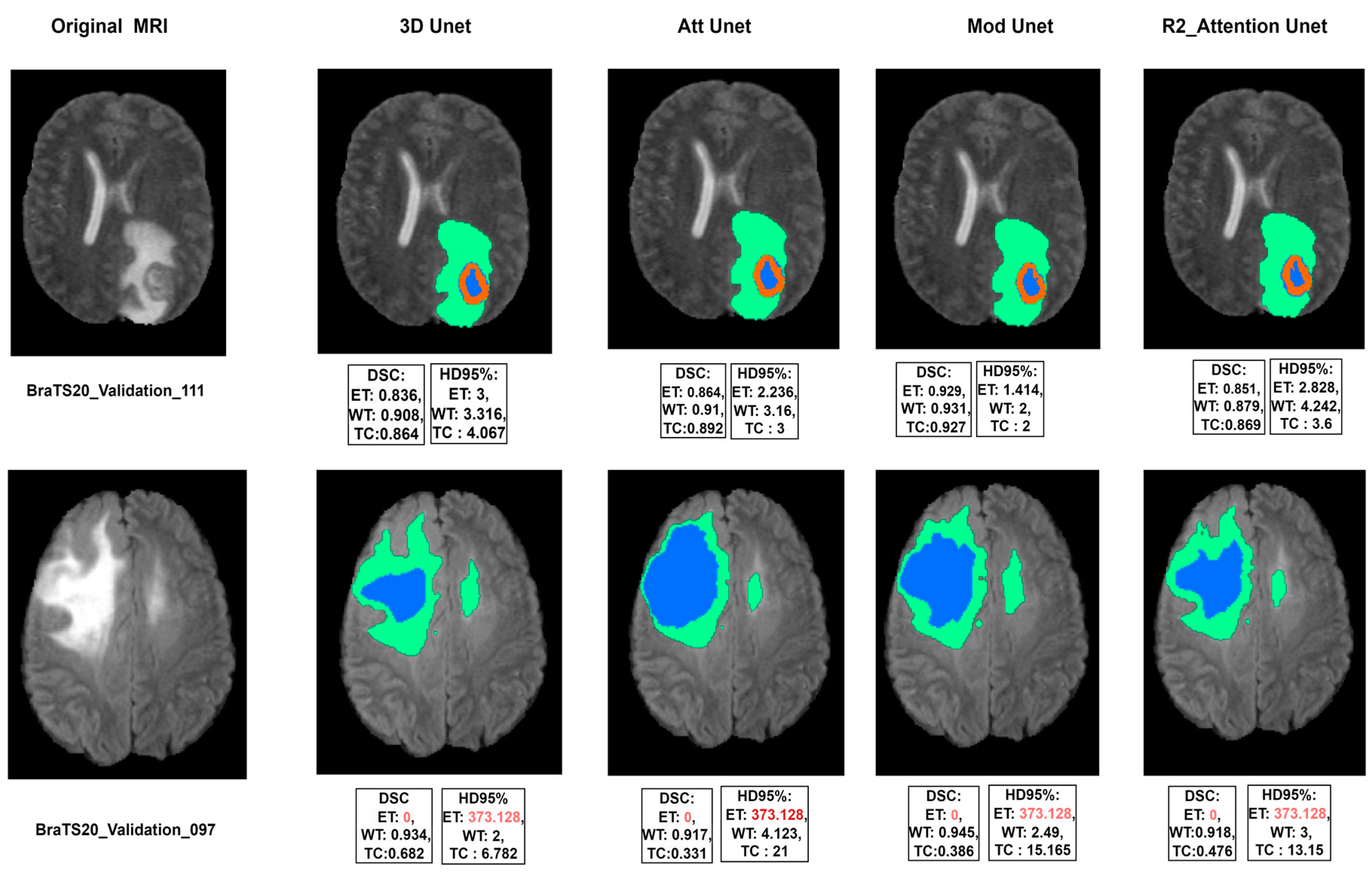
4. Experimental Results
Experimental Training Layout
- 3D U-Net: This architecture consists of four levels of convolutions in both the encoder and decoder. It was proposed in [96].
- Modified 3D U-Net: follows the same attributes as the previous model, but an extra level is added, so the encoder–decoder network uses five levels of convolutions.
- R2 Attention U-Net: Recurrent Residual Attention U-Net was proposed in [97], which adds the recurrent and residual blocks to the first 3D model.
5. Discussion
5.1. Limitations of this Research
5.2. Challenges
6. Conclusions
Author Contributions
Funding
Institutional Review Board Statement
Informed Consent Statement
Data Availability Statement
Conflicts of Interest
References
- Munsif, M.; Ullah, M.; Ahmad, B.; Sajjad, M.; Cheikh, F.A. Monitoring Neurological Disorder Patients via Deep Learning Based Facial Expressions Analysis. In Artificial Intelligence Applications and Innovations. AIAI 2022 IFIP WG 12.5 International Workshops; Springer International Publishing: Cham, Switzerland, 2022; pp. 412–423. [Google Scholar]
- Hussain, A.; Khan, A.; Yar, H. Efficient Deep learning Approach for Classification of Pneumonia using Resources Constraint Devices in Healthcare. In Proceedings of the 5th International Conference on Next Generation Computing, Bidholi Via-Prem Nagar, India, 20–21 December 2019; pp. 20–21. [Google Scholar]
- Li, J.P.; Khan, S.; Alshara, M.A.; Alotaibi, R.M.; Mawuli, C. DACBT: Deep learning approach for classification of brain tumors using MRI data in IoT healthcare environment. Sci. Rep. 2022, 12, 15331. [Google Scholar]
- Chopra, P.; Junath, N.; Singh, S.K.; Khan, S.; Sugumar, R.; Bhowmick, M. Cyclic GAN Model to Classify Breast Cancer Data for Pathological Healthcare Task. Biomed Res. Int. 2022, 2022, 6336700. [Google Scholar] [CrossRef] [PubMed]
- Haq, A.U.; Li, J.P.; Khan, I.; Agbley, B.L.Y.; Ahmad, S.; Uddin, M.I.; Zhou, W.; Khan, S.; Alam, I. DEBCM: Deep Learning-Based Enhanced Breast Invasive Ductal Carcinoma Classification Model in IoMT Healthcare Systems. IEEE J. Biomed. Health Inform. 2022, 1–12. [Google Scholar] [CrossRef] [PubMed]
- Agbley, B.L.Y.; Li, J.P.; Haq, A.U.; Bankas, E.K.; Mawuli, C.B.; Ahmad, S.; Khan, S.; Khan, A.R. Federated Fusion of Magnified Histopathological Images for Breast Tumor Classification in the Internet of Medical Things. IEEE J. Biomed. Health Inform. 2023, 1–12. [Google Scholar] [CrossRef]
- Haq, A.U.; Li, J.P.; Ahmad, S.; Khan, S.; Alshara, M.A.; Alotaibi, R.M. Diagnostic approach for accurate diagnosis of COVID-19 employing deep learning and transfer learning techniques through chest X-ray images clinical data in E-healthcare. Sensors 2021, 21, 8219. [Google Scholar] [CrossRef]
- Lu, S.-Y.; Zhang, Z.; Zhang, Y.-D.; Wang, S.-H. CGENet: A Deep Graph Model for COVID-19 Detection Based on Chest CT. Biology 2022, 11, 33. [Google Scholar] [CrossRef] [PubMed]
- Khan, J.; Khan, G.A.; Li, J.P.; AlAjmi, M.F.; Haq, A.U.; Khan, S.; Ahmad, N.; Parveen, S.; Shahid, M.; Ahmad, S. Secure smart healthcare monitoring in industrial internet of things (iiot) ecosystem with cosine function hybrid chaotic map encryption. Sci. Program 2022, 2022, 8853448. [Google Scholar] [CrossRef]
- Fazil, M.; Khan, S.; Albahlal, B.M.; Alotaibi, R.M.; Siddiqui, T.; Shah, M.A. Attentional Multi-Channel Convolution With Bidirectional LSTM Cell Toward Hate Speech Prediction. IEEE Access 2023, 11, 16801–16811. [Google Scholar] [CrossRef]
- Khan, S.; Fazil, M.; Sejwal, V.K.; Alshara, M.A.; Alotaibi, R.M.; Kamal, A.; Baig, A.R. BiCHAT: BiLSTM with deep CNN and hierarchical attention for hate speech detection. J. King Saud Univ. Inf. Sci. 2022, 34, 4335–4344. [Google Scholar] [CrossRef]
- Khan, S.; Kamal, A.; Fazil, M.; Alshara, M.A.; Sejwal, V.K.; Alotaibi, R.M.; Baig, A.R.; Alqahtani, S. HCovBi-Caps: Hate Speech Detection Using Convolutional and Bi-Directional Gated Recurrent Unit With Capsule Network. IEEE Access 2022, 10, 7881–7894. [Google Scholar] [CrossRef]
- Morrow, M.; Waters, J.; Morris, E. MRI for breast cancer screening, diagnosis, and treatment. Lancet 2011, 378, 1804–1811. [Google Scholar] [CrossRef]
- Zhao, M.; Cao, X.; Zhou, M.; Feng, J.; ** using 3D Generative Adversarial Networks with increased receptive field. Neuroimage 2020, 207, 116389. [Google Scholar] [CrossRef] [PubMed]
- Teki, S.M.; Varma, M.K.; Yadav, A.K. Brain tumour segmentation using U-net based adversarial networks. Trait. Du Signal 2019, 36, 353–359. [Google Scholar] [CrossRef]
- Chen, J.; Lu, Y.; Yu, Q.; Luo, X.; Adeli, E.; Wang, Y.; Lu, L.; Yuille, A.L.; Zhou, Y. TransUNet: Transformers Make Strong Encoders for Medical Image Segmentation. ar**v 2021, ar**v:2102.04306. [Google Scholar]
- Milletari, F.; Navab, N.; Ahmadi, S.-A. V-Net: Fully Convolutional Neural Networks for Volumetric Medical Image Segmentation. In Proceedings of the 2016 Fourth International Conference on 3D Vision (3DV), Stanford, CA, USA, 5–28 October 2016; pp. 565–571. [Google Scholar]
- Alom, M.Z.; Yakopcic, C.; Hasan, M.; Taha, T.M.; Asari, V.K. Recurrent residual U-Net for medical image segmentation. J. Med. Imaging 2019, 6, 014006. [Google Scholar] [CrossRef]
- Fatemeh, Z.; Nicola, S.; Satheesh, K.; Eranga, U. Ensemble U-net-based method for fully automated detection and segmentation of renal masses on computed tomography images. Med. Phys. 2020, 47, 4032–4044. [Google Scholar] [CrossRef] [PubMed]
- Feng, X.; Wang, C.; Cheng, S.; Guo, L. Automatic Liver and Tumor Segmentation of CT Based on Cascaded U-Net. In Proceedings of 2018 Chinese Intelligent Systems Conference; Springer: Singapore, 2019; pp. 155–164. [Google Scholar]
- Valanarasu, J.M.J.; Sindagi, V.A.; Hacihaliloglu, I.; Patel, V.M. KiU-Net: Overcomplete Convolutional Architectures for Biomedical Image and Volumetric Segmentation. IEEE Trans. Med. Imaging 2021, 41, 965–976. [Google Scholar] [CrossRef]
- Zhang, J.; Lv, X.; Zhang, H.; Liu, B. AResU-Net: Attention residual U-Net for brain tumor segmentation. Symmetry 2020, 12, 721. [Google Scholar] [CrossRef]
- Lecun, Y.; Bottou, L.; Bengio, Y.; Haffner, P. Gradient-based learning applied to document recognition. Proc. IEEE 1998, 86, 2278–2324. [Google Scholar] [CrossRef]
- Dice, L.R. Measures of the Amount of Ecologic Association Between Species. Ecology 1945, 26, 297–302. [Google Scholar] [CrossRef]
- Fidon, L.; Li, W.; Garcia-Peraza-Herrera, L.C.; Ekanayake, J.; Kitchen, N.; Ourselin, S.; Vercauteren, T. Generalised Wasserstein Dice Score for Imbalanced Multi-class Segmentation Using Holistic Convolutional Networks. In Brainlesion: Glioma, Multiple Sclerosis, Stroke and Traumatic Brain Injuries; Springer International Publishing: Cham, Switzerland, 2018; pp. 64–76. [Google Scholar]
- Jaccard, P. The distribution of the flora in the alpine zone.1. New Phytol. 1912, 11, 37–50. [Google Scholar] [CrossRef]
- Abraham, N.; Khan, N.M. A Novel Focal Tversky Loss Function With Improved Attention U-Net for Lesion Segmentation. In Proceedings of the 2019 IEEE 16th International Symposium on Biomedical Imaging (ISBI 2019), Venice, Italy, 8–11 April 2019; pp. 683–687. [Google Scholar]
- Kervadec, H.; Bouchtiba, J.; Desrosiers, C.; Granger, E.; Dolz, J.; Ben Ayed, I. Boundary loss for highly unbalanced segmentation. In Proceedings of the 2nd International Conference on Medical Imaging with Deep Learning, London, UK, 8–10 July 2019; Volume 102, pp. 285–296. Available online: https://proceedings.mlr.press/v102/kervadec19a.html (accessed on 20 November 2022).
- Gerig, G.; Jomier, M.; Chakos, M. Valmet: A New Validation Tool for Assessing and Improving 3D Object Segmentation. In Medical Image Computing and Computer-Assisted Intervention—MICCAI 2001; Springer: Berlin/Heidelberg, Germany, 2001; pp. 516–523. [Google Scholar]
- Nai, Y.H.; Teo, B.W.; Tan, N.L.; O’Doherty, S.; Stephenson, M.C.; Thian, Y.L.; Chiong, E.; Reilhac, A. Comparison of metrics for the evaluation of medical segmentations using prostate MRI dataset. Comput. Biol. Med. 2021, 134, 104497. [Google Scholar] [CrossRef]
- Taha, A.A.; Hanbury, A. Metrics for evaluating 3D medical image segmentation: Analysis, selection, and tool. BMC Med. Imaging 2015, 15, 29. [Google Scholar] [CrossRef]
- Menze, B.H.; Jakab, A.; Bauer, S.; Kalpathy-Cramer, J.; Farahani, K.; Kirby, J.; Burren, Y.; Porz, N.; Slotboom, J.; Wiest, R. The multimodal brain tumor image segmentation benchmark (BRATS). IEEE Trans. Med. Imaging 2014, 34, 1993–2024. [Google Scholar] [CrossRef]
- Isensee, F.; Jäger, P.F.; Full, P.M.; Vollmuth, P.; Maier-Hein, K.H. nnU-Net for brain tumor segmentation. In International MICCAI Brainlesion Workshop; Springer: Berlin/Heidelberg, Germany, 2020; pp. 118–132. [Google Scholar]
- Sahayam, S.; Nenavath, R.; Jayaraman, U.; Prakash, S. Brain tumor segmentation using a hybrid multi resolution U-Net with residual dual attention and deep supervision on MR images. Biomed. Signal Process. Control 2022, 78, 103939. [Google Scholar] [CrossRef]
- Maji, D.; Sigedar, P.; Singh, M. Attention Res-UNet with Guided Decoder for semantic segmentation of brain tumors. Biomed. Signal Process. Control 2022, 71, 103077. [Google Scholar] [CrossRef]
- Ellis, D.G.; Aizenberg, M.R. Trialing u-Net Training Modifications for Segmenting Gliomas Using Open Source Deep Learning Framework. In International MICCAI Brainlesion Workshop; Springer: Berlin/Heidelberg, Germany, 2020; pp. 40–49. [Google Scholar]
- Qamar, S.; Ahmad, P.; Shen, L. Hi-net: Hyperdense Inception 3d Unet for Brain Tumor Segmentation. In International MICCAI Brainlesion Workshop; Springer: Berlin/Heidelberg, Germany, 2020; pp. 50–57. [Google Scholar]
- Cirillo, M.D.; Abramian, D.; Eklund, A. Vox2Vox: 3D-GAN for Brain Tumour Segmentation. In International MICCAI Brainlesion Workshop; Springer: Berlin/Heidelberg, Germany, 2020; pp. 274–284. [Google Scholar]
- Ahmad, P.; Qamar, S.; Shen, L.; Saeed, A. Context aware 3D UNet for Brain Tumor Segmentation. In International MICCAI Brainlesion Workshop; Springer: Berlin/Heidelberg, Germany, 2020; pp. 207–218. [Google Scholar]
- Agarwala, S.; Sharma, S.; Uma Shankar, B. A-UNet: Attention 3D UNet architecture for multiclass segmentation of Brain Tumor. In Proceedings of the 2022 IEEE Region 10 Symposium (TENSYMP), Mumbai, India, 1–3 July 2022; pp. 1–5. [Google Scholar]
- Raza, R.; Bajwa, U.I.; Mehmood, Y.; Anwar, M.W.; Jamal, M.H. dResU-Net: 3D deep residual U-Net based brain tumor segmentation from multimodal MRI. Biomed. Signal Process. Control 2023, 79, 103861. [Google Scholar] [CrossRef]
- AboElenein, N.M.; Songhao, P.; Afifi, A. IRDNU-Net: Inception residual dense nested u-net for brain tumor segmentation. Multimed. Tools Appl. 2022, 81, 24041–24057. [Google Scholar] [CrossRef]
- Ghaffari, M.; Sowmya, A.; Oliver, R. Automated Brain Tumour Segmentation Using Cascaded 3D Densely-Connected U-Net BT—Brainlesion: Glioma, Multiple Sclerosis, Stroke and Traumatic Brain Injuries; Springer International Publishing: Cham, Switzerland, 2021; pp. 481–491. [Google Scholar]
- Wang, W.; Chen, C.; Ding, M.; Yu, H.; Zha, S.; Li, J. Transbts: Multimodal brain tumor segmentation using transformer. In Proceedings of the International Conference on Medical Image Computing and Computer-Assisted Intervention, Strasbourg, France, 27 September 2021; pp. 109–119. [Google Scholar]
- Henry, T.; Carré, A.; Lerousseau, M.; Estienne, T.; Robert, C.; Paragios, N.; Deutsch, E. Brain Tumor Segmentation with Self-Ensembled, Deeply-Supervised 3D U-Net Neural Networks: A BraTS 2020 Challenge Solution. In International MICCAI Brainlesion Workshop; Springer: Berlin/Heidelberg, Germany, 2020; pp. 327–339. [Google Scholar]
- Zuo, Q.; Chen, S.; Wang, Z. R2AU-Net: Attention recurrent residual convolutional neural network for multimodal medical image segmentation. Secur. Commun. Netw. 2021, 2021, 6625688. [Google Scholar] [CrossRef]
- Chen, L.C.; Papandreou, G.; Kokkinos, I.; Murphy, K.; Yuille, A.L. DeepLab: Semantic Image Segmentation with Deep Convolutional Nets, Atrous Convolution, and Fully Connected CRFs. IEEE Trans. Pattern Anal. Mach. Intell. 2018, 40, 834–848. [Google Scholar] [CrossRef]
- Peiris, H.; Chen, Z.; Egan, G.; Harandi, M. Reciprocal adversarial learning for brain tumor segmentation: A solution to BraTS challenge 2021 segmentation task. ar**v 2022, ar**v:2201.03777. [Google Scholar]
- Hussain, Z.; Gimenez, F.; Yi, D.; Rubin, D. Differential Data Augmentation Techniques for Medical Imaging Classification Tasks. AMIA Annu. Symp. Proc. AMIA Symp. 2017, 2017, 979–984. [Google Scholar]
- Goodfellow, I.; Bengio, Y.; Courville, A. Deep Learning? Nature 2016, 29, 1–73. [Google Scholar]
- Yi, X.; Walia, E.; Babyn, P. Generative adversarial network in medical imaging: A review. Med. Image Anal. 2019, 58, 101552. [Google Scholar] [CrossRef]
- Papernot, N.; McDaniel, P.; Goodfellow, I.; Jha, S.; Celik, Z.B.; Swami, A. Practical Black-Box Attacks against Machine Learning. In Proceedings of the 2017 ACM on Asia Conference on Computer and Communications Security, Abu Dhabi, United Arab Emirates, 2–6 April 2017; pp. 506–519. [Google Scholar] [CrossRef]


| Ref. | Model | DSC | ||
|---|---|---|---|---|
| ET | WT | TC | ||
| [87] | Modified U-Net | 0.7412 | 0.8988 | 0.8086 |
| [88] | HI-Net | 0.741 | 0.906 | 0.842 |
| [89] | Vox-to-vox | 0.75 | 0.892 | 0.791 |
| [41] | Residual Mobile U-Net | 0.832 | 0.913 | 0.881 |
| [84] | nnU-Net architecture with augmentation and modification | 0.82 | 0.889 | 0.85 |
| [90] | Dense U-Net | 0.791 | 0.891 | 0.847 |
| [91] | Attention 3D U-Net | 0.78 | 0.92 | 0.87 |
| [92] | Residual U-Net | 0.82 | 0.86 | 0.84 |
| [93] | Inception Residual Dense Nested U-Net | 0.819 | 0.88 | 0.876 |
| [94] | Cascaded 3D Dense U-Net | 0.78 | 0.901 | 0.83 |
| [95] | Trans U-Net (TransBTS) | 0.787 | 0.909 | 0.817 |
| [68] | Deep V-Net | 0.689 | 0.861 | 0.779 |
| Activation Function | Leaky-ReLU |
|---|---|
| Epochs | 200 |
| Loss function | Dice loss |
| Optimizer | Adam |
| Model | DSC | HD95% | Parameters | Time | ||||
|---|---|---|---|---|---|---|---|---|
| ET | WT | TC | ET | WT | TC | |||
| 3D U-Net [96] | 0.779 | 0.881 | 0.827 | 27.23 | 7.788 | 8.278 | 23 M | 6 h (1.2 s/sample) |
| Modified U-Net | 0.781 | 0.905 | 0.807 | 26.607 | 5.785 | 18.545 | 26 M | 10 h (3.8 s/sample) |
| Attention U-Net [44] | 0.778 | 0.878 | 0.827 | 26.662 | 7.794 | 8.305 | 23.2 M | 6.2 h (1.7 s/sample) |
| R2 Attention U-Net [97] | 0.7426 | 0.8784 | 0.7993 | 36.653 | 9.228 | 9.95 | 22 M | 5.8 h (0.8 s/sample) |
Disclaimer/Publisher’s Note: The statements, opinions and data contained in all publications are solely those of the individual author(s) and contributor(s) and not of MDPI and/or the editor(s). MDPI and/or the editor(s) disclaim responsibility for any injury to people or property resulting from any ideas, methods, instructions or products referred to in the content. |
© 2023 by the authors. Licensee MDPI, Basel, Switzerland. This article is an open access article distributed under the terms and conditions of the Creative Commons Attribution (CC BY) license (https://creativecommons.org/licenses/by/4.0/).
Share and Cite
Yousef, R.; Khan, S.; Gupta, G.; Siddiqui, T.; Albahlal, B.M.; Alajlan, S.A.; Haq, M.A. U-Net-Based Models towards Optimal MR Brain Image Segmentation. Diagnostics 2023, 13, 1624. https://doi.org/10.3390/diagnostics13091624
Yousef R, Khan S, Gupta G, Siddiqui T, Albahlal BM, Alajlan SA, Haq MA. U-Net-Based Models towards Optimal MR Brain Image Segmentation. Diagnostics. 2023; 13(9):1624. https://doi.org/10.3390/diagnostics13091624
Chicago/Turabian StyleYousef, Rammah, Shakir Khan, Gaurav Gupta, Tamanna Siddiqui, Bader M. Albahlal, Saad Abdullah Alajlan, and Mohd Anul Haq. 2023. "U-Net-Based Models towards Optimal MR Brain Image Segmentation" Diagnostics 13, no. 9: 1624. https://doi.org/10.3390/diagnostics13091624