
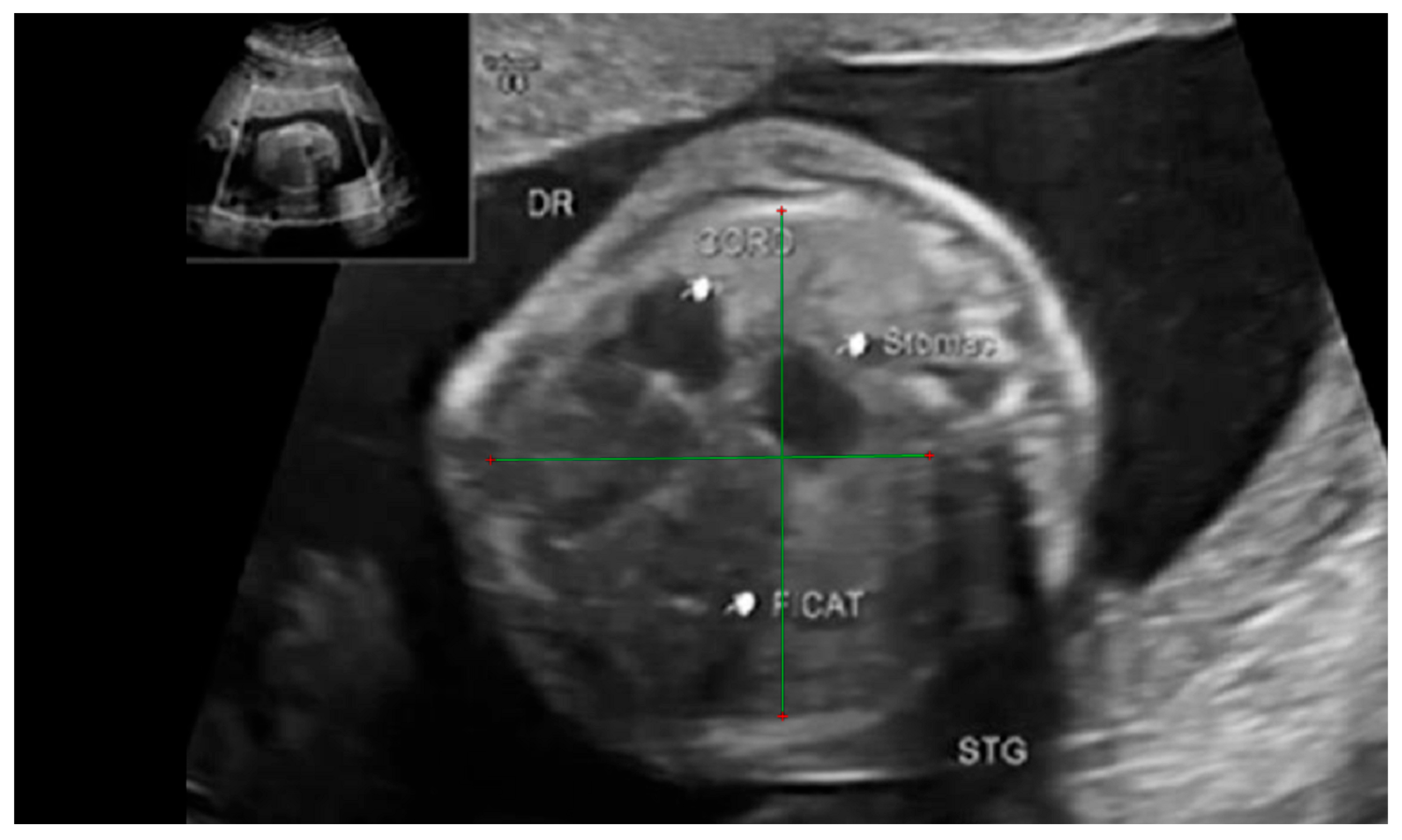
Thoracic Biometry in Patients with Congenital Diaphragmatic Hernia, a Magnetic Resonance Imaging Study
 ,
,
Abstract
:1. Introduction
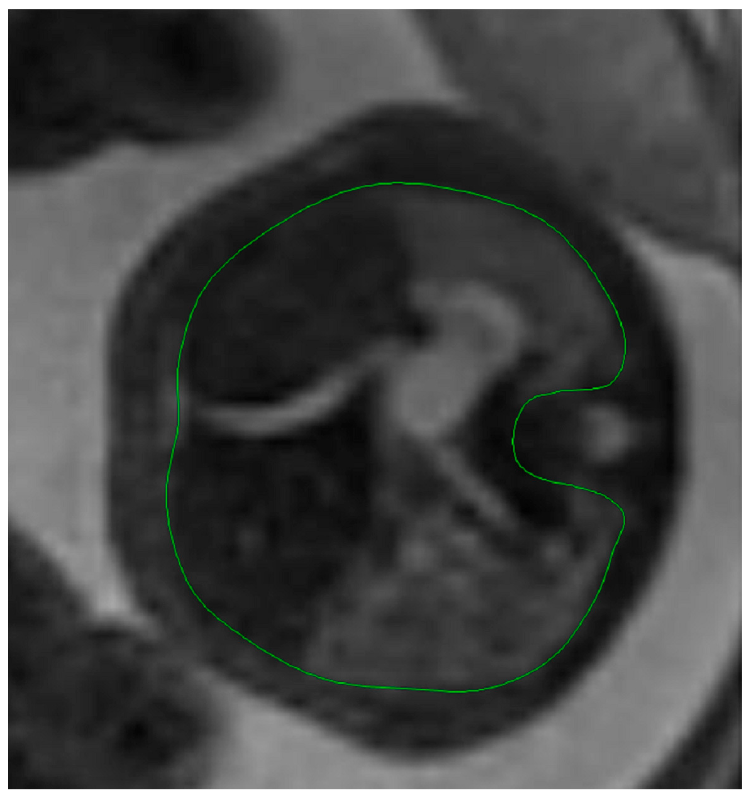
2. Materials and Methods
5. Conclusions
Author Contributions
Funding
Institutional Review Board Statement
Informed Consent Statement
Data Availability Statement
Conflicts of Interest
References
- Tisekar, O.R.; Ak, A.K. Hypoplastic Lung Disease; StatPearls Publishing: StatPearls, FL, USA, 2021. [Google Scholar]
- Weerakkody, Y. Pulmonary Hypoplasia|Radiology Reference Article|Radiopaedia.org. Available online: https://radiopaedia.org/articles/pulmonary-hypoplasia?lang=us (accessed on 15 March 2024).
- The Fetal Medicine Foundation. Available online: https://fetalmedicine.org/education/fetal-abnormalities/thorax/lung-agenesis-hypoplasia (accessed on 15 March 2024).
- Themes, U.F.O. Ultrasound Evaluation of the Fetal Thorax; Radiologykey: Minneapolis, MN, USA, 2019. [Google Scholar]
- Dumpa, V.; Chandrasekharan, P. Congenital Diaphragmatic Hernia; StatPearls: Treasure Island, FL, USA, 2021. [Google Scholar]
- Chakraborty, R.K.; Modi, P.; Sharma, S. Pulmonary Sequestration; StatPearls: Treasure Island, FL, USA, 2021. [Google Scholar]
- Kasprian, G. MRI of the Pathological Fetal Thorax. In Fetal MRI; Prayer, D., Ed.; Springer: Berlin/Haidelberg, Germany, 2011. [Google Scholar] [CrossRef]
- Schittny, J.C. Development of the lung. Cell Tissue Res. 2017, 367, 427–444. [Google Scholar] [CrossRef] [PubMed]
- Lauria, M.; Gonik, B.; Romero, R. Pulmonary hypoplasia: Pathogenesis, diagnosis, and antenatal prediction. Obstet. Gynecol. 1995, 86, 466–475. [Google Scholar] [CrossRef] [PubMed]
- Avena-Zampieri, C.L.; Hutter, J.; Rutherford, M.; Milan, A.; Hall, M.; Egloff, A.; Lloyd, D.F.; Nanda, S.; Greenough, A.; Story, L. Assessment of the fetal lungs in utero. Am. J. Obstet. Gynecol. MFM 2022, 4, 100693. [Google Scholar] [CrossRef]
- Hedrick, H.L. Management of prenatally diagnosed congenital diaphragmatic hernia. Semin. Pediatr. Surg. 2013, 22, 37–43. [Google Scholar] [CrossRef]
- Kalache, K.D.; Espinoza, J.; Chaiworapongsa, T.; Londono, J.; Schoen, M.L.; Treadwell, M.C.; Lee, W.; Romero, R. Three- dimensional ultrasound fetal lung volume measurement: A systematic study comparing the multiplanar method with the rotational (VOCAL) technique. Ultrasound Obstet. Gynecol. 2003, 21, 111–118. [Google Scholar] [CrossRef] [PubMed]
- Kasprian, G.; Balassy, C.; Brugger, P.C.; Prayer, D. MRI of normal and pathological fetal lung development. Eur. J. Radiol. 2006, 57, 261–270. [Google Scholar] [CrossRef] [PubMed]
- Jani, J.; Nicolaides, K.H.; Keller, R.L.; Benachi, A.; Peralta, C.F.A.; Favre, R.; Moreno, O.; Tibboel, D.; Lipitz, S.; Eggink, A.; et al. Observed to expected lung area to head circumference ratio in the prediction of survival in fetuses with isolated diaphragmatic hernia. Ultrasound Obstet. Gynecol. 2007, 30, 67–71. [Google Scholar] [CrossRef]
- Chin, T.W.; Kataria, N. Pediatric Pulmonary Hypoplasia Treatment Management: Medical Care, Surgical Care, Consultations. EMedicine 2023. Available online: https://emedicine.medscape.com/article/1005696-treatment?form=fpf (accessed on 16 March 2024).
- Sefidbakht, S.; Dehdashtian, A.; Bagheri, F.; Rahimirad, N.; Keshavarz, P.; Bijan, B. Standard Normal Fetal Lung Volume by MRI Measurement. Iran. J. Radiol. 2020, 17. [Google Scholar] [CrossRef]
- Osada, H.; Kaku, K.; Masuda, K.; Iitsuka, Y.; Seki, K.; Sekiya, S. Quantitative and Qualitative Evaluations of Fetal Lung with MR Imaging. Radiology 2004, 231, 887–892. [Google Scholar] [CrossRef]
- Rypens, F.; Metens, T.; Rocourt, N.; Sonigo, P.; Brunelle, F.; Quere, M.P.; Guibaud, L.; Maugey-Laulom, B.; Durand, C.; Avni, F.E.; et al. Fetal Lung Volume: Estimation at MR Imaging—Initial Results. Radiology 2001, 219, 236–241. [Google Scholar] [CrossRef]
- Meyers, M.L.; Garcia, J.R.; Blough, K.L.; Zhang, W.; Cassady, C.I.; Mehollin-Ray, A.R. Fetal Lung Volumes by MRI: Normal Weekly Values From 18 Through 38 Weeks’ Gestation. Am. J. Roentgenol. 2018, 211, 432–438. [Google Scholar] [CrossRef] [PubMed]
- Lian, X.; Xu, Z.; Zheng, L.; Zhu, Z.; Ejiwale, T.; Kumar, A.; Cai, P.; He, S.; Liu, S.; Zhang, Y.; et al. Reference range of fetal thorax using two-dimensional and three-dimensional ultrasound VOCAL technique and application in fetal thoracic malformations. BMC Med. Imaging 2021, 21, 34. [Google Scholar] [CrossRef] [PubMed]
- Cerbelle, V.; Le Duc, K.; Lejeune, S.; Mur, S.; Lerisson, H.; Drumez, E.; Sfeir, R.; Bigot, J.; Verpillat, P.; Boukhris, R.; et al. Fetal Lung Volume Appears to Predict Respiratory Morbidity in Congenital Diaphragmatic Hernia. J. Clin. Med. 2023, 12, 1508. [Google Scholar] [CrossRef] [PubMed]
- Gonçalves, A.N.; Correia-Pinto, J.; Nogueira-Silva, C. Imagiological methods for prediction of fetal pulmonary hypoplasia: A systematic review. J. Matern. Fetal Neonatal Med. 2021, 34, 1459–1468. [Google Scholar] [CrossRef] [PubMed]
- Nimrod, C.; Davies, D.; Iwanicki, S.; Harder, J.; Persaud, D.; Nicholson, S. Ultrasound prediction of pulmonary hypoplasia. Obstet. Gynecol. 1986, 68, 495–498. [Google Scholar] [PubMed]
- Fong, K.; Ohlsson, A.; Zalev, A. Fetal thoracic circumference: A prospective cross-sectional study with real-time ultrasound. Am. J. Obstet. Gynecol. 1988, 158, 1154–1160. [Google Scholar] [CrossRef] [PubMed]
- Chitkara, U.; Rosenberg, J.; Chervenak, F.A.; Berkowitz, G.S.; Levine, R.; Fagerstrom, R.M.; Walker, B.; Berkowitz, R.L. Prenatal sonographic assessment of the fetal thorax: Normal values. Am. J. Obstet. Gynecol. 1987, 156, 1069–1074. [Google Scholar] [CrossRef]
- Vintzileos, A.M.; Campbell, W.A.; Rodis, J.F.; Nochimson, D.J.; Pinette, M.G.; Petrikovsky, B.M. Comparison of six different ultrasonographic methods for predicting lethal fetal pulmonary hypoplasia. Am. J. Obstet. Gynecol. 1989, 161, 606–612. [Google Scholar] [CrossRef]
- Laudy, J.A.M.; Wladimiroff, J.W. The fetal lung 2: Pulmonary hypoplasia. Ultrasound Obstet. Gynecol. 2000, 16, 482–494. [Google Scholar] [CrossRef]
- Yoshimura, S.; Masuzaki, H.; Gotoh, H.; Fukuda, H.; Ishimaru, T. Ultrasonographic prediction of lethal pulmonary hypoplasia: Comparison of eight different ultrasonographic parameters. Am. J. Obstet. Gynecol. 1996, 175, 477–483. [Google Scholar] [CrossRef]
- Ruano, R.; Marques, M.; Alvares, J.; Papanna, R.; Moise, K.; Tannuri, U.; Zugaib, M. Fetal Pulmonary Response After Fe- toscopic Tracheal Occlusion for Severe Isolated Congenital Diaphragmatic Hernia. Obstet. Gynecol. 2012, 119, 93–101. [Google Scholar] [CrossRef]
- Prayer, F.; Metzelder, M.; Krois, W.; Brugger, P.C.; Gruber, G.M.; Weber, M.; Scharrer, A.; Rokitansky, A.; Langs, G.; Prayer, D.; et al. Three-dimensional reconstruction of defects in congenital diaphragmatic hernia: A fetal MRI study. Ultrasound Obstet. Gynecol. 2019, 53, 816–826. [Google Scholar] [CrossRef]
- Van der Veeken, L.; Russo, F.M.; De Catte, L.; Gratacos, E.; Benachi, A.; Ville, Y.; Nicolaides, K.; Berg, C.; Gardener, G.; Persico, N.; et al. Fetoscopic endoluminal tracheal occlusion and reestablishment of fetal airways for congenital diaphragmatic hernia. Gynecol. Surg. 2018, 15, 9. [Google Scholar] [CrossRef]
- Perrone, E.E.; Deprest, J.A. Fetal endoscopic tracheal occlusion for congenital diaphragmatic hernia: A narrative review of the history, current practice, and future directions. Transl. Pediatr. 2021, 10, 1448–1460. [Google Scholar] [CrossRef]
- Harrison, M.R.; Adzick, N.; Bullard, K.M.; Farrell, J.A.; Howell, L.J.; Rosen, M.A.; Sola, A.; Goldberg, J.D.; Filly, R.A. Correction of congenital diaphragmatic hernia in utero VII: A prospective trial. J. Pediatr. Surg. 1997, 32, 1637–1642. [Google Scholar] [CrossRef]
- Deprest, J. Prenatal treatment of severe congenital diaphragmatic hernia: There is still medical equipoise. Ultrasound Obstet. Gynecol. 2020, 56, 493–497. [Google Scholar] [CrossRef] [PubMed]


| Variable | Minimum | Maximum | Mean | Std. Deviation | p Anderson–Darling | Interpretation |
|---|---|---|---|---|---|---|
| Observed thoracic tr mm | 42.00 | 77.00 | 59.21 | 9.35 | 0.8132 | Gaussian distribution |
| Observed thoracic ap mm | 30.00 | 65.00 | 44.00 | 10.39 | 0.0556 | Gaussian distribution |
| Observed thoracic circumference mm | 147.00 | 286.00 | 217.58 | 40.64 | 0.6961 | Gaussian distribution |
| Observed thoracic area mm2 | 1219.00 | 4656.00 | 2765.53 | 1037.80 | 0.0721 | Gaussian distribution |
| Observed thoracic volume mL | 31.42 | 177.79 | 84.60 | 50.38 | 0.0040 | non-Gaussian distribution |
| Observed lung-to-thoracic volume ratio | 0.05 | 0.39 | 0.18 | 0.08 | 0.0407 | non-Gaussian distribution |
| Expected thoracic tr mm | 40.80 | 66.30 | 54.74 | 7.95 | 0.1238 | Gaussian distribution |
| Expected thoracic ap mm | 29.40 | 56.40 | 43.79 | 8.83 | 0.1289 | Gaussian distribution |
| Expected thoracic circumference mm | 144.60 | 254.30 | 204.09 | 34.23 | 0.1457 | Gaussian distribution |
| Expected thoracic area mm2 | 1565.00 | 4599.00 | 3070.32 | 1010.04 | 0.0578 | Gaussian distribution |
| Expected total pulmonary volume (mL) VOCAL study | 13.54 | 64.67 | 38.45 | 16.99 | 0.0589 | Gaussian distribution |
| Expected thoracic volume | 27.10 | 114.23 | 70.22 | 28.75 | 0.0761 | Gaussian distribution |
| Total pulmonary volume index VOCAL comparison | 0.08 | 0.69 | 0.37 | 0.14 | 0.2561 | Gaussian distribution |
| Expected lung-to-thoracic volume ratio | 0.50 | 0.57 | 0.54 | 0.02 | 0.1867 | Gaussian distribution |
| Observed total pulmonary volume (mL) | 1.6800 | 31.4700 | 14.0316 | 7.3288 | 0.9550 | Gaussian distribution |
| Expected total pulmonary volume (mL) mean MRI studies | 15.2444 | 64.0786 | 39.7768 | 15.2639 | 0.1321 | Gaussian distribution |
| Total pulmonary volume index MRI comparison | 0.0709 | 0.6281 | 0.3494 | 0.1344 | 0.4444 | Gaussian distribution |
| Variable 1 | Variable 2 | p Student | Significance |
|---|---|---|---|
| Observed thoracic tr mm | Expected thoracic tr mm | 0.0002 | HS |
| Observed thoracic ap mm | Expected thoracic ap mm | 0.8797 | NS |
| Observed thoracic circumference mm | Expected thoracic circumference mm | 0.0005 | HS |
| Observed thoracic area mm2 | Expected thoracic area mm2 | 0.0042 | S |
| Observed thoracic volume mL | Expected thoracic volume | 0.0518 | NS |
| Observed lung-to-thoracic volume ratio | Expected lung-to-thoracic volume ratio | 0.0000 | HS |
| Observed total pulmonary volume (mL) | Expected total pulmonary volume (mL) mean MRI studies | 0.0000 | HS |
| Observed total pulmonary volume (mL) | Expected total pulmonary volume (mL) VOCAL study | 0.0000 | HS |
| Total pulmonary volume index MRI comparison | Total pulmonary volume index VOCAL comparison | 0.0035 | S |
| Expected total pulmonary volume (mL) mean MRI studies | Expected total pulmonary volume (mL) VOCAL study | 0.0112 | S |
Disclaimer/Publisher’s Note: The statements, opinions and data contained in all publications are solely those of the individual author(s) and contributor(s) and not of MDPI and/or the editor(s). MDPI and/or the editor(s) disclaim responsibility for any injury to people or property resulting from any ideas, methods, instructions or products referred to in the content. |
© 2024 by the authors. Licensee MDPI, Basel, Switzerland. This article is an open access article distributed under the terms and conditions of the Creative Commons Attribution (CC BY) license (https://creativecommons.org/licenses/by/4.0/).
Share and Cite
Neștianu, E.G.; Popescu, S.; Alexandru, D.O.; Giurcăneanu, L.; Vlădăreanu, R. Thoracic Biometry in Patients with Congenital Diaphragmatic Hernia, a Magnetic Resonance Imaging Study. Diagnostics 2024, 14, 641. https://doi.org/10.3390/diagnostics14060641
Neștianu EG, Popescu S, Alexandru DO, Giurcăneanu L, Vlădăreanu R. Thoracic Biometry in Patients with Congenital Diaphragmatic Hernia, a Magnetic Resonance Imaging Study. Diagnostics. 2024; 14(6):641. https://doi.org/10.3390/diagnostics14060641
Chicago/Turabian StyleNeștianu, Erick George, Septimiu Popescu, Dragoș Ovidiu Alexandru, Laura Giurcăneanu, and Radu Vlădăreanu. 2024. "Thoracic Biometry in Patients with Congenital Diaphragmatic Hernia, a Magnetic Resonance Imaging Study" Diagnostics 14, no. 6: 641. https://doi.org/10.3390/diagnostics14060641