Review

miR-125 in Breast Cancer Etiopathogenesis: An Emerging Role as a Biomarker in Differential Diagnosis, Regenerative Medicine, and the Challenges of Personalized Medicine

by
Roberto Piergentili
1,
Enrico Marinelli
2,
Gaspare Cucinella
3,
Alessandra Lopez
3,
Gabriele Napoletano
4,
Giuseppe Gullo
3,† and
Simona Zaami
4,*,†
1
Institute of Molecular Biology and Pathology, Italian National Research Council (CNR-IBPM), 00185 Rome, Italy
2
Department of Medico-Surgical Sciences and Biotechnologies, “Sapienza” University of Rome, 04100 Latina, Italy
3
Department of Obstetrics and Gynecology, Villa Sofia Cervello Hospital, University of Palermo, 90146 Palermo, Italy
4
Department of Anatomical, Histological, Forensic and Orthopedic Sciences, Section of Forensic Medicine, “Sapienza” University of Rome, 00161 Rome, Italy
*
Author to whom correspondence should be addressed.
These authors contributed equally to this work.
Non-Coding RNA 2024, 10(2), 16; https://doi.org/10.3390/ncrna10020016
Submission received: 15 December 2023 / Revised: 10 February 2024 / Accepted: 19 February 2024 / Published: 21 February 2024

Abstract

:
Breast Cancer (BC) is one of the most common cancer types worldwide, and it is characterized by a complex etiopathogenesis, resulting in an equally complex classification of subtypes. MicroRNA (miRNA or miR) are small non-coding RNA molecules that have an essential role in gene expression and are significantly linked to tumor development and angiogenesis in different types of cancer. Recently, complex interactions among coding and non-coding RNA have been elucidated, further shedding light on the complexity of the roles these molecules fulfill in cancer formation. In this context, knowledge about the role of miR in BC has significantly improved, highlighting the deregulation of these molecules as additional factors influencing BC occurrence, development and classification. A considerable number of papers has been published over the past few years regarding the role of miR-125 in human pathology in general and in several types of cancer formation in particular. Interestingly, miR-125 family members have been recently linked to BC formation as well, and complex interactions (competing endogenous RNA networks, or ceRNET) between this molecule and target mRNA have been described. In this review, we summarize the state-of-the-art about research on this topic.

1. Introduction

Breast Cancer (BC) is the most commonly diagnosed cancer type, accounting for one in seven cancer diagnoses [1]. Available data show that incidence and mortality in developed countries have been declining over time, while they have increased in low-income countries [2]. In 2020, there were about 2.3 million new cases of BC globally and about 685,000 deaths from this disease, with large geographical variations reported in individual countries and world regions. BC incidence rates are highest in developed countries, whereas develo** countries have a disproportionately high share of BC deaths [3]. In the United States, BC alone is expected to account for more than 30% of all new cancers in women [1]. The 2018 GLOBOCAN (Global Cancer Data) shows that age-standardized incidence rates (ASIR) of BC are strongly and positively associated with the Human Development Index (HDI) [4]. The HDI is a statistical composite index developed by the United Nations Development Programme’s (UNDP) Human Development Report Office to measure life expectancy at birth, education, and national per capita incomes. The above-mentioned study focused on BC expanding and completing the HDI data by considering additional indexes. Data reported by Sharma indicate that the high incidence and high survival rates in developed countries probably reflect BC early detection, likely due to better cancer infrastructures available (e.g., hospitals), systematic screening programs (e.g., breast mammograms), and more efficient BC treatment in these countries, which low HDI countries do not possess.

2. Clinical Features of BC

2.1. Main Risk Factors of BC

A variety of risk factors for BC have been well-established by epidemiologic studies and include ethnicity as well as behavioral variables, such as sedentary lifestyle or increased alcohol consumption. Overdrinking can, in fact, elevate estrogen-related hormone levels in the blood and trigger estrogen receptor pathways [5]. In addition, endogenous and exogenous estrogens are associated with an increased risk of BC. Endogenous estrogen is usually produced by the ovary in premenopausal women, and ovariectomy can reduce the risk of BC. The main sources of exogenous estrogen are oral contraceptives and hormone replacement therapy (HRT) [6]. Certain female reproductive factors, such as younger age at menarche, low parity, late menopause, and older age at first full-term pregnancy, may influence BC risk through long-term effects on sex hormone levels or by other biological mechanisms, although recent studies suggest that triple negative BC may have a distinct etiology [7]. Over the past decades, the incidence of pregnancy-associated BC (PABC) has been on the rise as well [8]. Lastly, having extremely dense breast tissue is also significantly associated with increased BC risk compared to having scattered dense breast tissue [9].
Nearly one-fourth of all BC cases are related to family medical history: women whose mothers or sisters have BC are more prone to develo** this disease [10], indicating a strong genetic basis in its etiology. Indeed, the inherited susceptibility to BC is attributed to mutations in BC-related genes such as BRCA1 and BRCA2 [11]. Additional genes that, during the years, have been implicated in BC pathogenesis include PTEN [12], TP53 [13], CDH1 [14], STK11 [15], CHEK2 [16], PALB2 [17], ATM [18], RAD51C [19], RAD51D [20], BARD1 [21], NF1 [22], BRIP1 [23], CASP8, CTLA4, NBN, and, possibly, CYP19A1, TERT and XRCC3 [24]. A summary of the most frequently mutated genes in BC, their function and their relation to BC is summarized in Table 1.

2.2. BC Characterization and Patient Management

BC is commonly diagnosed via ultrasonography [29,30]. Mammography screening for malignancy is commonly used as well to detect the disease [31], while breast magnetic resonance imaging (MRI) is used in conjunction with mammography as a support tool [32]. In this case, MRI can be helpful in deciding whether to have a breast-conserving mastectomy or surgery [33,34]. A biopsy is performed when mammograms, other imaging tests, or a physical exam show a breast change that may be identified as a possible cancerous mass. Computerized tomography (CT) scans, MRIs, ultrasound, and positron emission tomography (PET) scans may also provide information as to cancer extension and position. Laboratory tests of cancer cells (from biopsy or surgery) and blood tests can also be used to help stage some types of cancer [35]. Recently developed techniques for BC diagnosis include Digital Breast Tomosynthesis (DBT), which is a subset of the mammography procedure [36], and the Contrast-enhanced digital mammography (CEDM), which represents the angiogenic pattern of the masses and allows depicting the anatomical information of the tissue [37]. Each of these methods has different subdivisions and their advantages and disadvantages have been discussed in the literature [38,39,40]. Although there are ways to improve these methods, it should be kept in mind that simultaneously combining multiple imaging techniques would significantly improve BC early detection [41].
The stage of a cancer is helpful in assessing its extension and spreading. The tumor-node-metastasis (TNM) staging system (Table 2) is currently the most widespread method to stage BC. However, staging for BC can be very complex; many different factors should be accounted for before the cancer stage can be confirmed to outline the most suitable therapeutic approach. The TNMEIO system was suggested by the European Institute of Oncology (EIO) to include tumor characteristics affecting treatment decisions in the TNM system [42].
Perou and Sorlie proposed the “Molecular Classification” terminology in BC for the first time in 2000 with a comprehensive study showing the differences in gene expression of different BC specimens. In this study, breast tissue samples were divided into different sub-groups according to variable gene expression, i.e., ER+/luminal-like, basal-like, Erb-B2+ and normal breast [43,44].
The complexity of BC staging is also reflected in the different histological classifications used to identify its various subtypes. This has led to defining different BC types, such as ductal carcinoma (in situ or invasive), medullary carcinoma, lobular carcinoma (in situ or infiltrating), tubular and mucinous carcinoma. Expression analysis of additional molecular markers such as the estrogen receptor (ER), progesterone receptor (PR) and HER2/neu proteins can provide further, highly valuable knowledge for the oncologist. Once the status of these proteins is known, the prognosis can be reasonably predicted, and more appropriate therapies may be chosen for treatment [45]. Recently, the eighth edition of the American Joint Commission of Cancer (AJCC) staging system for BC approved major changes in the classification system, adopting an anatomy-based and histology-based subdivision built on the original TNM staging system and adding various biomarkers to refine the prognostic information for better selection of therapy with improved outcome [46]. This finer classification should contribute to better characterizing the molecular and anatomical features of each patient and delineating a therapeutic pathway accordingly. Thus, it is crucial for oncologists to have the most comprehensive description of BC, both from a histological and a molecular standpoint.
BC therapy involves a multidisciplinary approach relying on surgery, radiotherapy, chemotherapy, hormone therapy, immunotherapy, neoadjuvant and adjuvant therapy. Effective BC therapy must aim for maximum therapeutic efficacy [47]. There is increasing recognition that the care of a BC patient depends on highly individualized clinical features, including the stage at presentation, the biological subset of BC, the genetic factors that may underlie BC risk, the genomic signatures that advise treatment recommendations, the extent of response before surgery in patients who receive neoadjuvant therapy, and patient preferences. This customized approach to treatment requires a concerted, multidisciplinary effort shared among patients and radiology, pathology, genetics, and surgical, medical and radiation oncology providers to minimize adverse effects and preserve quality of life as much as possible [48].
The search for predictive biomarkers useful to draw further distinctions among BC subtypes is an active field of research that includes genomic, proteomic and/or machine learning approaches. In recent years, epigenetic biomarkers have gained growing attention, especially micro-RNAs (miRNA or miR) that have been predicted (and in some cases, validated) as very promising BC markers [49,50,51] for early detection as well [52,53,54]; notably, some studies also concentrated on circulating miR, opening the way towards a minimally invasive diagnostic approach [55,56,57,58].

3. Epigenetics of BC and the Role of miR-125

3.1. microRNA Nomenclature

MicroRNAs are short (20–25 nucleotides), single-stranded, non-coding RNA molecules whose main function is gene expression control, mainly silencing. They exert this downregulation by binding the 3′ end of target mRNA(s) through sequence homology and promoting either their degradation or impairing their translation [59]. Over 2500 miR have been estimated to be encoded in the human genome, regulating over 60% of human genes [60]. In addition, thanks to imperfect sequence pairing, they can also bind multiple targets, thus amplifying their intracellular action.
Beyond the number identifier (ID, usually higher for miR described chronologically later), additional nomenclature rules are established to identify unequivocally each miR [61,62]. miRs with almost identical sequence are identified by a progressive lowercase letter after the identification number (miR-XXXa, miR-XXXb, etc.), while miRs with identical sequence but map** to different genomic locations are indicated by a progressive number separated from the ID number by a dash (e.g., miR-XXX-1, miR-XXX-2, etc.). To further distinguish molecules of different species, an additional three-letter code and a dash may be added at the beginning of the miR name (i.e., hsa-miR-XXX indicates a human–Homo sapiens–miR). Finally, the mature, single-stranded miR can be obtained either from the 5′ end or the 3′ end of its double-stranded miR precursor (pre-miR). Notably, sometimes both strands can—separately—be used for mRNA regulation, resulting in 5p and 3p miR forms if both are present and functional in the cell and the two miR are roughly equivalent in their intracellular amount; instead, if both are present but one is significantly more abundant than the other, then the rarer one has an asterisk at the end of its name (e.g., miR-XXX-5p*).

3.2. Role of miR in BC

Increasing evidence shows that miRs represent a central hub of gene expression control in human carcinogenesis, and, from this perspective, BC is not an exception [63,64].
Significantly, miR-21 has been shown to be responsible for the development of multidrug resistance [65], and it modulates the resistance of BC cells to doxorubicin by targeting PTEN [66]. In addition, miR-21 also plays a central role in BC proliferation and metastasis by targeting LZTFL1 [67]. Additional miR-21 targets involved in cell proliferation, metastasis, epithelial-to-mesenchymal transition (EMT), and apoptosis in BC include IGFBP3, TPM1, PCD4, and TGF-beta1 [68].
Another player in BC etiopathogenesis is miR-106a, which promotes cancer progression through the downregulation of RAF-1 [69], P53, BAX, and RUNX3 and the upregulation of Bcl-2 and ABCG2; it also confers cisplatin resistance upon its upregulation [70,71].
Upregulation of miR-155 causes telomere fragility through its action on TRF1, a component of the shelterin complex [72]. Interestingly, this miR, together with miR-10b, miR-34a and miR-141, is also a possible candidate for building a panel of circulating miR useful for non-invasive detection of this tumor [73].
Conversely, downregulated miR-141 has been reported to be a typical feature of BC, where its target is ANP32E [74], which, in turn, induces tumorigenesis in triple-negative BC (TNBC) cells by upregulating E2F1 [75]. Instead, high miR-141-3p expression is typical of grade III BC compared to grade II and, together with miR-181b1-5p and miR-23b-3p, it is a useful marker not only to discriminate between malignant and benign breast tissues but might also help in distinguishing TNBC from other molecular subtypes of BC [76].
The let-7 family of miR are tumor suppressors in several cancers, including BC, and their members can also be detected as circulating biomarkers [77]. Let-7 action involves the control of ERCC6 expression [78], and its overexpression could inhibit BC cell proliferation.
Another circulating marker of BC is miR-335 [79], which exerts its effects by simultaneously regulating the known BRCA1 activators ERα, IGF1R, SP1 and the repressor ID4, including a feedback regulation of miR-335 expression by estrogens [80]. Its overexpression causes decreased cell viability and increased apoptosis, while other findings show it to negatively regulate the HGF/c-Met pathway, thus affecting cell scattering, migration, and invasion [81].
Another downregulated miR in BC is miR-126 [82]. Its targets include VEGFA and PIK3R2 [83]. It also reduces trastuzumab resistance by targeting PIK3R2 and regulating the AKT/mTOR signaling pathway [84] and controls cell invasion by targeting ADAM9 [85].
Tumor suppressor miR-199a/b-3p inhibits migration and invasion of BC cells by downregulating the PAK4/MEK/ERK signaling pathway [86]. Overexpression of miR-199a-3p targets the c-Met and mTOR pathways, increases doxorubicin sensitivity and causes G1 phase arrest, thus reducing cell invasion and promoting doxorubicin-induced apoptosis [87]. This miR also confers resistance to cisplatin treatment by downregulating TFAM [88] and, at the same time, promotes BC development and metastasis under hypoxic conditions by controlling the regulatory axis consisting of HIF-1, SNHG1, and TFAM [89]. The same study mentioned above [87] also shows that in TNBC patients, additional circulating miR (i.e., miR-19a/b-3p, miR-25-3p, miR-22-3p, miR-210-3p and miR-93-5p) are deregulated as well and control several molecular pathways involved in drug resistance, making them amenable to be used as BC biomarkers, together with let-7a-5p, miR-100-5p and miR-101-3p, identified in another study [90].
Tumor suppressor miR-101 is downregulated as well in BC, and its targets include POMP, Stmn1, DNMT3A, EYA1, VHL, SOX2, Jak2 and MCL-1 (reviewed in [91]). For this reason, it plays a major role in the control of several cancer-related cellular processes, such as proliferation, apoptosis, angiogenesis, drug resistance, invasion, and metastasis. Overexpression of miR-101-3p can inhibit the migration of BC cells into the brain endothelium, a frequent and late event in BC patients, by inducing COX-2/MMP1 signaling, which can degrade the inter-endothelial junctions (claudin-5 and VE-cadherin) [92]. Jiang and collaborators showed that the suppression of the oncogene EZH2 in BC by miR-101-3p is potentiated in the presence of syn-cal14.1a, a synthetic peptide derived from Californiconus californicus (a sea snail), thus inhibiting cell migration, invasion, and proliferation [93]. Additional data come from the work of Toda and collaborators, who performed an RNA-sequence-based microRNA expression signature in BC and identified other dysregulated miRs in BC (e.g., miR-99a-5p/-3p, miR-101-5p/-3p, miR-126-5p/-3p, miR-143-5p/-3p, and miR-144-5p/-3p) and found that miR-101-5p controls the expression of seven putative oncogenes (i.e., HMGB3, ESRP1, GINS1, TPD52, SRPK1, VANGL1 and MAGOHB) [94].
Finally, miR-9 is known to exert critical functions in the initiation and progression of BC. Its upregulation—together with that of miR-221/222, miR-373 and miR-10b—is linked to highly malignant invasive EMT and cancer stem cell production [95]. Conversely, its downregulation can lead to improved overall survival, smaller tumors, earlier stages, and ER-positive cancers due to the enrichment of estrogen response genes [96]. Gwak and collaborators showed that miR-9 is highly expressed in HER2+ and TNBC subtypes compared with luminal subtypes, tumors with a high tumor stage or histologic grade, and tumors displaying the CD44+/CD24− phenotype, vimentin expression, and E-cadherin loss [97]. Interestingly, Shen and collaborators showed miR-9-5p, together with miR-195-5p and miR-203a-3p, to be a part of the extracellular vesicle (EV)-encapsulated miR (enabling cancer cell–cell communication in tumor pathogenesis and response to therapies) excreted upon docetaxel treatment [98]. As for its targets, genes identified so far include FOXO1 [99], STARD13 [100], LIFR [101], elf5A2 [102], HMGA2, EGR1, and IGFBP3 [68] and PDGFRbeta [103]. In turn, its expression is activated by MYC and MYCN [104,105].
A summary of the miR involved in BC and their function is summarized in Table 3.
All together, these data point to the involvement of several miRs in BC formation, development, metastasis, and drug resistance, showing that at the molecular level it is crucial to identify which pathways are altered and why, for example, the same gene may be deregulated because of the alteration of diverse miR. Knowing which miR is altered may greatly affect therapeutic approaches, especially in terms of avoiding cross-effects due to off-target actions. Thus, there is a necessity to identify (hopefully, all) the players in BC pathogenesis in a patient-specific way. In this perspective, the miR-125 family of miR has gained increasing relevance and attention in BC research, thanks to the numerous publications released over the last few years. In light of the numerous targets of these miRs and the multitude of pathways potentially altered inside the cell upon their dysregulation, in the next few years, miR-125 is likely to become central to understanding BC biology.

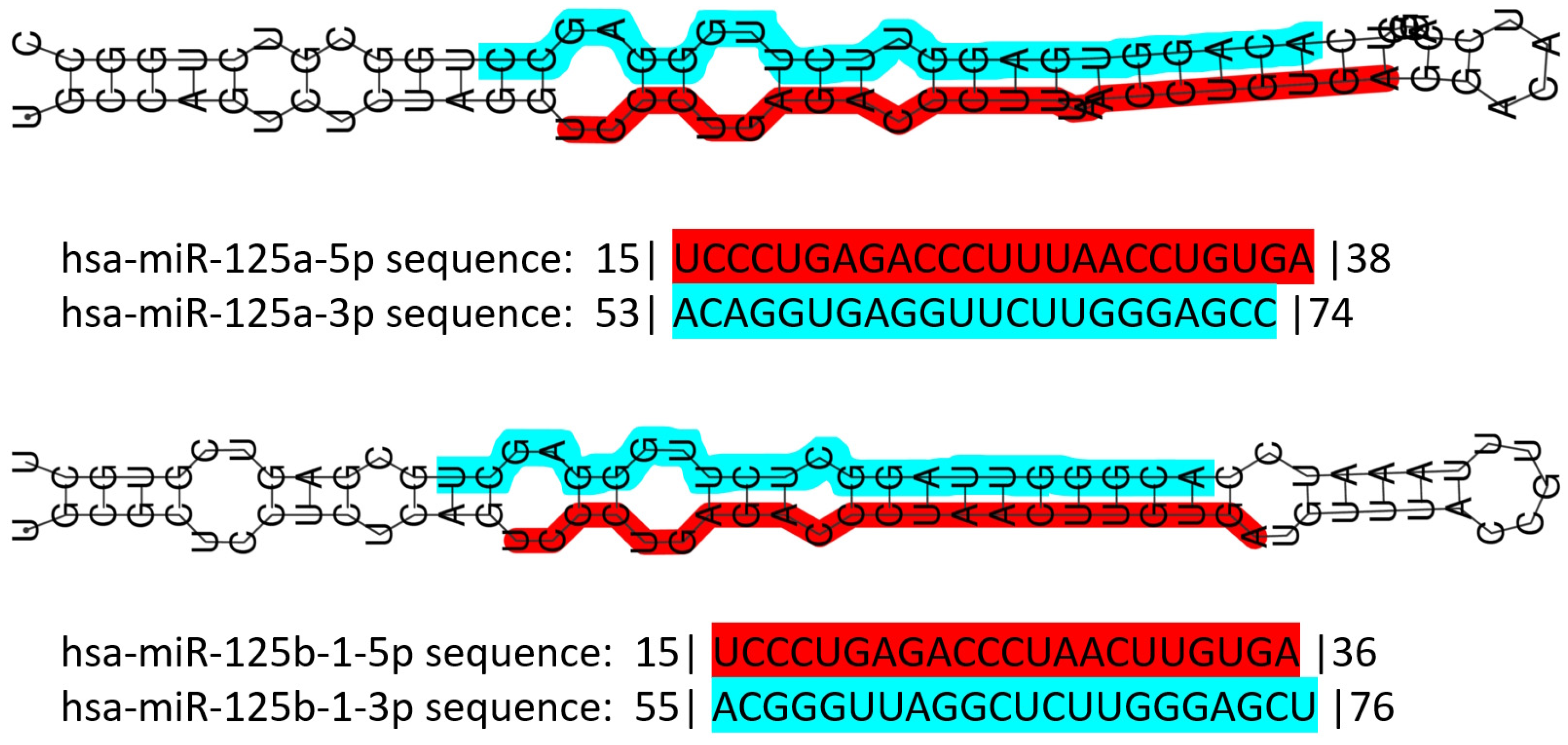
3.3. The miR-125 Family: Molecular Organization and Roles in Human Pathology

miR-125 is a highly conserved family of microRNAs whose members have also been found in nematodes (named lin-4 in 1993, the first miR described ever) [106]. The miR-125 family in H. sapiens includes three members, namely miR-125a, miR-125b-1 and miR-125b-2. The MIR125A gene maps to chromosome 19q13.41 [107], and miR-125a is part of a transcribed cluster of miR, together with miR-99b and let-7e [108]. The MIR125B1 gene maps to chromosome 11q24.1, and in this locus, it is part of a cluster including the LET7A2 and MIR100 genes [108,109]. These miRs are inside the third intron of the MIR100HG gene [110]. Finally, the MIR125B2 gene maps to chromosome 21q21.1, where it is included in a cluster together with the MIR99A and LET7C genes [108,109], inside the sixth intron of the MIR99AHG gene [110]. miR-125a and miR-125b differ only by a central diuridine insertion and a U-to-C change in miR-125a [111]. All members of the family show both 5p and 3p forms (Figure 1).
The miR-125 family is involved in several cell metabolic pathways controlling differentiation, proliferation, apoptosis, metastasis formation, drug resistance and immune system function because of the targeting of mRNAs related to these cellular processes [114] (Figure 2). miR-125 molecules have a complex behavior inside the cell, which mirrors their expression pattern in different tissues/cell types [112,113], their ample variety of targets [112,113], the intracellular role of their targets, and the way miR and mRNAs are either up- or down-regulated upon expression.
The role of miR-125 family members has been extensively demonstrated in the muscle. It interacts with insulin-like growth factor II (IGF-II) to regulate myoblast differentiation in vitro and muscle regeneration in vivo [116], and with TRAF6 to prevent atrophy [117]. It is also involved in the proliferation and migration of vascular smooth muscle cells induced by platelet-derived growth factor BB [118]. In cardiac muscles, miR-125 participates in the development of the heart in embryonic mammals (reviewed in [119]); it regulates muscle-enriched transcription factors in cardiac and skeletal myocytes [120]; it can modulate cardiac progenitor cell proliferation and migration potential [121]; and it regulates cardiomyocytes proliferation and apoptosis under oxidative stress conditions [122]. Cardiac-specific miR-125b deficiency has recently been shown to induce perinatal death and cardiac hypertrophy [123].
miR-125 is one of the most abundant microRNAs in the central nervous system (CNS) in both mice and men [124]. In humans, miR-125b promotes neuronal differentiation in human cells by repressing at least ten target mRNAs involved in those pathways [125,126]. It also regulates dendritic spine morphology and synaptic maturation [127], it is implicated in synaptic plasticity [128], promotes astrogliogenesis, and is involved in astrogliosis and glial cell proliferation [129]. Its deregulation has also been linked to CNS tumor formation and growth, such as pediatric low-grade glioma [130]; it regulates cell growth arrest and apoptosis of human neuroblastoma- and medulloblastoma-derived cell lines [131,132]; it inhibits cell apoptosis through p53 and p38MAPK-independent pathways in glioblastoma cells [133]; and, in glioma, it targets BMF [134].
In the immune system, miR-125 regulates hematopoiesis, inflammation, and immune cell function. miR-125a controls stem cell homeostasis during hematopoiesis [135,136,137,138] and plays a role in immune cell identity [138]. miR-125-5p targeting IL-6R regulates macrophage inflammatory response and intestinal epithelial cell apoptosis in ulcerative colitis through the JAK1/STAT3 and NF-κB pathways [139]. miR-125b-1-3p is expressed in hMSCs-Ad exosomes and can promote T lymphocyte apoptosis and alleviate atherosclerosis (AS) by down-regulating BCL11B expression, thus providing potential molecular targets for the clinical treatment of AS [140].
All together, these data emphasize the multiple roles of miR-125 family members in cell proliferation and differentiation in numerous body locations.

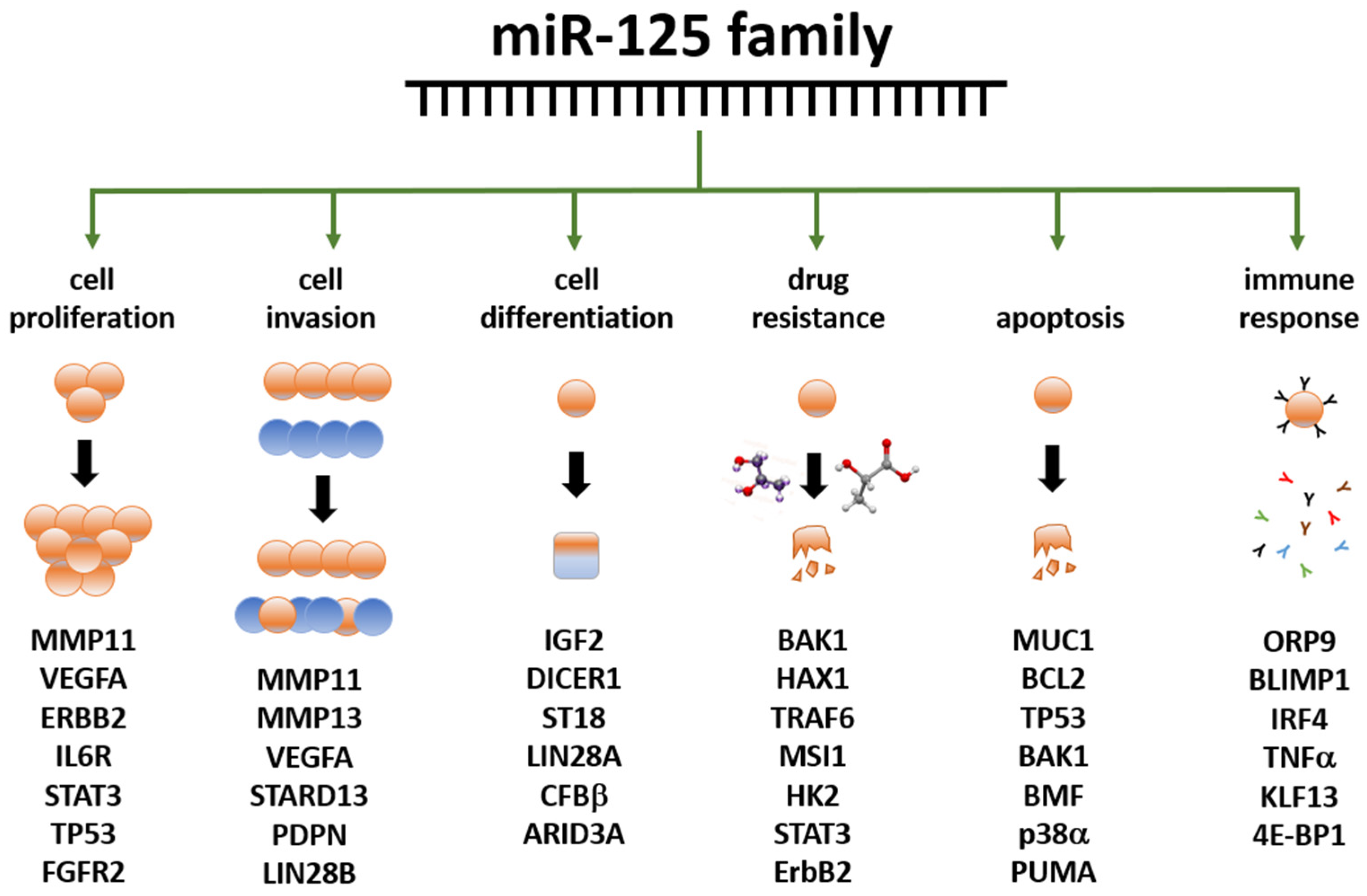
3.4. miR-125 and Cancer

Studying the role of miR-125 in cancer is an important research area; beyond the above-mentioned tumors of the CNS, this noncoding RNA is indeed deregulated in several other tumors [141]. Hematological cancers are the best-characterized malignancies in which miR-125 role is well established; due to the rather conclusive amount of findings available, we redirect the reader to specific and comprehensive reviews [142,143]. Additional organs affected by miR-125-related cancers include the ovary, bladder, liver, skin, bone, lung, pancreas, prostate, thyroid, stomach, colon and kidney. A summary of these cancers, known targets and related bibliographic references are reported in Table 4. A detailed description of the role of miR-125 family members in BC is reported in the next section.

3.5. Role of miR-125 in BC

A relatively small amount of research is currently available on the role of miR-125 in BC. The reports showing its altered expression in these malignancies started to be published more than 20 years ago, and the research is still running in search of an affordable diagnostic panel based on this noncoding RNA [183,184,185,186,187,188]. Among the targets first identified, it is worth mentioning ERBB2 and ERBB3 [189] mRNA. In 2011, Zhang and colleagues demonstrated the action of miR-125b on the regulation of the ETS1 proto-oncogene in human invasive BC [190]. Rajabi et al. found that miR-125b, downregulated in BC, can reduce the expression of MUC1 (an oncoprotein), whose silencing causes DNA damage-induced apoptosis in cancer cells [191]. Tang and collaborators studied the effects of miR-125 deregulation on metastasis formation, finding that miR-125b induces metastasis by targeting STARD13 mRNA in MCF-7 and MDA-MB-231 BC cells [192], in contrast with the tumor suppressive action described before. Using the same BC cell lines, Metheetrairut and collaborators showed that forced expression of miR-125b results in radiosensitivity, as seen by reduced clonogenic survival, enhanced apoptotic activity and enhanced senescence post-ionizing radiation treatment. Moreover, re-expression of c-JUN in MDA-MB-231 cells promoted radioresistance and abrogated miR-125-mediated radiosensitization, suggesting that overexpression of miR-125b causes sensibilization to γ-irradiation and indicating this miR as a possible target for adjuvant therapy [193]. In contrast, Wang et al. found an association between miR-125b expression and chemoresistance [194], again indicating an oncogenic role for this miR. In line with these last results, Zhou and collaborators found that miR-125b confers the resistance of BC cells to paclitaxel through suppression of pro-apoptotic Bcl-2 antagonist killer 1 (Bak1) expression [195]. He and collaborators studied the expression of miR-125a-5p/3p and miR-125b in 143 pairs of BC and normal adjacent tissues, finding that miR-125a-5p and miR-125b were significantly down-regulated in BC tissue samples and that the expression level of miR-125a-5p was significantly higher in younger patients (<35 years) than in older ones, and a gradual reduction in miR-125a-5p expression was observed in BC tissue samples correlated to increasing age [196]. Recently, a paper showed the oncosuppressor role of miR-125b via the inhibition of proliferation, migration, and invasion of BC cells through targeting MMP11 protein expression [197]. A summary of the data reported above is illustrated in Table 5.

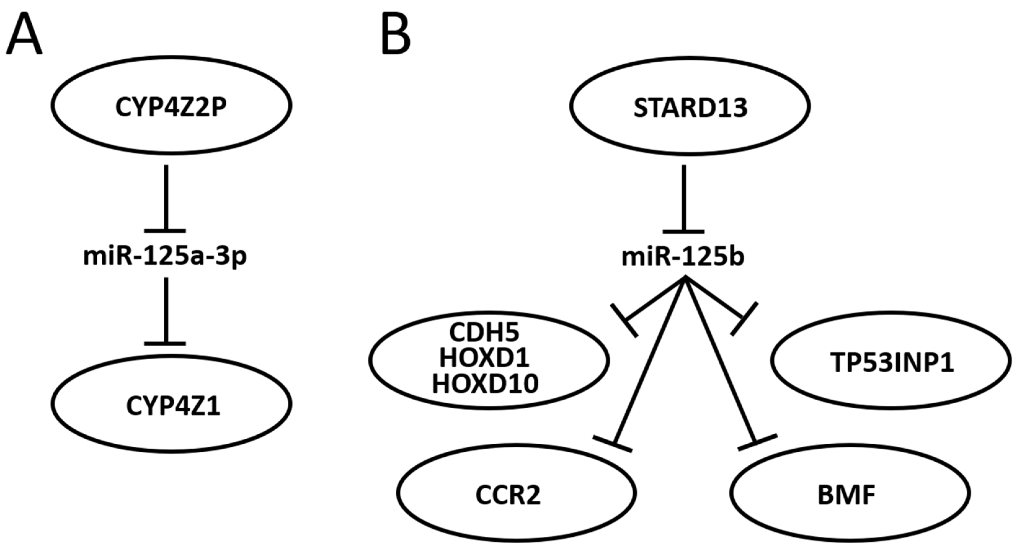
3.6. Further Mining miR-125 Function in BC: Competing Endogenous RNA Networks (ceRNET)

A fundamental way to control gene expression through miRs has been elucidated in recent years, consisting of the so-called ceRNET. In fact, miRs have been shown to act as controllers of target mRNAs by altering their half-life or translation. However, they are also controlled, in many cases, by other long non-coding RNAs (lncRNA) or even other mRNA, which “sponge” miR through sequence homology, avoiding their interaction with mRNA targets [198]. In other words, lncRNA and mRNA compete for binding miR; these two molecules form a competing endogenous RNA (ceRNA) couple. If the lncRNA efficiently sponges the miR, then miR inhibitory action is not accomplished, and the target mRNA is regularly translated. In this case, the lncRNA, inhibitor of an inhibitor, has a function resembling that of an enhancer of gene expression. Hence, if the mRNA encodes an oncoprotein, the lncRNA has an oncogenic effect, while the miR has an oncosuppressive role. The same, with opposite effects, occurs in the case of the mRNA coding an oncosuppressor. The three molecules, taken together, form what is currently known as a regulatory axis, and the sum of many axes creates the ceRNET. Here, lncRNA and mRNA constitute the nodes of the network, while miR represent their connections. A growing number of research works have been published in recent years outlining the increasing structure and complexity of the ceRNET in BC (see [199] and references therein), including the action of pseudogenes in this phenomenon. In fact, Welch and collaborators found that 309 pseudogenes exhibit significant differential expression among BC subtypes, and their expression pattern allows recognizing tumor samples from normal samples and discriminating the basal subtype from the luminal and Her2 subtypes; of them, 177 transcribed pseudogenes possess binding sites for co-expressed miRs that are also predicted to target their parent genes [200]. Recently, in a work by Zhu and collaborators, the authors took advantage of the data available in the exoRbase database and derived it from the exosomes of human BC samples [201]. Their study allowed for the identification of a ceRNA network including 19 mRNA nodes, 2 lncRNA nodes, 8 circular RNA nodes, and 41 miR connections. KEGG enrichment analysis showed that differentially expressed mRNA in the regulatory network is mainly enriched in the p53 signaling pathway.
Research centered around portions of a miR-125-centered ceRNET has been expanding steadily. The miR-125 interactions with the mRNA described in the previous section are therefore likely to become axes of the growing BC ceRNET as well, as soon as the appropriate lncRNA is identified in the pathway. However, some axes have already been described, and some of them, being interconnected, can be used to build a basic version of this network (Figure 3).
In 2004, Rieger and collaborators discovered a new human cytochrome P450 (CYP), termed CYP4Z1, which is specifically expressed in mammary gland and breast carcinoma [202]. They also found a transcribed pseudogene, named CYP4Z2P, that codes for a truncated CYP protein (340 amino acids vs. 505) with 96% identity to CYP4Z1. Both CYPs are highly expressed in BC, although the expression level of CYP4Z2P is approximately 20 times lower than that of CYP4Z1 in mammary tissues and barely expressed elsewhere. Later, it was shown that increased expression of CYP4Z1 promotes tumor angiogenesis and growth in human BC [203] and that CYP4Z2P 3′-UTR is involved in promoting BC angiogenesis through the VEGF/VEGFR2 pathway [204]. In 2015, Zheng et al. showed that the action of CYP4Z2P 3′-UTR is sponging several miRs, including miR-125a-3p, and that this pseudogene acts as a ceRNA with respect to CYP4Z1 mRNA, enhancing its expression levels [205]. They also showed that tumor angiogenesis is promoted by overexpression of the CYP4Z2P and CYP4Z1-3′UTRs, which significantly increased the activation of the ERK1/2 and PI3K/Akt pathways through the induction of their phosphorylation. The same group also showed a number of interactions later: (i) deregulation of these ceRNA also confers tamoxifen resistance in BC through the enhancement of the transcriptional activity of ERα via its phosphorylation dependent on cyclin-dependent kinase 3 (CDK3) [206]; (ii) downregulation of CYP4Z1 or CYP4Z2P through 3′-UTR binding promotes cell apoptosis, mirroring the functions and modulating the expression of human telomerase reverse transcriptase (hTERT) [207]; (iii) transcriptional factor six2 activates these CYPs ceRNET by directly binding to their promoters, thus activating the downstream PI3K/Akt and ERK1/2 pathways and consequently being involved not only in chemoresistance but also regulating the stemness of BC cells [208].
STARD13 (StAR-related lipid transfer domain protein 13, also known as deleted in liver cancer 2 protein (DLC-2)) is a Rho GTPase-activating protein (Rho GAP) that selectively activates RhoA and CDC42 and suppresses cell growth by inhibiting actin stress fiber assembly in hepatocellular carcinoma (HCC) [209]; this protein is ubiquitously expressed in normal tissues and downregulated in HCC. In mice, STARD13 promotes angiogenesis through the actions of RhoA [210]. Its role is well established in BC as well, where it acts as a tumor suppressor gene [211], regulates cell motility and invasion [212], endothelial differentiation [103], metastasis formation [213,214], cell migration [215], and apoptosis [216]. It has also been shown that STARD13 exerts its function in BC through its participation in many ceRNETs, such as the one involving a positive TGF-β/miR-9 regulatory loop mediated by the STARD13/YAP axis [217], the one involving hsa-miR-21-3p [218], or even the more complex network that involves five different miRs and that controls YAP/TAZ nuclear accumulation and transcriptional activity via modulation of Hippo and Rho-GTPase/F-actin signaling pathways [219]. A direct link between miR-125 and STARD13 expression has been described, too. Li and coworkers showed that CDH5, HOXD1, and HOXD10 encode putative STARD13 ceRNA and display concordant patterns with STARD13 in different metastatic potential BC cell lines and tissues; in addition, they also show that the 3′ UTR of STARD13 mRNA can bind miR-125b (and also miR-9 and miR-10b), indicating that this mRNA may participate in multiple pathways simultaneously [220], thus confirming their previous study about this interaction [192] and showing that the transcripts of the tumor suppressor genes CDH5, HOXD1, and HOXD10 inhibit BC metastasis in vitro and in vivo by competing with STARD13 mRNA for these three miR. Interestingly, CDH5, HOXD1 and HOXD10, along with STARD13, are BC players also in a different ceRNET, competing for a different set of miRs, indicating that STARD13′s role in BC is very complex. In 2017, Hu et al. discovered another ceRNET axis in which STARD13 and miR-125b control CCR2 (cysteine–cysteine chemokine receptor 2) expression levels [213]. In this case, the authors found that the CCR2 3′ UTR harbors three miR-125 binding sites that both inhibit MDA-MB-231 and MCF-7 cell metastasis by repressing epithelial-mesenchymal transition (EMT) in vitro and suppress BC metastasis in vivo through competition with STARD13 in a miR-125b-dependent and protein-coding-independent manner. Another component of the same ceRNET is TP53INP1 (tumor protein p53-inducible nuclear protein 1). TP53INP1 is an antiproliferative and proapoptotic protein involved in cell stress response that acts as a dual regulator of transcription and autophagy and is modulated by p53 in response to stress; it also interacts with kinases HIPK2 and PKCδ, which phosphorylate p53, creating a positive feedback loop between p53 and TP53INP1 [221]. TP53INP1 is also involved in SPARC (secreted protein acidic and rich in cysteine)-mediated-promotive effects on cancer cell migration and metastasis [222]. In 2018, Zheng et al. found a ceRNA interaction between STARD13 and TP53INP1 mediated by competitively binding to miR-125b in BC [223]. In this case, STARD13 promotes upregulation of TP53INP1, causing the inhibition of BC cell metastasis through competitive binding to miR-125b thanks to the inhibition of SPARC gene expression. Later, Guo and co-workers also found a ceRNET axis in BC involving miR-125b, STARD13 and BMF (Bcl-2-modifying factor) mRNA [216]. BMF is a member of the BCL2 protein family and controls apoptosis in several cell types [224]. The authors [216] found that miR-125b directly binds the 3′ UTR and thus downregulates BMF expression, and that STARD13, sponging miR-125b, upregulates BMF in BC both in vitro and in vivo. All together, these results suggest novel therapies for BC treatment and aid in selecting adequate drugs, depending on the molecular biology of the tumor, from a perspective aiming at the goal of personalized medicine. Indeed, a recent study showed that tanshinone IIA (an effective component extracted from Salvia miltiorrhiza that regulates the stemness of tumor cells) attenuates this phenotype in BC cells by downregulating miR-125b levels and upregulating its target gene STARD13 expression, while miR-125b overexpression or STARD13 knockdown impairs the inhibitory effects of tanshinone IIA on the stemness of BC cells [225].

4. Discussion

BC is a heterogeneous disease; thus, patients that are histologically diagnosed with the same cancer type might have different molecular characteristics, genetic mutations or tumor microenvironments that can deeply influence the prognosis or treatment response. Consequently, the challenge in personalized medicine is to distinguish these diverse molecular characteristics, separate patients accordingly, and treat them using a tailored approach that considers all these features. Personalized medicine might profoundly improve patient outcomes thanks to diagnostic tests capable of identifying specific biomarkers, thus enabling doctors to select the most effective treatment for each patient, reduce the risk of adverse reactions and increase the likelihood of a successful outcome.
To better understand the complexities and implications from the ethical, legal and social perspectives of personalized early detection and prevention of BC, it is necessary to rely on recommendations and evidence-based criteria issued by scientific and policy institutions, e.g., the European Collaborative on Personalized Early Detection and Prevention of Breast Cancer (ENVISION) in its 2020 consensus statement [226]. Such guidance is all the more essential, in fact, when highly innovative practices and techniques are applied, whose potential growth may outpace our current ethics and legal criteria [227,228]. As ENVISION points out, in fact, there is no denying that a great deal of progress has been made in evidence-based personalized interventions capable of maximizing the benefits and mitigating the downsides of currently available BC screening and prevention programs. Such progress has resulted in substantial research innovations for assessing an individual woman’s risk of develo** BC and relies on key factors such as the implementation of risk stratification models in BC prevention studies, achieving an effective degree of benefit–harm balance of risk-stratified early detection approaches, and the evaluation of the acceptability and feasibility of programs aimed at prevention and screening. Such a degree of innovation needs to be transposed into health outcomes for all; to achieve that, it is of utmost importance to devise and put in place a systematic approach for the assessment of risk-based programs, to be implemented along with thorough counseling being provided to patients in a highly targeted and tailored fashion [229]. In light of such needs, the classification of patients in the most precise way, at the molecular level, should be prioritized in order to better understand the biological features of the tumor to be treated. In this context, miR-125 and its targets are emerging as promising biomarkers in BC classification.
It is noteworthy that, depending on the study, miR-125 has been described as having either oncogenic or oncosuppressive roles. However, this should not be surprising. We recall that, for their very nature, the action of any miR is strictly connected with that of its targets; thus, if its target is an oncogene, then miR-125 acts as an oncosuppressor, and vice versa (see Table 4 and Table 5). However, the situation is further complicated for at least three reasons: cell changes in space (i.e., different regions of the same tumoral mass, which influence the cell response according to its diverse neighborhood), time (i.e., how the biology of the tumor changes over time), and miR-125 regulation. Firstly, different cancers, and sometimes also different stages of the same cancer, or even different populations of the same tumoral mass, have different metabolic needs [230]. Therefore, in the presence of both the same miR and corresponding mRNA target, the effect on the tumoral cell metabolism may significantly vary, with different spatial effects of miR on the transcriptome. Second, the evolution of cancer during time is associated with alterations in the cancer cell proteome; new genes are activated, other genes are suppressed or lost (for example, through aneuploidy [231] or copy number variations [232,233]) and also as a response to the internal and external microenvironmental interactions [234,235,236]. This is obviously true for every single cancer subpopulation (the space variable described before). Third, we recall here the organization of the miR-125b genes: this miR is transcribed from two different loci in the genome, which are under two different promoters [108]. Consequently, it is quite straightforward to hypothesize that the two copies of miR-125b may not be fully interchangeable in their function despite their sequence identity, since they may be transcribed under different cellular conditions and, thus, bind different targets at the time of miR-125b expression. In addition, the two clusters where miR-125b is embedded vary in their contents; the transcription of the cluster as a single pri-miR using the same promoter suggests that these diverse miR need to act in concert, thus the two different clusters, upon expression, likely modify the host cell proteome in different ways. It is therefore possible to theorize that such inconsistencies in the role of miR-125 (oncogene vs. oncosuppressor) might just reflect differences in the cells analyzed or in the analytical protocols applied, rather than real contradictions. Moreover, additional variables might be taken into account to explain these inconsistencies; as mentioned earlier, the same cell in a different microenvironment could respond differently to both internal (e.g., mutations, nutrient shortages, oxidative stress) and external (cell–cell interactions, response to immune system attack) stimuli, thus further encouraging the molecular characterization of each patient is arguably becoming a priority. Indeed, some support for this explanation is available for BC cell lines used to verify miR-125 function. In fact, miR-125 has been shown to be expressed in MCF-7 spheroids but not in MDA-MB-231 spheroids; in addition, the unique cluster of miRs found in each cell type is reportedly associated with their chemoresistance properties and cancer progression, most likely influencing the maintenance of the spheroid-enriched cancer stem cell properties [237]. Similar differences in different BC cell lines have also been reported by Ahram and coworkers who compared MDA-MB-453, MCF-7 and T47D cells, finding that miR-125b is highly expressed in T47D cells and slightly downregulated in MDA-MB-453 cells, with all the predictable consequences related to their target fate [238]. All together, these data underline the importance and complexity of the expression of miR-125 family members in the etiopathogenesis of BC and the need to characterize this biomarker further and better in BC patients.

5. Conclusions

It is noteworthy that, at present, the role of miR-125 in BC is quite underestimated in clinical practice. A search on the website clinicaltrial.gov performed in November 2023, using BC and microRNA as keywords, retrieved only 21 hits; of them, only one (ID: NCT04778202) is aimed at studying ‘miR 125a-5p and miR 143-3p as non-invasive biomarkers in the diagnosis of BC and the relationship between miR expression and histopathological features as tumor stage, grade, molecular subtypes’; for this trial, however, recruiting has not started yet [239]. No clinical trial is presently planned to study miR-125 as a possible therapeutic agent to control gene regulation in selected patients with altered expression of known target genes, despite its growing importance (see Figure 2 and Figure 3, and Table 5). Yet, by its very nature, this molecule is remarkably challenging in its clinical use. The usual approach to silence or enhance the function of a dysregulated miR basically relies on two approaches: (i) to restore miR expression with tumor suppressing activity (gain of function) or (ii) to block miR with oncogenic activity inhibiting its function (loss of function) [240]. Such approaches, however, are not fully applicable to miR-125, especially if it is a direct target of the therapy. For the silencing, both strands (5p and 3p) have a biological function in the cell, so the risk of inhibiting one strand by upregulating the other is high. For the enhancement obtained, for example, through the ectopic expression of an artificial construct, the fact that miR-125 is co-transcribed with other miR complicates this approach because all co-expressed miR need to be characterized, quantified, and possibly re-regulated. For this reason, directly targeting the miR would be, in our view, very complicated. Instead, it would be easier to target the locus activity harboring miR-125 so that co-expressed miRs are synchronously regulated. Naturally, this requires a profound knowledge of the locus organization, including the presence of enhancers, silencers, other regulatory elements, and chromatin modifications, and the study on miR-125 is, unfortunately, not so advanced in this perspective. At the moment, however, miR-125 has a potentially great impact on clinical practice as a biomarker, either in biopsies or as a circulating molecule, and the technology is fully proficient to perform such kinds of analyses.
Personalized medicine undoubtedly constitutes a broad-ranging breakthrough with huge potential to change healthcare to its very core. At the same time, such a potential will likely bring about a sea-change in that the current sets of ethical, legal and policy-making standards that provide us guidance today may be outpaced and ultimately inadequate. Therefore, new criteria need to be devised if we are to rely on equitable, effective healthcare for all in the long term. These criteria need to be supported by scientific discoveries, and likely miR-125 will be highly relevant and meaningful over the next few years in BC diagnosis and treatment.

Author Contributions

Conceptualization, R.P., G.G. and S.Z.; methodology, R.P., G.G., G.C. and A.L.; validation, A.L., G.N. and S.Z.; formal analysis, R.P., G.G., G.C. and S.Z.; investigation, R.P., G.G., G.C., A.L. and E.M.; resources, R.P., G.G., G.C., G.N. and S.Z.; data curation, R.P., G.G., G.C. and S.Z.; writing—original draft preparation, R.P., G.G., G.C., G.N., E.M. and S.Z.; writing—review and editing, R.P., G.G., G.C., A.L. and S.Z.; visualization, R.P., G.G., G.C., G.N., E.M. and S.Z.; supervision, R.P., G.G., E.M. and S.Z.; project administration, R.P., G.G., G.C., G.N., E.M. and S.Z. All authors have read and agreed to the published version of the manuscript.

Funding

This research received no external funding.

Institutional Review Board Statement

Not applicable.

Informed Consent Statement

Not applicable.

Data Availability Statement

Not applicable.

Conflicts of Interest

The authors declare no conflicts of interests.

References

  1. Siegel, R.L.; Miller, K.D.; Wagle, N.S.; Jemal, A. Cancer Statistics, 2023. CA Cancer J. Clin. 2023, 73, 17–48. [Google Scholar] [CrossRef] [PubMed]
  2. Winters, S.; Martin, C.; Murphy, D.; Shokar, N.K. Breast Cancer Epidemiology, Prevention, and Screening. Prog. Mol. Biol. Transl. Sci. 2017, 151, 1–32. [Google Scholar] [PubMed]
  3. Arnold, M.; Morgan, E.; Rumgay, H.; Mafra, A.; Singh, D.; Laversanne, M.; Vignat, J.; Gralow, J.R.; Cardoso, F.; Siesling, S.; et al. Current and Future Burden of Breast Cancer: Global Statistics for 2020 and 2040. Breast 2022, 66, 15–23. [Google Scholar] [CrossRef] [PubMed]
  4. Sharma, R. Global, Regional, National Burden of Breast Cancer in 185 Countries: Evidence from GLOBOCAN 2018. Breast Cancer Res. Treat. 2021, 187, 557–567. [Google Scholar] [CrossRef]
  5. Jung, S.; Wang, M.; Anderson, K.; Baglietto, L.; Bergkvist, L.; Bernstein, L.; van den Brandt, P.A.; Brinton, L.; Buring, J.E.; Heather Eliassen, A.; et al. Alcohol Consumption and Breast Cancer Risk by Estrogen Receptor Status: In a Pooled Analysis of 20 Studies. Int. J. Epidemiol. 2016, 45, 916–928. [Google Scholar] [CrossRef]
  6. Key, T. Sex Hormones and Risk of Breast Cancer in Premenopausal Women: A Collaborative Reanalysis of Individual Participant Data from Seven Prospective Studies. Lancet Oncol. 2013, 14, 1009–1019. [Google Scholar] [CrossRef]
  7. Coughlin, S.S. Epidemiology of Breast Cancer in Women. Adv. Exp. Med. Biol. 2019, 1152, 9–29. [Google Scholar] [PubMed]
  8. Galati, F.; Magri, V.; Arias-Cadena, P.A.; Moffa, G.; Rizzo, V.; Pasculli, M.; Botticelli, A.; Pediconi, F. Pregnancy-Associated Breast Cancer: A Diagnostic and Therapeutic Challenge. Diagnostics 2023, 13, 604. [Google Scholar] [CrossRef]
  9. Bodewes, F.T.H.; van Asselt, A.A.; Dorrius, M.D.; Greuter, M.J.W.; de Bock, G.H. Mammographic Breast Density and the Risk of Breast Cancer: A Systematic Review and Meta-Analysis. Breast 2022, 66, 62–68. [Google Scholar] [CrossRef]
  10. Majeed, W.; Aslam, B.; Javed, I.; Khaliq, T.; Muhammad, F.; Ali, A.; Raza, A. Breast Cancer: Major Risk Factors and Recent Developments in Treatment. Asian Pac. J. Cancer Prev. 2014, 15, 3353–3358. [Google Scholar] [CrossRef]
  11. Petrucelli, N.; Daly, M.B.; Pal, T. BRCA1- and BRCA2-Associated Hereditary Breast and Ovarian Cancer. 4 September 1998 [Updated 21 September 2023]. In GeneReviews® [Internet]; Adam, M.P., Feldman, J., Mirzaa, G.M., Pagon, R.A., Wallace, S.E., Bean, L.J.H., Gripp, K.W., Amemiya, A., Eds.; University of Washington: Seattle, WA, USA, 1993–2024; Available online: https://www.ncbi.nlm.nih.gov/books/NBK1247/ (accessed on 15 October 2023).
  12. Carbognin, L.; Miglietta, F.; Paris, I.; Dieci, M.V. Prognostic and Predictive Implications of PTEN in Breast Cancer: Unfulfilled Promises but Intriguing Perspectives. Cancers 2019, 11, 1401. [Google Scholar] [CrossRef] [PubMed]
  13. Shahbandi, A.; Nguyen, H.D.; Jackson, J.G. TP53 Mutations and Outcomes in Breast Cancer: Reading beyond the Headlines. Trends Cancer 2020, 6, 98–110. [Google Scholar] [CrossRef] [PubMed]
  14. Corso, G.; Veronesi, P.; Sacchini, V.; Galimberti, V. Prognosis and Outcome in CDH1-Mutant Lobular Breast Cancer. Eur. J. Cancer Prev. 2018, 27, 237–238. [Google Scholar] [CrossRef] [PubMed]
  15. Beggs, A.D.; Latchford, A.R.; Vasen, H.F.A.; Moslein, G.; Alonso, A.; Aretz, S.; Bertario, L.; Blanco, I.; Bülow, S.; Burn, J.; et al. Peutz–Jeghers Syndrome: A Systematic Review and Recommendations for Management. Gut 2010, 59, 975–986. [Google Scholar] [CrossRef] [PubMed]
  16. Apostolou, P.; Papasotiriou, I. Current Perspectives on CHEK2 Mutations in Breast Cancer. Breast Cancer Targets Ther. 2017, 9, 331–335. [Google Scholar] [CrossRef] [PubMed]
  17. Nepomuceno, T.C.; Carvalho, M.A.; Rodrigue, A.; Simard, J.; Masson, J.Y.; Monteiro, A.N.A. PALB2 Variants: Protein Domains and Cancer Susceptibility. Trends Cancer 2021, 7, 188–197. [Google Scholar] [CrossRef] [PubMed]
  18. Stucci, L.S.; Internò, V.; Tucci, M.; Perrone, M.; Mannavola, F.; Palmirotta, R.; Porta, C. The ATM Gene in Breast Cancer: Its Relevance in Clinical Practice. Genes 2021, 12, 727. [Google Scholar] [CrossRef]
  19. Li, N.; McInerny, S.; Zethoven, M.; Cheasley, D.; Lim, B.W.X.; Rowley, S.M.; Devereux, L.; Grewal, N.; Ahmadloo, S.; Byrne, D.; et al. Combined Tumor Sequencing and Case-Control Analyses of RAD51C in Breast Cancer. J. Natl. Cancer Inst. 2019, 111, 1332–1338. [Google Scholar] [CrossRef]
  20. Chen, X.; Li, Y.; Ouyang, T.; Li, J.; Wang, T.; Fan, Z.; Fan, T.; Lin, B.; ** Review. Cureus 2023, 15, e44061. [Google Scholar] [CrossRef]
  21. Abdul Halim, A.A.; Andrew, A.M.; Mohd Yasin, M.N.; Abd Rahman, M.A.; Jusoh, M.; Veeraperumal, V.; Rahim, H.A.; Illahi, U.; Abdul Karim, M.K.; Scavino, E. Existing and Emerging Breast Cancer Detection Technologies and Its Challenges: A Review. Appl. Sci. 2021, 11, 10753. [Google Scholar] [CrossRef]
  22. Iranmakani, S.; Mortezazadeh, T.; Sajadian, F.; Ghaziani, M.F.; Ghafari, A.; Khezerloo, D.; Musa, A.E. A Review of Various Modalities in Breast Imaging: Technical Aspects and Clinical Outcomes. Egypt. J. Radiol. Nucl. Med. 2020, 51, 57. [Google Scholar] [CrossRef]
  23. Veronesi, U.; Viale, G.; Rotmensz, N.; Goldhirsch, A. Rethinking TNM: Breast Cancer TNM Classification for Treatment Decision-Making and Research. Breast 2006, 15, 3–8. [Google Scholar] [CrossRef]
  24. Eliyatkin, N.; Yalcin, E.; Zengel, B.; Aktaş, S.; Vardar, E. Molecular Classification of Breast Carcinoma: From Traditional, Old-Fashioned Way to A New Age, and A New Way. J. Breast Health 2015, 11, 59–66. [Google Scholar] [CrossRef]
  25. Perou, C.M.; Sørile, T.; Eisen, M.B.; Van De Rijn, M.; Jeffrey, S.S.; Ress, C.A.; Pollack, J.R.; Ross, D.T.; Johnsen, H.; Akslen, L.A.; et al. Molecular Portraits of Human Breast Tumours. Nature 2000, 406, 747–752. [Google Scholar] [CrossRef]
  26. Sinn, H.P.; Kreipe, H. A Brief Overview of the WHO Classification of Breast Tumors, 4th Edition, Focusing on Issues and Updates from the 3rd Edition. Breast Care 2013, 8, 149–154. [Google Scholar] [CrossRef]
  27. Giuliano, A.E.; Connolly, J.L.; Edge, S.B.; Mittendorf, E.A.; Rugo, H.S.; Solin, L.J.; Weaver, D.L.; Winchester, D.J.; Hortobagyi, G.N. Breast Cancer-Major Changes in the American Joint Committee on Cancer Eighth Edition Cancer Staging Manual. CA Cancer J. Clin. 2017, 67, 290–303. [Google Scholar] [CrossRef]
  28. Fisusi, F.A.; Akala, E.O. Drug Combinations in Breast Cancer Therapy. Pharm. Nanotechnol. 2019, 7, 3–23. [Google Scholar] [CrossRef]
  29. Burstein, H.J.; Curigliano, G.; Thürlimann, B.; Weber, W.P.; Poortmans, P.; Regan, M.M.; Senn, H.J.; Winer, E.P.; Gnant, M.; Aebi, S.; et al. Customizing Local and Systemic Therapies for Women with Early Breast Cancer: The St. Gallen International Consensus Guidelines for Treatment of Early Breast Cancer 2021. Ann. Oncol. 2021, 32, 1216–1235. [Google Scholar] [CrossRef] [PubMed]
  30. Li, X.; Dai, A.; Tran, R.; Wang, J. Identifying MiRNA Biomarkers for Breast Cancer and Ovarian Cancer: A Text Mining Perspective. Breast Cancer Res. Treat. 2023, 201, 5–14. [Google Scholar] [CrossRef] [PubMed]
  31. Li, J.; Zhang, H.; Gao, F. Identification of MiRNA Biomarkers for Breast Cancer by Combining Ensemble Regularized Multinomial Logistic Regression and Cox Regression. BMC Bioinform. 2022, 23, 434. [Google Scholar] [CrossRef] [PubMed]
  32. Davey, M.G.; Davies, M.; Lowery, A.J.; Miller, N.; Kerin, M.J. The Role of MicroRNA as Clinical Biomarkers for Breast Cancer Surgery and Treatment. Int. J. Mol. Sci. 2021, 22, 8290. [Google Scholar] [CrossRef] [PubMed]
  33. Jang, J.Y.; Kim, Y.S.; Kang, K.N.; Kim, K.H.; Park, Y.J.; Kim, C.W. Multiple MicroRNAs as Biomarkers for Early Breast Cancer Diagnosis. Mol. Clin. Oncol. 2021, 14, 1–9. [Google Scholar] [CrossRef] [PubMed]
  34. Garrido-Palacios, A.; Rojas Carvajal, A.M.; Núñez-Negrillo, A.M.; Cortés-Martín, J.; Sánchez-García, J.C.; Aguilar-Cordero, M.J. MicroRNA Dysregulation in Early Breast Cancer Diagnosis: A Systematic Review and Meta-Analysis. Int. J. Mol. Sci. 2023, 24, 8270. [Google Scholar] [CrossRef] [PubMed]
  35. Sharifi, Z.; Talkhabi, M.; Taleahmad, S. Identification of Potential MicroRNA Diagnostic Panels and Uncovering Regulatory Mechanisms in Breast Cancer Pathogenesis. Sci. Rep. 2022, 12, 20135. [Google Scholar] [CrossRef]
  36. Khadka, V.S.; Nasu, M.; Deng, Y.; Jijiwa, M. Circulating MicroRNA Biomarker for Detecting Breast Cancer in High-Risk Benign Breast Tumors. Int. J. Mol. Sci. 2023, 24, 7553. [Google Scholar] [CrossRef]
  37. Nguyen, T.H.N.; Nguyen, T.T.N.; Nguyen, T.T.M.; Nguyen, L.H.M.; Huynh, L.H.; Phan, H.N.; Nguyen, H.T. Panels of Circulating MicroRNAs as Potential Diagnostic Biomarkers for Breast Cancer: A Systematic Review and Meta-Analysis. Breast Cancer Res. Treat. 2022, 196, 1–15. [Google Scholar] [CrossRef]
  38. Huynh, K.Q.; Le, A.T.; Phan, T.T.; Ho, T.T.; Pho, S.P.; Nguyen, H.T.; Le, B.T.; Nguyen, T.T.; Nguyen, S.T. The Diagnostic Power of Circulating MiR-1246 in Screening Cancer: An Updated Meta-Analysis. Oxid. Med. Cell. Longev. 2023, 2023, 8379231. [Google Scholar] [CrossRef]
  39. Tiberio, P.; Gaudio, M.; Belloni, S.; Pindilli, S.; Benvenuti, C.; Jacobs, F.; Saltalamacchia, G.; Zambelli, A.; Santoro, A.; De Sanctis, R. Unlocking the Potential of Circulating MiRNAs in the Breast Cancer Neoadjuvant Setting: A Systematic Review and Meta-Analysis. Cancers 2023, 15, 3424. [Google Scholar] [CrossRef]
  40. Naeli, P.; Winter, T.; Hackett, A.P.; Alboushi, L.; Jafarnejad, S.M. The Intricate Balance between MicroRNA-Induced MRNA Decay and Translational Repression. FEBS J. 2023, 290, 2508–2524. [Google Scholar] [CrossRef] [PubMed]
  41. Friedman, R.C.; Farh, K.K.H.; Burge, C.B.; Bartel, D.P. Most Mammalian MRNAs Are Conserved Targets of MicroRNAs. Genome Res. 2009, 19, 92–105. [Google Scholar] [CrossRef] [PubMed]
  42. Ambros, V.; Bartel, B.; Bartel, D.P.; Burge, C.B.; Carrington, J.C.; Chen, X.; Dreyfuss, G.; Eddy, S.R.; Griffiths-Jones, S.; Marshall, M.; et al. A Uniform System for MicroRNA Annotation. RNA 2003, 9, 277–279. [Google Scholar] [CrossRef] [PubMed]
  43. Griffiths-Jones, S.; Grocock, R.J.; van Dongen, S.; Bateman, A.; Enright, A.J. MiRBase: MicroRNA Sequences, Targets and Gene Nomenclature. Nucleic Acids Res. 2006, 34, D140–D144. [Google Scholar] [CrossRef] [PubMed]
  44. Dziechciowska, I.; Dąbrowska, M.; Mizielska, A.; Pyra, N.; Lisiak, N.; Kopczyński, P.; Jankowska-Wajda, M.; Rubiś, B. MiRNA Expression Profiling in Human Breast Cancer Diagnostics and Therapy. Curr. Issues Mol. Biol. 2023, 45, 9500–9525. [Google Scholar] [CrossRef] [PubMed]
  45. Loh, H.Y.; Norman, B.P.; Lai, K.S.; Rahman, N.M.A.N.A.; Alitheen, N.B.M.; Osman, M.A. The Regulatory Role of MicroRNAs in Breast Cancer. Int. J. Mol. Sci. 2019, 20, 4940. [Google Scholar] [CrossRef] [PubMed]
  46. Najjary, S.; Mohammadzadeh, R.; Mokhtarzadeh, A.; Mohammadi, A.; Kojabad, A.B.; Baradaran, B. Role of MiR-21 as an Authentic Oncogene in Mediating Drug Resistance in Breast Cancer. Gene 2020, 738, 144453. [Google Scholar] [CrossRef] [PubMed]
  47. Wang, Z.X.; Lu, B.B.; Wang, H.; Cheng, Z.X.; Yin, Y.M. MicroRNA-21 Modulates Chemosensitivity of Breast Cancer Cells to Doxorubicin by Targeting PTEN. Arch. Med. Res. 2011, 42, 281–290. [Google Scholar] [CrossRef] [PubMed]
  48. Wang, H.; Tan, Z.; Hu, H.; Liu, H.; Wu, T.; Zheng, C.; Wang, X.; Luo, Z.; Wang, J.; Liu, S.; et al. MicroRNA-21 Promotes Breast Cancer Proliferation and Metastasis by Targeting LZTFL1. BMC Cancer 2019, 19, 738. [Google Scholar] [CrossRef] [PubMed]
  49. Shi, Y.; Ye, P.; Long, X. Differential Expression Profiles of the Transcriptome in Breast Cancer Cell Lines Revealed by Next Generation Sequencing. Cell. Physiol. Biochem. 2017, 44, 804–816. [Google Scholar] [CrossRef]
  50. Mohmmed, E.A.; Shousha, W.G.; EL-Saiid, A.S.; Ramadan, S.S. A Clinical Evaluation of Circulating MiR-106a and Raf-1 as Breast Cancer Diagnostic and Prognostic Markers. Asian Pac. J. Cancer Prev. 2021, 22, 3513–3520. [Google Scholar] [CrossRef]
  51. You, F.; Luan, H.; Sun, D.; Cui, T.; Ding, P.; Tang, H.; Sun, D. MiRNA-106a Promotes Breast Cancer Cell Proliferation, Clonogenicity, Migration, and Invasion Through Inhibiting Apoptosis and Chemosensitivity. DNA Cell Biol. 2019, 38, 198–207. [Google Scholar] [CrossRef]
  52. You, F.; Li, J.; Zhang, P.; Zhang, H.; Cao, X. MiR106a Promotes the Growth of Transplanted Breast Cancer and Decreases the Sensitivity of Transplanted Tumors to Cisplatin. Cancer Manag. Res. 2020, 12, 233–246. [Google Scholar] [CrossRef]
  53. Dinami, R.; Ercolani, C.; Petti, E.; Piazza, S.; Ciani, Y.; Sestito, R.; Sacconi, A.; Biagioni, F.; Le Sage, C.; Agami, R.; et al. MiR-155 Drives Telomere Fragility in Human Breast Cancer by Targeting TRF1. Cancer Res. 2014, 74, 4145–4156. [Google Scholar] [CrossRef]
  54. Roth, C.; Rack, B.; Müller, V.; Janni, W.; Pantel, K.; Schwarzenbach, H. Circulating MicroRNAs as Blood-Based Markers for Patients with Primary and Metastatic Breast Cancer. Breast Cancer Res. 2010, 12, R90. [Google Scholar] [CrossRef]
  55. Li, P.; Xu, T.; Zhou, X.; Liao, L.; Pang, G.; Luo, W.; Han, L.; Zhang, J.; Luo, X.; ** Breast Cancer Microenvironment Through Single-Cell Omics. Front. Immunol. 2022, 13, 868813. [Google Scholar] [CrossRef] [PubMed]
  56. Boo, L.; Ho, W.Y.; Ali, N.M.; Yeap, S.K.; Ky, H.; Chan, K.G.; Yin, W.F.; Satharasinghe, D.A.; Liew, W.C.; Tan, S.W.; et al. Phenotypic and MicroRNA Transcriptomic Profiling of the MDA-MB-231 Spheroid-Enriched CSCs with Comparison of MCF-7 MicroRNA Profiling Dataset. PeerJ 2017, 5, e3551. [Google Scholar] [CrossRef] [PubMed]
  57. Ahram, M.; Mustafa, E.; Zaza, R.; Abu Hammad, S.; Alhudhud, M.; Bawadi, R.; Zihlif, M. Differential Expression and Androgen Regulation of MicroRNAs and Metalloprotease 13 in Breast Cancer Cells. Cell Biol. Int. 2017, 41, 1345–1355. [Google Scholar] [CrossRef]
  58. Search for: Breast Cancer, Other Terms: Micro RNA, Micro RNA|Card Results|ClinicalTrials.Gov. Available online: https://clinicaltrials.gov/search?cond=Breast%20Cancer&term=Micro%20RNA&intr=micro%20RNA (accessed on 2 November 2023).
  59. Lima, J.F.; Cerqueira, L.; Figueiredo, C.; Oliveira, C.; Azevedo, N.F. Anti-MiRNA Oligonucleotides: A Comprehensive Guide for Design. RNA Biol. 2018, 15, 338–352. [Google Scholar] [CrossRef]
Figure 1. Schematic representation of human miR-125 illustrating the structure of the double-stranded pre-miR of both miR-125a and miR-125b, and their sequence. Color codes: red highlight for the 5p form, blue highlight for the 3p form. Numbers before and after the sequences indicate the number of nucleotides trimmed away from the mature miR. Data retrieved and partially modified from miRTarBase v9 update 2022 [112,113].
Ncrna 10 00016 g001
Figure 2. Summary of the main functions exerted by the miR-125 family members in human biology. Examples of miR-125 target genes are reported below each function. Data partly retrieved from [114,115] The listed genes at the bottom are involved in the control of the cellular functions below the green arrows; they are controlled, either directly or indirectly, by miR-125 family members, either by up- or down-regulation. Further details have been laid out in the article’s body.
Ncrna 10 00016 g002
Figure 3. Schematic representation of two miR-125-centered ceRNET in BC. (A): a simple ceRNET having only one axis, where a pseudogene (CYP4Z2P) mRNA inhibits miR-125a-3p action on the target (CYP4Z1) mRNA by sponging it, thus enhancing CYP4Z1 expression. (B): a more complex ceRNET in which the interaction between miR-125b and STARD13 mRNA controls the expression of multiple target mRNA. See text for references and further explanations.
Ncrna 10 00016 g003
Table 1. Main genes associated with BC formation and development. Data in columns 3–4 partly retrieved from [25,26,27,28]. Estimated risk refers to the probability to develop a BC in presence of a mutation in the corresponding gene. Abbreviations: TNBC—triple negative breast cancer; BC—breast cancer; n/a: data not available (e.g., low risk gene, insufficient data available, non-specific effect); refs—bibliographic references.
GeneFunction(s)Estimated RiskBC TypeRefs
BRCA1DNA repair
transcription regulation
cell cycle regulation
chromatin remodeling
55–65% by age 70TNBC
luminal B
[11,25,27,28]
BRCA2DNA repair
DNA replication
transcription regulation
cell cycle regulation
mitophagy
~45% by age 70TNBC
luminal B
[11,25,28]
PALB2DNA repairAll women: RR 2.3, 95% CI 1.4–3.9 < 50 years: RR 3.0, 95% CI 1.4–5.5n/a[17,25,28]
PTENcell survival
cell growth
85% lifetimeluminal A
luminal B
[12,25,26,27,28]
TP53cell cycle regulation25% by age 74all[13,25,26,27,28]
CDH1cell adhesion39% lifetimeluminal A[14,25,26,27,28]
STK11cell cycle regulation32% by age 60n/a[15,25,28]
CHEK2DNA repair
cell cycle regulation
apoptosis
Female: RR 1.70, 95% CI 1.3–2.2 Male: RR 10.3, 95% CI 3.5–30.0n/a[16,25,26,27,28]
BRIP1DNA repairAll women: RR 2.0, 95%n/a[23,25]
ATMDNA repairRR 2.37, 95% CI 1.5–3.8n/a[18,25,26,27,28]
Table 2. TNM (tumor-node-metastasis) staging for BC. A given BC can be identified by any combination of T-N-M parameters, based on patient’s clinical status.
TumorNodeMetastasis
Txno primary tumor informationNxnot assessableMxnot assessed
T0no primary tumor evidenceN0no clinically positive nodesM0no evidence
TIScarcinoma in situ (primary sites)N1single, ipsilateral, size < 3 cmM1metastasis present at distance
T1size < 2 cmN2asingle, ipsilateral, size 3–6 cm
T2size 2 to 4 cmN2bmultiple, ipsilateral, size < 6 cm
T3size > 4 cmN3massive/ipsilateral/bilateral/controlateral
T4size > 4 cm, pterygoid muscle, base of tongue or skin involvedN3aipsilateral node(s), one more than 6 cm
N3bbilateral
N4controlateral
Table 3. List of miR playing a direct role in BC. Target genes are those for which the miR/mRNA interaction is direct (usually, at the mRNA 3′ UTR), thus indirect interactions (e.g., other proteins of the same metabolic axis) are not reported in this table; additional miR studied only as BC biomarkers are collectively reported in the bottom row; see text for additional explanations. Abbreviations: n/a—data not available.
miR NameTarget Gene(s)Affected Cellular FunctionsRefs
miR-21PTENdrug resistance[65,66]
miR-21LZTFL1proliferation and metastasis[67]
miR-21IGFBP3
TPM1
PCD4
TGF-β1
proliferation, metastasis, epithelial-to-mesenchymal transition (EMT), apoptosis[68]
miR-106aRAF-1invasion and proliferation[69]
miR-106aP53
BAX
RUNX3
Bcl-2
ABCG2
proliferation, colony-forming capacity, migration, invasion, apoptosis, sensitivity to cisplatin[70,71]
miR-155TRF1telomere fragility[72]
miR-141ANP32Emigration and invasion[74]
let-7ERCC6proliferation, apoptosis[78]
miR-335ERα
IGF1R
SP1
ID4
proliferation, apoptosis[80]
miR-335c-Metcell scattering, migration, and invasion[81]
miR-126VEGFA
PIK3R2
angiogenesis, tumor genesis and growth[83]
miR-126PIK3R2trastuzumab resistance[84]
miR-199a/b-3pPAK4migration and invasion[86]
miR-199a-3pmTOR
c-Met
cell cycle progression, doxorubicin sensitivity, apoptosis[87]
miR-199a-3pTFAMresistance to cisplatin[88]
miR-199a-3pTFAMangiogenesis and metastasis under hypoxia[89]
miR-101POMP
Stmn1
DNMT3A
EYA1
VHL
SOX2
Jak2
MCL-1
proliferation, apoptosis, angiogenesis, drug resistance, invasion, metastasis[91]
miR-101-3pCOX-2migration, metastasis[92]
miR-101-3pEZH2migration, invasion, proliferation[93]
miR-101-5pGINS1DNA replication[94]
miR-9FOXO1proliferation, migration, invasion[99]
miR-9STARD13EMT, metastasis[100]
miR-9LIFRmetastasis[101]
miR-9elf5A2resistance to doxorubicin[102]
miR-9HMGA2
EGR1
IGFBP3
proliferation, metastasis, EMT, apoptosis[68]
miR-9PDGFRβvasculogenesis[103]
miR-200PDGFRβvasculogenesis[103]
let-7a-5p
miR-9-5p
miR-10b
miR-21
miR-22-3p
miR-23b-3p
miR-25-3p
miR-29
miR-34a
miR-93-5p
miR-99a-5p/-3p
miR-100-5p
miR-101-3p
miR-101-5p
miR-126-5p/-3p
miR-141-3p
miR-143-5p/-3p
miR-144-5p/-3p
miR-145
miR-155
mir-181b1-5p
miR-195-5p
miR-199a-5p
miR-200a
miR-203
miR-203a-3p
miR-205
miR-210-3p
miR-221/222
miR-373
n/abiomarkers[73,76,87,90,94,95,97,98]
Table 4. Summary of the affected organs and mRNA targets of miR-125 family members in human cancers. Data regarding BC is reported in Section 3.5. Abbreviations: CNS—central nervous system; refs—references; n/a—data not available in the cited reference(s). In the “notes” column, data refers to reported anomalies in miR-125 regulation, to its action on specific pathways, or to additional data that might explain its role in the specific cancer; in case nothing is relevant—beyond the identified target genes—we report “none.” Reported sources can be broadly divided into two classes: those investigating deregulated miR in cancer samples (for which target identification is usually absent) and those investigating miR-125 functional role(s), for which the main aim of the study is reported in the first three columns.
miROrganTarget(s)NotesRefs
125CNSn/aderegulated, pediatric[130]
125CNSn/aderegulated[131,132]
125CNSp53, p38MAPKnone[133]
125CNSBMFnone[134]
125aovaryn/aEMT negative regulator[144]
125bovaryBCL3none[145]
125bovaryn/aserum biomarker[146]
125bbladderE2F3none[147]
125bbladdern/aurine biomarker[148]
125-3pbladdern/ahypoxia regulated[149]
125bladdern/asurvival predictor[150]
125aliverMMP11, VEGFnone[151]
125bliverMcl-1, IL6Rnone[152]
125bliverLin28B2none[153]
125liverPokemonnone[154]
125liverTRAF6none[155]
125liverhexokinase IInone[156]
125liverFOXM1none[157]
125skinNCAMnone[158]
125skinc-Junnone[159]
125bskinMMP13none[160]
125bskinSTAT3none[161]
125skinn/aderegulated[162]
125bboneSTAT3none[163,164]
125boneErbB2none[165]
125boneBAP1none[166]
125lungn/asurvival predictor[167]
125lungEGFRnone[168]
125lungHER2trastuzumab resistance[169]
125lungMMP13none[170]
125pancreasn/aderegulated[171,172]
125pancreasNEDD9none[173]
125prostaten/aderegulated[174,175,176]
125prostateBAK1none[177]
125prostatep53, PUMAnone[178]
125bthyroidFoxp3cisplatin sensitivity[179]
125bstomachPPP1CA-Rbnone[180]
125a-5pcolonBCL2, BCL2L12, MCL1none[181]
125bkidneyn/asurvival predictor[182]
Table 5. Role of miR-125 in BC formation and development. In columns 2 and 4, reg. stands for regulation; an arrow pointing upwards means upregulation, while an arrow pointing downwards means downregulation; each arrow describes the miR/target regulation reported to its left. Note that in BC models, miR-125 members may be either up- or down-regulated, indicating either an oncogenic or oncosuppressive role for this molecule, in that context. Possible interpretations of these contradictory data are reported in the Discussion. In columns 3–4, n/a stands for ‘data not available’ or ‘not applicable’. In column 6, ‘blood samples’ means that circulating miR have been studied. In column 7, ref. stands for reference(s).
miRReg.TargetReg.Cellular FunctionCell LineRef.
miR-125a
miR-125b

ERBB2
ERBB3

migration
invasion
SKBR3[189]
miR-125bETS1proliferationBC samples[190]
miR-125bMUC1apoptosisBT-549
ZR-75-1
[191]
miR-125bSTARD13metastasisMCF-7
MDA-MB-231
[192]
miR-125n/an/aradioresistanceMCF-7
MDA-MB-231
[193]
miR-125bn/an/achemoresistance
proliferation
apoptosis
blood samples[194]
miR-125bBAK1chemoresistance
apoptosis
MDA-435
MDA-436
MDA-231
MCF7
SKBR3
[195]
miR-125a-5p
miR-125b

n/an/aage-dependent BC formationBC samples[196]
miR-125bMMP11proliferation
migration
invasion
T47D
SKBR3
[197]
Disclaimer/Publisher’s Note: The statements, opinions and data contained in all publications are solely those of the individual author(s) and contributor(s) and not of MDPI and/or the editor(s). MDPI and/or the editor(s) disclaim responsibility for any injury to people or property resulting from any ideas, methods, instructions or products referred to in the content.

