
Terbium and Europium Chlorocyananilate-Based 2D Coordination Polymers

 , , , , , , and
Abstract
:
1. Introduction
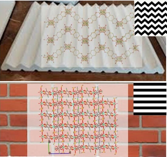
2. Results
3. Conclusions
4. Materials and Methods
4.1. Cell Determination from X-ray Diffraction Data
4.2. Ab Initio Crystal Structure Solution from X-ray Diffraction Data
Supplementary Materials
Author Contributions
Funding
Institutional Review Board Statement
Informed Consent Statement
Data Availability Statement
Acknowledgments
Conflicts of Interest
References
- Maslennikova, V.V.; Filatov, S.N.; Orlov, A.V.; Surin, N.M.; Svidchenko, E.A.; Chistyakov, E.M. Luminescent Coatings Based on (3-Aminopropyl)triethoxysilane and Europium Complex β-Diketophosphazene. Polymers 2022, 14, 728. [Google Scholar] [CrossRef]
- Chistyakov, E.M.; Tupikov, A.S.; Buzin, M.I.; Borisov, R.S.; Kireev, V.V. Preparation of films based on β-diketophosphazene and different amines and study their properties. Mater. Chem. Phys. 2019, 223, 353–359. [Google Scholar] [CrossRef]
- Nikovskii, I.A.; Chistyakov, E.M.; Tupikov, A.S. Phosphazene-Containing Ligands and Complexes on Their Base. Russ. J. Gen. Chem. 2018, 88, 474–494. [Google Scholar] [CrossRef]
- James, S.L. Metal-organic frameworks. Chem. Soc. Rev. 2003, 32, 276–288. [Google Scholar] [CrossRef] [PubMed]
- Zhou, H.C.J.; Kitagawa, S. Metal-Organic Frameworks (MOFs). Chem. Soc. Rev. 2014, 43, 5415–5418. [Google Scholar] [CrossRef] [PubMed]
- Oggianu, M.; Manna, F.; Sahadevan, S.A.; Avarvari, N.; Abhervé, A.; Mercuri, M.L. Metal-Organic Framework vs. Coordination Polymer—Influence of the Lanthanide on the Nature of the Heteroleptic Anilate/Terephtalate 3D Network. Crystals 2022, 12, 763. [Google Scholar] [CrossRef]
- Oggianu, M.; Monni, N.; Mameli, V.; Cannas, C.; Sahadevan, S.A.; Mercuri, M.L. Designing magnetic nanomofs for biomedicine: Current trends and applications. Magnetochemistry 2020, 6, 39. [Google Scholar] [CrossRef]
- Ashoka Sahadevan, S.; Monni, N.; Oggianu, M.; Abhervé, A.; Marongiu, D.; Saba, M.; Mura, A.; Bongiovanni, G.; Mameli, V.; Cannas, C.; et al. Heteroleptic NIR-Emitting YbIII/Anilate-Based Neutral Coordination Polymer Nanosheets for Solvent Sensing. ACS Appl. Nano Mater. 2020, 3, 94–104. [Google Scholar] [CrossRef]
- Ashoka Sahadevan, S.; Manna, F.; Abhervé, A.; Oggianu, M.; Monni, N.; Mameli, V.; Marongiu, D.; Quochi, F.; Gendron, F.; Le Guennic, B.; et al. Combined Experimental/Theoretical Study on the Luminescent Properties of Homoleptic/Heteroleptic Erbium(III) Anilate-Based 2D Coordination Polymers. Inorg. Chem. 2021, 60, 17765–17774. [Google Scholar] [CrossRef] [PubMed]
- Sahadevan, S.A.; Monni, N.; Abhervé, A.; Cosquer, G.; Oggianu, M.; Ennas, G.; Yamashita, M.; Avarvari, N.; Mercuri, M.L. Dysprosium Chlorocyanoanilate-Based 2D-Layered Coordination Polymers. Inorg. Chem. 2019, 58, 13988–13998. [Google Scholar] [CrossRef] [PubMed]
- Junggeburth, S.C.; Diehl, L.; Werner, S.; Duppel, V.; Sigle, W.; Lotsch, B.V. Ultrathin 2D coordination polymer nanosheets by surfactant-mediated synthesis. J. Am. Chem. Soc. 2013, 135, 6157–6164. [Google Scholar] [CrossRef] [PubMed]
- Wang, X.; Zhou, J.; Fu, H.; Li, W.; Fan, X.; **n, G.; Zheng, J.; Li, X. MOF derived catalysts for electrochemical oxygen reduction. J. Mater. Chem. A 2014, 2, 14064–14070. [Google Scholar] [CrossRef]
- Kitagawa, S.; Kawata, S. Coordination compounds of 1,4-dihydroxybenzoquinone and its homologues. Structures and properties. Coord. Chem. Rev. 2002, 224, 11–34. [Google Scholar] [CrossRef]
- Benmansour, S.; Gómez-Claramunt, P.; Vallés-García, C.; Mínguez Espallargas, G.; Gómez García, C.J. Key Role of the Cation in the Crystallization of Chiral Tris(Anilato)Metalate Magnetic Anions. Cryst. Growth Des. 2016, 16, 518–526. [Google Scholar] [CrossRef]
- Mencel, K.; Kinzhybalo, V.; Jakubas, R.; Zarȩba, J.K.; Szklarz, P.; Durlak, P.; Drozd, M.; Piecha-Bisiorek, A. 0D Bismuth(III)-Based Hybrid Ferroelectric: Tris(acetamidinium) Hexabromobismuthate(III). Chem. Mater. 2021, 33, 8591–8601. [Google Scholar] [CrossRef]
- Souto, M.; Perepichka, D.F. Electrically conductive covalent organic frameworks: Bridging the fields of organic metals and 2D materials. J. Mater. Chem. C 2021, 9, 10668–10676. [Google Scholar] [CrossRef]
- Chakraborty, G.; Park, I.H.; Medishetty, R.; Vittal, J.J. Two-Dimensional Metal-Organic Framework Materials: Synthesis, Structures, Properties and Applications. Chem. Rev. 2021, 121, 3751–3891. [Google Scholar] [CrossRef]
- Atzori, M.; Artizzu, F.; Marchiò, L.; Loche, D.; Caneschi, A.; Serpe, A.; Deplano, P.; Avarvari, N.; Mercuri, M.L. Switching-on luminescence in anilate-based molecular materials. Dalt. Trans. 2015, 44, 15786–15802. [Google Scholar] [CrossRef]
- Gómez-Claramunt, P.; Benmansour, S.; Hernández-Paredes, A.; Cerezo-Navarrete, C.; Rodríguez-Fernández, C.; Canet-Ferrer, J.; Cantarero, A.; Gómez-García, C.J. Tuning the structure and properties of lanthanoid coordination polymers with an asymmetric anilato ligand. Magnetochemistry 2018, 4, 6. [Google Scholar] [CrossRef]
- Ashoka Sahadevan, S.; Monni, N.; Abhervé, A.; Marongiu, D.; Sarritzu, V.; Sestu, N.; Saba, M.; Mura, A.; Bongiovanni, G.; Cannas, C.; et al. Nanosheets of Two-Dimensional Neutral Coordination Polymers Based on Near-Infrared-Emitting Lanthanides and a Chlorocyananilate Ligand. Chem. Mater. 2018, 30, 6575–6586. [Google Scholar] [CrossRef]
- Zhang, S.; Sunami, Y.; Hashimoto, H. Mini review: Nanosheet technology towards biomedical application. Nanomaterials 2017, 7, 246. [Google Scholar] [CrossRef] [PubMed]
- Zhao, Y.; Wei, C.; Chen, X.; Liu, J.; Yu, Q.; Liu, Y.; Liu, J. Drug Delivery System Based on Near-Infrared Light-Responsive Molybdenum Disulfide Nanosheets Controls the High-Efficiency Release of Dexamethasone to Inhibit Inflammation and Treat Osteoarthritis. ACS Appl. Mater. Interfaces 2019, 11, 11587–11601. [Google Scholar] [CrossRef] [PubMed]
- Hofmann, D.W.M. Fast estimation of crystal densities. Acta Crystallogr. Sect. B Struct. Sci. 2002, 58, 489–493. [Google Scholar] [CrossRef]
- Benmansour, S.; Gómez-García, C.J. Lanthanoid-anilato complexes and lattices. Magnetochemistry 2020, 6, 71. [Google Scholar] [CrossRef]
- Benmansour, S.; Pérez-Herráez, I.; López-Martínez, G.; Gómez García, C.J. Solvent-modulated structures in anilato-based 2D coordination polymers. Polyhedron 2017, 135, 17–25. [Google Scholar] [CrossRef]
- Benelli, C.; Gatteschi, D. Magnetism of lanthanides in molecular materials with transition-metal ions and organic radicals. Chem. Rev. 2002, 102, 2369–2387. [Google Scholar] [CrossRef]
- Giménez-Agullõ, N.; Depipaõn, C.S.; Adriaenssens, L.; Filibian, M.; Martínez-Belmonte, M.; Escudero-Adán, E.C.; Carretta, P.; Ballester, P.; Galán-Mascarõs, J.R. Single-molecule-magnet behavior in the family of [ln(oetap)2] double-decker complexes (ln=lanthanide, oetap=octa(ethyl)tetraazaporphyrin). Chem.—A Eur. J. 2014, 20, 12817–12825. [Google Scholar] [CrossRef] [PubMed]
- Clemente-Juan, J.M.; Coronado, E.; Gaita-Ariñoa, A. Magnetic polyoxometalates: From molecular magnetism to molecular spintronics and quantum computing. Chem. Soc. Rev. 2012, 41, 7464–7478. [Google Scholar] [CrossRef]
- Altomare, A.; Burla, M.C.; Camalli, M.; Cascarano, G.L.; Giacovazzo, C.; Guagliardi, A.; Moliterni, A.G.G.; Polidori, G.; Spagna, R. SIR97: A new tool for crystal structure determination and refinement. J. Appl. Crystallogr. 1999, 32, 115–119. [Google Scholar] [CrossRef]
- Farrugia, L.J. WinGX and ORTEP for Windows: An update. J. Appl. Crystallogr. 2012, 45, 849–854. [Google Scholar] [CrossRef]
- Coelho, A.A. Indexing of powder diffraction patterns by iterative use of singular value decomposition. J. Appl. Crystallogr. 2003, 36, 86–95. [Google Scholar] [CrossRef]
- Rietveld, H.M. A profile refinement method for nuclear and magnetic structures. J. Appl. Crystallogr. 1969, 2, 65–71. [Google Scholar] [CrossRef]
- Cheary, R.W.; Coelho, A. Fundamental parameters approach to x-ray line-profile fitting. J. Appl. Crystallogr. 1992, 25, 109–121. [Google Scholar] [CrossRef]


| Species | Symmetry | a, Å | b, Å | c, Å | α, ° | β, ° | γ, ° | V, Å3 |
|---|---|---|---|---|---|---|---|---|
| 1′ | Triclinic | 10.12 | 10.68 | 10.36 | 73.3 | 88.0 | 60.0 | 920.7 |
| 2′ | Triclinic | 10.13 | 10.70 | 10.36 | 73.4 | 88.0 | 59.9 | 922.5 |
| Compound | (1) | (2) |
|---|---|---|
| Formula | C33H36Cl3N3O18S6Tb2 | C33H36Cl3Eu2N3O18S6 |
| Few | 1379.20 | 1365.28 |
| Crystal color | Red | Red |
| Sample size (mm3) | 0.02 × 0.04 × 0.07 | 0.02 × 8.0 × 10.0 |
| Temperature (K) | 150 (2) | 298 (2) |
| Wavelength (Å) | 1.54184 | 1.54184 |
| Crystal system, Z | Monoclinic, 2 | Triclinic, 1 |
| Space group | P21/n | P-1 |
| a (Å) | 9.6868 (2) | 9.704 (1) |
| b (Å) | 16.3511 (3) | 9.710 (1) |
| c (Å) | 15.1558 (3) | 14.146 (1) |
| α (°) | 90 | 84.003 (6) |
| β (°) | 93.683 (2) | 97.145 (6) |
| γ (°) | 90 | 78.526 (5) |
| V (Å3) | 2395.57 (8) | 1284.6 (4) |
| ρcalc (g.cm−3) | 1.912 | 1.764 |
| μ(CuKα) (mm−1) | 18.94 | 19.92 |
| θ range (°) | 3.981–73.587 | 2.5–52.5 |
| Data collected | 19515 | 5001 |
| Data unique | 4767 | - |
| Data observed | 4408 | - |
| Number of parameters/restraints | 388/65 | 58/9 |
| R(int) | 0.0486 | - |
| R1(F), a I > 2σ(I) | 0.0490 | (Rp) 0.0491 |
| wR2(F2), b all data | 0.1253 | (Rwp) 0.0755 |
| S(F2), c all data | 1.106 | 6.13 |
Disclaimer/Publisher’s Note: The statements, opinions and data contained in all publications are solely those of the individual author(s) and contributor(s) and not of MDPI and/or the editor(s). MDPI and/or the editor(s) disclaim responsibility for any injury to people or property resulting from any ideas, methods, instructions or products referred to in the content. |
© 2023 by the authors. Licensee MDPI, Basel, Switzerland. This article is an open access article distributed under the terms and conditions of the Creative Commons Attribution (CC BY) license (https://creativecommons.org/licenses/by/4.0/).
Share and Cite
Oggianu, M.; Abhervé, A.; Marongiu, D.; Quochi, F.; Galán-Mascarós, J.R.; Bertolotti, F.; Masciocchi, N.; Avarvari, N.; Mercuri, M.L. Terbium and Europium Chlorocyananilate-Based 2D Coordination Polymers. Molecules 2023, 28, 6453. https://doi.org/10.3390/molecules28186453
Oggianu M, Abhervé A, Marongiu D, Quochi F, Galán-Mascarós JR, Bertolotti F, Masciocchi N, Avarvari N, Mercuri ML. Terbium and Europium Chlorocyananilate-Based 2D Coordination Polymers. Molecules. 2023; 28(18):6453. https://doi.org/10.3390/molecules28186453
Chicago/Turabian StyleOggianu, Mariangela, Alexandre Abhervé, Daniela Marongiu, Francesco Quochi, José Ramón Galán-Mascarós, Federica Bertolotti, Norberto Masciocchi, Narcis Avarvari, and Maria Laura Mercuri. 2023. "Terbium and Europium Chlorocyananilate-Based 2D Coordination Polymers" Molecules 28, no. 18: 6453. https://doi.org/10.3390/molecules28186453